
Cutaneous Larva Migrans in a Girl
Hervina
1*
1
Departement of Dermatology and Venereology, RSUD Dr. R.M Djoelham Binjai Hospital – Medan, Indonesia
Keyword : Cutaneus Larva Migrans, a girl, 18-year-old
Abstract: Cutaneous larvae migrants (CLM) is a skin disease which is a linear or winding and progressive winding
inflammation caused by invasion of hookworm larvae originating from dogs and cats.
1
Cutaneous larvae
migrants (CLM) are skin disorders caused by larvae hookworm. The main causes are hookworm larvae
originating from dogs and cats, namely Ancylostoma braziliense and Ancylostoma caninum. In East Asia,
CLM is generally caused by gnatostomas in pig and cat animals
An 18-year-old girl came to the skin clinic and sex center at the DR. R.M Djoelham Binjai with complaints
of red pimples forming an elongated line that meanders in the skin accompanied by itching, this has been
experienced since ± 1 month ago.
This skin disorder is also known as creeping eruption, creeping verminous dermatitis, stray larvae, migraine
dermatosis, sandworm disease, plumber 's itch, duck hunter' s itch.
3
CLM has a wide distribution, mostly in
warm and humid tropical and subtropical climates.
One case of CLM has been reported based on clinical findings in the form of very itchy lesions on the
surface of the abdomen and upper thighs with tunnel-shaped images of serpiginous papules similar to the
skin. Patients were given single dose of albendazole 400 mg for 3 days and oral antihistamines. On the fifth
day after therapy, the lesion undergoes spontaneous resolution and symptoms disappear.
1 INTRODUCTION
Cutaneous larvae migrants (CLM) is a skin disease
which is a linear or winding and progressive
winding inflammation caused by invasion of
hookworm larvae originating from dogs and cats.
Cutaneous larvae migrants (CLM) are skin disorders
caused by larvae hookworm. The main causes are
hookworm larvae originating from dogs and cats,
namely Ancylostomabraziliense and
Ancylostomacaninum. In East Asia, CLM is
generally caused by gnatostomas in pig and cat
animals. In some cases Uncinariastenocephala
(hookworm from European dogs) and
Bunostomumphlebotomum (hookworm from a type
of cattle) can be found (Baple et al, 2015). Creeping
Eruption is a typical skin disorder in the form of a
straight or winding line, progressive, due to stray
larvae (Siregar, 2013). This CLM has been known
since 1874 then in 1929 it was known that this
disease was associated with subcutaneous migration
of Ancylostoma larvae (Eckert, 2005).
Hygiene or hygiene factors play an important
role in the spread of disease (Eckert, 2005). These
skin disorders are transmitted through direct contact
with sand or soil contaminated with animal waste
containing arvafilariform (infective larvae). Larvae
can penetrate the surface of the skin, migrate along
the epidermis and leave a linear or serpiginous
characteristic rash commonly known as 'creeping
eruption'. Most larvae cannot develop into an adult
form or invade the deeper layers of the skin. The
larva can die on its own in a few weeks to several
months (Aisah, 2010; Supplee et al, 2013).
2 CASE REPORT
An 18-year-old girl came to the skin clinic and sex
center at the DR. R.M Djoelham Binjai with
complaints of red pimples forming an elongated line
that meanders in the skin accompanied by itching,
this has been experienced since ± 1 month ago.
From the results of the history of the patient, it was
found that the initial complaint arose ± 1 month ago
with the initial condition felt itchy, then the rash
started with a small wound, the longer it expanded to
form a winding and very itchy tunnel especially at
Hervina, .
Cutaneous Larva Migrans in a Girl.
DOI: 10.5220/0009990003990401
In Proceedings of the 2nd International Conference on Tropical Medicine and Infectious Disease (ICTROMI 2019), pages 399-401
ISBN: 978-989-758-469-5
Copyright
c
2020 by SCITEPRESS – Science and Technology Publications, Lda. All rights reserved
399
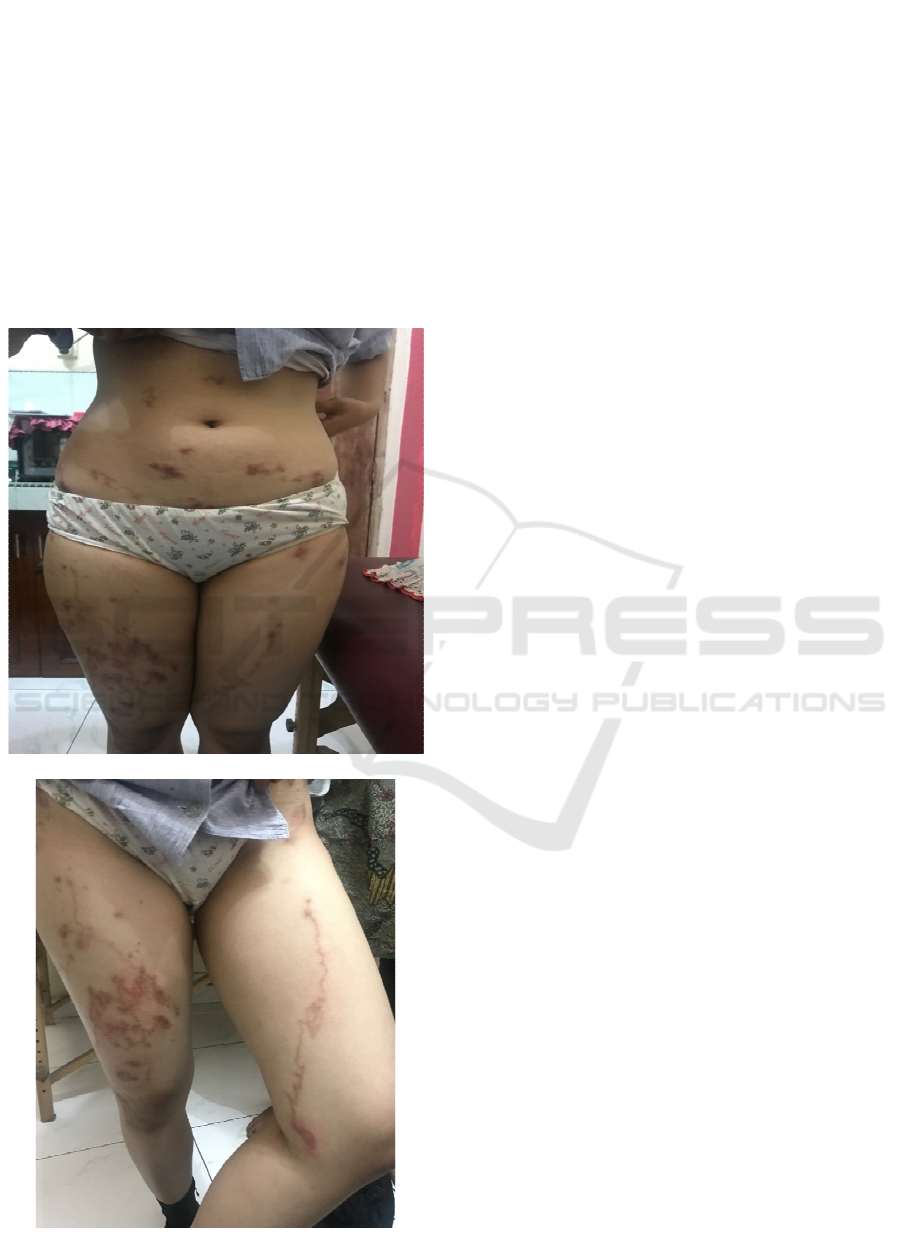
night. then a winding line appears in the thigh
section,
The patient has experienced this disorder for the
first time, there is no history of drug use, and a
history of food allergies.
Physical examination found the patient looked
good with good nutrition. Its generalization status is
within normal limits. Dermatological status in the
abdominal region, inferior limb region.In this patient
Albendazole 2 x 400 mg was given for 3 days,
Cetirizine 10 mg (if itchy), Ethyl Chloride (spray)
was given 1 x 1. 2 weeks later the rash in the patient
experienced healing.
Figure: Creeping Eruption
3 DISCUSSION
Cutaneous larvae migrants (CLM) are typical skin
disorders in the form of linear or winding,
embossed, and progressive inflammation, caused by
invasion of hookworm larvae originating from dogs
and cats, namely Ancylostomabraziliense and
Ancylostomacaninum. This skin disorder is also
known as creeping eruption, creeping verminous
dermatitis, stray larvae, migraine dermatosis,
sandworm disease, plumber 's itch, duck hunter' s
itch. (Siregar, 2013) CLM has a wide distribution,
mostly in warm and humid tropical and subtropical
climates. Hygiene and sanitation factors play an
important role. (Siregar, 2013;Padmavaty et al,2015)
Humans are incidental hosts, infections occur due to
direct contact between the skin and sand or soil
contaminated with animal waste containing
filariform larvae (infective larvae) hookworms.
(Baple et al, 2015; Supplee et al, 2013).
Larval entry into the skin is usually accompanied
by itching and heat. Itching is usually more severe at
night. At first papules will appear, then followed by
a distinctive form, namely linear or winding lesions,
arising with a diameter of 3 mm, reddish.
Erythematous papule lesions suggest that larvae
have been on the skin for several hours or days.
Furthermore, this red papule spreads like a winding
thread, polycyclic, serpiginose, arises, and forms a
tunnel (burrow), reaching a length of several
millimeters to centimeters per day. Predilections on
the back of the hands, limbs, plantar, soles of the
feet, anus, buttocks and thighs, can also be found in
parts of the body that are often in direct contact with
sand or soil where larvae are located (Eckert, 2005).
A study in Brazil reported that the lesion length was
significantly related to duration or the duration of
infection, the average length of 2.7 mm per day, so it
can help estimate the time and place of exposure to
infection.
Another clinical manifestation is hookworm
folliculitis. Patients usually present with pruritic
folliculitis and creeping eruption. Folliculitis can be
in the form of 20-100 follicular papules and pustules
spread in certain areas, usually in the buttocks. It can
also be found 2-10 lesions in the form of linear
(burrow) or serpiginose tunnels of 1-5 centimeters in
the same or different locations. (Baple et al, 2015)
In
these patients the therapy given is in accordance
with the theory that first-line therapy is albendazole
(400-800 mg / day) single dose orally for three days
or anti-helminticivermectin (150-200 µg / kg body
weight) single dose. (Eckert, 2005) The cure rate
ICTROMI 2019 - The 2nd International Conference on Tropical Medicine and Infectious Disease
400

reaches 100 percent. However, because other safe
and sufficiently effective options are topical
tiabendazole and topical albendazole this drug is not
available in all countries, then other therapies with
ethyl chloride are given even though it is not
recommended. (Aisah, 2010)
4 CONCLUSION
Cutaneous larval migrans (CLM) is a skin disorder
caused by animal hookworm larvae originating from
dogs and cats, namely Ancylostomabraziliense and
Ancylostomacaninum. One case of CLM has been
reported based on clinical findings in the form of
very itchy lesions on the surface of the abdomen and
upper thighs with tunnel-shaped images of
serpiginous papules similar to the skin. Patients were
given single dose of albendazole 400 mg for 3 days
and oral antihistamines. On the fifth day after
therapy, the lesion undergoes spontaneous resolution
and symptoms disappear.
REFERENCES
Aisah S.. 2010.Ilmu penyakit kulit dan kelamin. Edisi ke-
6. Jakarta: Balai Penerbit FKUI
Baple K, Clayton J. 2015. Hookworm-related cutaneous
larva migrans acquired in the UK: Case report. BMJ
Case Rep.
Eckert J. 2005. Larva migrainsexterna or cutaneous larva
migrains, dalam :BienzKA,editor. Medical
microbiology. New york: Thiame Medical Publisher.
Falabela R, Barona MI. 2008. Update on skin
repigmentationtherapies in vitiligo. Pigment Cell
Melanoma Res. 22: 42–65.
Padmavathy L, Rao LL. 2015. Cutaneous larva migrans: A
case report. Indian J Med Microbiol. 23(2):135-6.
Radtke MA, Schafer I, Gajur A, Langenburch A, Augustin
A. 2009. Willingness to pay and quality of lifein
patients with vitiligo. Br J Dermatol. 161:134–9.
Siregar,R,S. 2013. Saripati Penyakit Kulit. Edisi 3.
Jakarta: Penerbit Buku Kedokteran EGC.
Supplee SJ, Gupta S, Alweis R. Creeping eruptions:
Cutaneous larva migrans. J Comm Hospital Intern
Med Perspectives. 2013;3:21833
Cutaneous Larva Migrans in a Girl
401
