
Perianal Giant Condyloma Acuminata in Men Who Have
Sex with Men with HIV
Eunice Gunawan
1*
, Syafria Zidni
1
, Retno Indar Widayati
1
, Puguh Riyanto
1
, Ika Pawitra Miranti
2
1,2,3,4
Departement of Dermatovenereology, Medical Faculty of Diponegoro University/ Dr. Kariadi Hospital, Semarang
5
Departement of Pathological Anatomy, Medical Faculty of Diponegoro University/ Dr. Kariadi Hospital, Semarang
*
Corresponding auhtor
Keywords: Giant Condyloma Acuminata, HIV, HPV, MSM
Abstract: Giant condyloma acuminata (GCA) is a large condylomacaused by the proliferation of squamous epithelial
cells in the presence of Human Papillomavirus (HPV) infection, mostly type 6 and 11. It is most commonly
seen in the genital, anal, and perianal regions. Sexual behavior of MSM further increases the risk of HIV
infection and other viral infections such as GCA. An unmarried Javanese 38-year-old man complained of a
wart on his perianal area since one year ago. Initially, the lesion was small and progressively enlarged tothe
size of a chicken’s egg. The patient was an MSM with multiple sexual partners. The patient was HIV-
positive and received antiretroviral therapy (ARV). The clinical finding showed a large cauliflower-like
growth tumor on the perianal area, 7x5 x 2centimeters in size, with a positive ace-to-whitening test.
Histopathological examination showedpapillomatosis, hyperkeratosis, parakeratosis, and koilocytosis, in
accordance with GCA diagnosis. The VCT result showed a CD4 of 51cell/ml. Therapy for this patient was
electrodesiccation and curettage. The diagnosis of GCA was established on the history, clinical findings,
and histopathological examination.Electrodesiccation and curettage and ARV therapy in perianal GCA
patient showed a satisfactory result. Regular examination after therapy is required to identify and prevent
recurrence or metastasize potential.
1 INTRODUCTION
Giant condyloma acuminata (GCA) is a large
condyloma caused by the proliferation of squamous
epithelial cells in the presence of Human
Papillomavirus (HPV) infection, mostly type 6 and
11. It is most commonly seen in the genital, anal,
and perianal regions.GCA-also is known as
Buschke-Lowenstein Tumor (BLT)-is classified as a
sexually transmitted infection due to the factthat
more than 90% of cases are transmitted through
sexual contact.GCA is a semi-malignant
verrucoustumor characterized by aggressive growth
into the underlying dermal structure. It is recurrent
in 30-70% of cases and can progress slowly into
exophytic, ulcerative, and cauliflower-like tumors
that can form abscesses and fistulas (Suarez et al,
2016; Rahmayunita et al, 2017).
Due to a variety of sexual behavior nowadays,
i.e., men who have sex with men (MSM) can further
increase the risk of HIV infection and other viral
infections such as GCA. The prevalence of HPV on
MSM with HIV increased sixty-fold compared to
men in the general population (Indriatmi et al,
2016).
The treatment for GCA is based on size, location,
and a number of lesions. The therapeutic options
include topical ointments, cryotherapy, laser
vaporization, electrosurgery, and surgical excision
(Suarez et al, 2016; Murtiastutik et al, 2008;
Mistrangelo et al, 2018).
The aim of this case report is to report a case of
perianal giant condyloma acuminata, which is one of
the sexually transmitted infections, on an HIV
patient with MSM sexual behavior treated with
electrodesiccation and curettage.
2 CASE
Anunmarried Javanese 38-year-old man came to
thedermatology and venereology clinic of Dr.
Kariadi Hospital Semarang. He complained of a
wart on the perianal area since one year ago at first
362
Gunawan, E., Zidni, S., Widayati, R., Riyanto, P. and Miranti, I.
Perianal Giant Condyloma Acuminata in Men Who Have Sex with Men with HIV.
DOI: 10.5220/0009989003620365
In Proceedings of the 2nd International Conference on Tropical Medicine and Infectious Disease (ICTROMI 2019), pages 362-365
ISBN: 978-989-758-469-5
Copyright
c
2020 by SCITEPRESS – Science and Technology Publications, Lda. All rights reserved

small and then grew to the size of a chicken’s egg.
Sometimes itchy but not painful and not quickly
bled. HIV screening result from two years ago was
positive. The patient takes ARV regularly,
consistingof Efavirenz 600mg, Lamivudine 300 mg,
and Tenofovir disoproxil 300 mg. The patient
hadnever complained of any wart before. He had a
history of sex with multiple men. The patient has
never used a condom during intercourse. The history
of injury on the genital was denied, the patient has
never received a blood transfusion, nor has he had a
history of injection drug use. None of his family
experienced the same complaint. The patientwas a
store employee. Health costwas covered by BPJS.
The social, economic status was below average.
From the physical examination, we found the
patient to be compos mentis, with a body height of
165 centimeters, and the bodyweight of 58
kilograms. The blood pressure was 110/70 mmHg,
pulse rate 88 times/minute, respiratory rate 20
times/minute, and the axillary temperature was 36.8 ̊
C. Dermatologic status founda large cauliflower-like
growth tumor covering all perianal area, 7x5 x 2
centimeters in size, with positiveacetowhitening test.
Laboratory examination found reactive VCT
examination with CD4 of 51 cells/ml, a serologic
examination of TPHA and VDRL werenegative, and
other blood tests were within average values. The
rectal examination and the colonoscopy showed the
anal canal mucosa was healthy and free of
tumor.The histopathological examination
showedhyperplastic stratified keratinized squamous
epithelium with papillomatosis, parakeratosis, and
koilocytosis. Dermis consisted of hyperemic fibrous
connective tissue along with scattered lymphocytes,
histiocytes, PMN leucocytes; there was no sign of
malignancy. And all of these histopathological
findings were in accordance with GCA diagnosis.
The diagnosis we established was giant perianal
condylomaacuminataon HIV patient.
Electrodesiccation and curettage therapy in the
lithotomy position and general anesthesia was
performed to the patient. Post-operative therapy was
Clindamycin 300 mg twice a day for seven days,
Mefenamic acid 500 mg three times a day, wound
care, and Fusidic acid cream 2% twice daily on post
electrodesiccation and curettage wound.The patient
also took anti-retroviral (ARV) medicine.
On the first week post-surgery, the wound healed
well. And on the fourth-month post-surgery, there
was no sign of recurrence.
3 DISCUSSION
The diagnosis of Giant condylomaacuminata(GCA)
was established fromanamnesis, physical
examination, and histopathological examination. An
unmarried Javanese 38-year-old man came to the
dermatology and venereology clinic of Dr.
KariadiHospital Semarang. He complained of a wart
on the perianal area since one year ago at first small
and then grew to the size of chicken’s
egg.Sometimes itchy but not painful and not quickly
bled.He had a history of sex with multiple men. The
literature mention that GCA is a verrucoustumor
characterized by aggressive growth and mostly seen
in the genital, anal, and perianal regions. GCA
caused by HPV infection, in which the risk factors
for HPV infection are related to sexual behavior, like
homosexuality, multiple sexual partners, poor
genital hygiene, and chronic genital infection.The
incidenceof HPV infection is common in active
sexual individuals. The prevalence of HPV infection
rises over the age of 25 years to 40 years and over.
(Suarez et al, 2016; Rahmayunita et al, 2017).
The patient’s HIV screening resultwas positive
from two years ago.The patient takes ARV regularly
consisting of Efavirenz 600mg, Lamivudine 300 mg,
and Tenofovirdisoproxil 300 mg. The literature
mention that HIV is one of immunocompromised
condition that also included in sexually transmitted
infection. In HIV patients, condylomaacuminatacan
be an opportunistic infection that often occurs in
stage 2,3, and 4 of HIV. The frequency of
condylomaacuminata in men and women is the
same, in which the sexual behavior of male who has
sex with male (MSM) increases the risk of HIV
infection and other viral infections such as HPV
infection. Infection is transmitted by sexual contact
with the initial lesion at the trauma site during
intercourse. Generally, CA does not cause any
complaints, but the CA on the perianal region
sometimes can cause irritation, pain, or bleeding.
(Indriatmi et al, 2016;Murtiastutik et al,2008)
From the physical examination, we founda
sizeable cauliflower-like growth tumor covering all
perianal area, 7x5 x 2 centimeters in size, with
positive acetowhitening test. According to the
literature, the clinical manifestations of GCA can
resemble cauliflower-like tumor, consisting of
papules or dermal and epidermal nodules on the
perineum, genitals, and anus.The literature mention
that GCA is not like a simple condyloma, GCA is a
large condyloma, although it is still controversial in
the determination of the size. Acetic acid is helpful
in visualizing lesions on the cervix and anus. This
Perianal Giant Condyloma Acuminata in Men Who Have Sex with Men with HIV
363
examination is necessary for unusual lesions or
subclinical lesions. (Indriatmi et al, 2016
; Atkinson
et al,2014;Murtiastutik et al,2008)
The histopathological examination showed
hyperplastic stratified keratinized squamous
epithelium with papillomatosis, parakeratosis, and
koilocytosis. Dermis consisted of hyperemic fibrous
connective tissue along with scattered lymphocytes,
histiocytes, PMN leucocytes; there was no sign of
malignancy. This finding is consistent with the
literature, where histopathological findings for
condylomaacuminata are characterized by
acanthosis and papillomatosis on Malpighi layer,
thickening, and elongation of rete ridges, with
parakeratosis on the cornified layer. On stratum
corneum, can be found mitotic cells, nucleus
koilocytosis, and mononuclear inflammatory cells
that infiltrated into the dermis, and no sign of
malignancy. The histopathology appearance of GCA
is similar to the simple condyloma and difficult to
differentiate, but it has to be distinguished from
squamous cell carcinoma. (Suarez et al, 2016;
Murtiastutik et al, 2008; Murtiastutik et al, 2008)
Management of this patientwas
electrodesiccation and curettage. Post-operative
therapy was Clindamycin 300 mg twice a day for
seven days, Mefenamic acid 500 mg three times a
day, wound care, and Fusidic acid cream 2% twice
daily on post electrodesiccation and curettage
wound. Based on the literature, there are several
options that are available to treat human
papillomavirus (HPV) related anogenital disease,
with the treatment of CA being the most widely
studied. Treatment of CA is generally not directed at
the treatment of HPV infection but rather at the
physical removal of lesions or stimulation of the
host immune response. Available therapeutic
modalities fall into two categories; The first one is
patient-applied, such as imiquimod, podofilox gel,
the newer polyphenon E ointment, and topical
cidofovir; The second is provider-administered,
including cryotherapy, surgical removal
(electrosurgery, curettage, excision, cryotherapy),
intralesional interferon (IFN), trichloroacetic acid
(TCA), and intralesionalcidofovir. While there are
many available treatment options for HPV-related
anogenital disease, comparative trials to evaluate the
efficacy of various treatment modalities are lacking,
and there is often a lack of consensus regarding best
practices among clinicians treating these diseases.
The treatment of GCA is determined by the size of
the lesion, the location, amount of the lesion, patient
preference, maintenance cost, comforts, side effects
and provider experience
(Rahmayunita et al, 2017;
Indriatmi et al, 2016;Gormley et al,2012).
The prognosis of this patient was quo ad vitam
and quo ad sanam dubia ad malam. Due to the
inability of the immune system against the entry of
pathogenic effectof HIV infection.Furthermore, any
STI that the patient suffers from can get worse,
recurrent or resistant, and the possibility of
developing another life-threatening infection is
higher.(Mudrikova et al,2008) Quo ad cosmeticam
wasdubiaadbonam. Due tothe complete removal of
all lesions and a low risk of scarring.
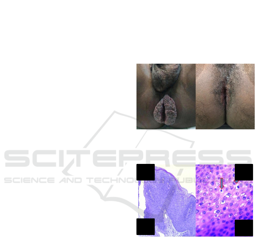
Figure 1. A.Pre-operative status of the disease. B.The
postoperative result after electrodesiccation and curettage
(four months post-treatment)
Figure 2. Histopathological Findings. A. Acanthosis,
Papillomatosis, Koilocytosis (100x).
B. Koilocytosis (400x) (H&E)
4 CONCLUSION
A case of perianal giant condyloma acuminata in an
MSM man with HIV.The management of this
patient was performed with electrodesiccation and
curettage. The prognosis of this patient was quo ad
vitam and quo ad sanam dubiaadmalam, and quo ad
cosmeticam dubia ad bonam.
A B
A
B
ICTROMI 2019 - The 2nd International Conference on Tropical Medicine and Infectious Disease
364

REFERENCES
Androphy EJ, Lowy DR. 2013. Human Papilloma Virus
Infection. In: Fitzpatrick TB, Johnson RA, Wolff K.
Color Atlas and Synopsis of Clinical
Dermatology.4
th
edition, United States: McGraw-Hill,
h 2878-90.
Atkinson AL, Pursell N, Sisay A. 2014. The giant
condyloma (Buschke-Lowenstein Tumor) in
immunocompromised patient. Case rep obstetgynecol,
pp.1-4
Gormley RH, Kovarik CI. 2012. Human – papillomavirus
related genital disease in the immunocompromised
host. Journal of the American Academy Dermatology.
Vol.66 (6):1-17.
Indriatmi W, Hanoko RP. 2016. Kondiloma Akuminatum.
In: Menaldi S, Bramono K, Indriatmi W, editor. Ilmu
penyakit kulit dan kelamin. 7
th
edition. Jakarta: FKUI.
p.481-3.
Mistrangelo M, Dal CI, Volpatto S, et al. 2018. Current
treatments for anal conylomata acuminate. Minerva
chir. 73(1):100-6.
Mudrikova T , Jaspers C, Ellerbroek P, Hoepelman A.
2008. HPV-related anogenital disease and HIV
infection: not always ‘ordinary’condylomata
acuminata. The Netherlands Journal of Medicine.
March, 66-3
Murtiastutik D, Barakbah J, Lumintang H, et al. 2008.
Kondiloma Akuminata. In: Infeksi Menular Seksual.
1
st
edition. Surabaya: Airlangga University Press. P.
170-9
Murtiastutik D, Barakbah J, Lumintang H, Martodihardjo
S. 2008. Kondiloma Akuminata. In: Infeksi Menular
Seksual. Surabaya: Airlangga University Press. p.165-
69.
Rahmayunita G, Wibawa LP, Suprapto N. 2017.
Pendekatan Diagnostik dan Penerapan Dermatoterapi
Berbasis Bukti. Jakarta : Indonesian University. p.210-
7.
Suarez-Ibarrola R, Heinze A, Sanchez-Sagastegu F, et al.
2016. Giant condylomaacuminatum in the genital,
perineal, and perianal region. 7:14-16
.
Perianal Giant Condyloma Acuminata in Men Who Have Sex with Men with HIV
365
