
Bullous Urticaria Pigmentosa in an Infant:
A Rare Form of Bullous Disorder
Lidwina Anissa
1*
, Sarah Mahri
1
, Rinadewi Astriningrum
1
, Triana Agustin
1
,
Rahadi Rihatmadja
1
, Githa Rahmayunita
1
1
Department of Dermatology and Venereology Faculty of Medicine Universitas Indonesia/
Dr. Cipto Mangunkusumo National General Hospital, Indonesia
*
Corresponding author
Keywords: Bullous Urticaria Pigmentosa
Abstract: Mastocytosis is a rare, sporadic, and a heterogeneous group of hematopoietic disorder, characterized by an
enormous number and accumulation of mast cells in one or more organ systems. The prevalence of
mastocytosis is challenging to determine due to underdiagnosis. Pediatric-onset mastocytosis which was
commonly diagnosed before two years of age is generally a benign disease. The course of pediatric-onset
mastocytosis is variable, from birth to the first year of life, with an average of 2.5 months. Cutaneous
mastocytosis may manifest as urticaria pigmentosa, diffuse cutaneous mastocytosis, and telangiectasia
macularis eruptive perstans. Bullous urticaria pigmentosa is a rare variant of urticaria pigmentosa. Blistering
is considered to be an effect of free mediator activity. The symptoms are mostly in proportion to the mast cell
degranulating activities in tissues, which may appear in the first year of life. Although systemic involvement
is rare in pediatric cutaneous mastocytosis, blistering may promote secondary infection and electrolyte
imbalance. We report a four-month-old infant with bullous urticaria pigmentosa. The symptoms had appeared
since the second day of life. Routine hematology examination revealed mild microcytic hypochromic anemia.
Skin biopsy from the lesional skin revealed diffuse dermal infiltration of mast cells, some showing granules
and scanty cytoplasm which supported the diagnosis of urticaria pigmentosa. The patient was managed with
antihistamines. In two-month-period of follow up, the development of new lesions is slowing.
1 INTRODUCTION
A mastocytosis is a heterogeneous group of myeloid
neoplasms with abnormal proliferation and
accumulation of mast cell in one or more organ
systems. (Asati DP et al., 2014)
During 2015-2019,
there are 12 new cases of cutaneous mastocytosis in
Pediatric Dermatology Division, Department of
Dermatology and Venereology Faculty of Medicine
Universitas Indonesia/ Dr. Cipto Mangunkusumo
National General Hospital. (Data Kunjungan
Poiklinik Dermatologi Pediatri Departemen Ilmu
kesehatan Kulit dan Kelamin FKUI/RSCM, 2015-
2019). (Asati DP et al., 2014;Barros et al., 2014)
reported two cases of congenital urticaria pigmentosa
in twin babies. (Barros et al., 2014) Year 2005
Working Conference on Mastocytosis’ classified
mastocytosis into three major categories: cutaneous
mastocytosis, systemic mastocytosis and
extracutaneous mast cell proliferation.
(Van Gysel D et
al., 2011) The cutaneous form consists of urticaria
pigmentosa, mastocytoma, diffuse cutaneous
mastocytosis, and telangiectasia macularis eruptive
perstans in their order of their frequency.
(Asati DP et
al., 2014
)
The diagnosis of cutaneous mastocytosis
(CM) is based on clinical and histological findings in
the skin, together with the absence of criteria that
would allow the diagnosis of systemic mastocytosis.
In cutaneous mastocytosis, the visible cutaneous
abnormalities are most often of major concern to the
patient and their family.
(Van Gysel D et al., 2011)
Urticaria pigmentosa is by far the most common
variant (70-90%) of childhood mastocytosis. The
lesions commonly appear in the first year of life and
maybe present at birth. The eruption consists of
slightly elevated, skin-colored, brown-red or yellow
macules, plaques or nodules.
(Van Gysel D et al., 2011)
Urticaria pigmentosa presenting with a vesicular and
bullous lesion as the predominant feature is a rare
entity. (Chintagunta SR et al.,2017)
.
Anissa, L., Mahri, S., Astriningrum, R., Agustin, T., Rihatmadja, R. and Rahmayunita, G.
Bullous Urticaria Pigmentosa in an Infant: A Rare Form of Bullous Disorder.
DOI: 10.5220/0009986302670271
In Proceedings of the 2nd International Conference on Tropical Medicine and Infectious Disease (ICTROMI 2019), pages 267-271
ISBN: 978-989-758-469-5
Copyright
c
2020 by SCITEPRESS – Science and Technology Publications, Lda. All rights reserved
267

The lesions occur in a generalized distribution but
tend to be of highest density on the trunk. (Van Gysel
D et al., 2011).
Less affected are palms, soles, scalp,
and face, as well as sun-exposed body areas.
(Chintagunta SR et al.,2017)
.
In the first two years, Urtica and erythema may
occur spontaneously or after stroking a lesion
(Darier’s sign), although Darier’s sign is not always
present in all patients.
(Van Gysel D et al., 2011;Castells
M et al., 2011)
All pediatric cutaneous forms of mastocytosis can
rarely present with acute mast cell degranulating
events, such as anaphylaxis, whole body flushing,
dyspnea, wheezing, vomiting, diarrhea and
sometimes cyanotic spells (Castells M et al.,
2011;Tharp MD et al., 2012)
The diagnosis of cutaneous mastocytosis requires a
history of new-onset skin lesions with or without
systemic symptoms. A physical examination with a
positive Darier's sign, supported with increasing
serum tryptase and dermal infiltration of mast cell on
skin biopsy is essential for building the diagnosis.
Analysis of c-kit mutations is recommended. In
addition to the skin biopsy, bone marrow studies are
recommended if the tryptase is significantly elevated,
severe systemic symptoms are present, if there is
associated organomegaly or if there is no significant
response to initial symptomatic therapy. Parents
should be explained carefully about the possibility of
evolution to a systemic form in a small number of
cases.(Castells M et al., 2011;Chintagunta SR et
al.,2017)
.
We report a case of bullous urticaria pigmentosa,
which is a rare clinical manifestation and also an
extreme form of cutaneous mastocytosis to raise
awareness about the differential diagnosis of vesicle
and bullae in the infancy period.
2 CASE
A four-month-old boy was referred to our hospital
presented with multiple patches and blisters, which
began on the second day after birth. The lesions were
first observed on the trunk and spread to the scalp and
limbs within several days. The lesions started as
erythematous patches all over his body, and several
evolved to the vesicle. The vesicles were easily
ruptured and became scars. He was the second child,
and his brother was healthy. His birth and
development were normal. He was breastfed and
supplemented with formula. There was no history of
drug ingestion both in mother and patient — no
current episode of facial flushing, dyspnea, and
diarrhea.
On physical examination, there were
erythematous and hyperpigmentation plaques,
vesicles, and erosions covered by crust (Figure 1). No
lymph node enlargement found. Darier’s sign was
negative. At the first visit, gram staining from the
erosions revealed moderate numbers of leukocytes
and Gram-positive coccus.
Laboratory examination revealed mild microcytic
hypochromic anemia (hemoglobin 11.9 g/dL, MCV
91.1 fL, and MCH 32 pg). Histopathology
examination revealed rete ridges elongation of the
epidermis and dermal perivascular and interstitial
infiltration of lymphocytes, histiocytes, neutrophils,
and mast cell, which came to a conclusion as
cutaneous mastocytosis (Figure 2).
The patient was given topical antibiotics for a
short period of time due to secondary infection on
several lesions and oral cetirizine to reduce the
symptoms. The patient was also referred to the
Pediatric Department to find systemic involvement of
mastocytosis. Bone marrow studies were not done
due to parents’ disapproval. In one month follow up
period, the patient showed a slower progression of
new lesions. No systemic symptoms reported.
However, education and counseling about the
possibility of systemic mastocytosis include the
symptoms are done to prevent life-threatening
systemic involvement.
ICTROMI 2019 - The 2nd International Conference on Tropical Medicine and Infectious Disease
268
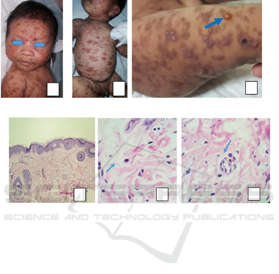
Figure 1. Clinical manifestation in patient. A. Lesions on face on the first visit, B. Lesion on the trunk on the first visit, C.
Bullae on the right thigh (blue arrow).
Figure 2. Histopathology findings. A. Epidermal rete ridges elongation (H&E,10x),
B. Interstitial mast cell infiltration (blue arrow, H&E, 100x), C. Perivascular mast cell infiltration (blue arrow, H&E,
100x)
3 DISCUSSION
There are many conditions which manifest as a blister
in the neonatal period. They can be caused by
infectious, traumatic, or inherited causes. Infectious
causes include bullous impetigo, staphylococcal
scalded skin syndrome, neonatal varicella,
candidiasis, etc.; meanwhile, non-infectious causes
include epidermolysis bullosa, epidermolytic
ichthyosis, incontinentia pigmenti, cutaneous
mastocytosis, Langerhans cell histiocytosis or
chronic bullous disease of the childhood.(Park MN et
al., 2014).
The diagnosis was established by excluding other
differential diagnoses. Since there was no sign of
infection, infectious etiologies were excluded. The
discrete distribution of lesion which was located on
non-traumatic region excluded epidermolysis
bullosa. Epidermolysis ichthyosis and incontinentia
pigmenti were excluded due to the difference in the
nature of the disease. Lastly, we excluded other types
of cutaneous mastocytosis. The clinical entity which
mimics initial clinical presentation, in this case, is
diffuse cutaneous mastocytosis. Diffuse cutaneous
mastocytosis (DCM) can be manifested initially as
two types: one with a minimal blistering and large
area of nodular and leathery skin and one with
extensive blistering and/or exfoliation, usually
accompanied by erythrodermic appearance. (Tharp
MD et al., 2012). Both features are not fulfilled in this
case; therefore, DCM was excluded. Telangiectasia
macularis eruptive perstants (TMEP) is the least
common form of cutaneous mastocytosis and rarely
manifests in childhood. The typical lesions are
telangiectatic macules in a tan or brown background
and may co-exist with urticaria pigmentosa. (Costa
DLM et al., 2011). In this patient, there was no
telangiectasia as well as telangiectatic macules, so
TMEP was also excluded.
The pathogenesis of pediatric cutaneous
mastocytosis is not well understood, and most
children do not present with mutations of c-kit in bone
A
C
A
B
C
B
Bullous Urticaria Pigmentosa in an Infant: A Rare Form of Bullous Disorder
269

marrow mast cells. (Castells M et al., 2011) An earlier
study by (Verzijl et al., 2007) found that a quarter of
pediatric patients presenting urticaria pigmentosa had
a D816V codon mutation. Unfortunately, genetic
testing has not been available yet in Indonesia.
Blistering of urticaria pigmentosa may happen
and is considered to be exaggerated of Darier's sign.
This is caused by the release of a mediator (mainly
chymase) upon mast cell degranulation, which binds
and cleaves the dermo-epidermal junction (DEJ). The
DEJ is slowly stabilizing over the first two years of
life, resulting in a reduction of vesiculobullous lesion
by the age of 3. (Briley LD et al., 2008)
In this case, the histopathology examination
showed diffuse dermal infiltration of mast cells which
verified the clinical diagnosis of urticaria pigmentosa.
Due to the presence of vesicles and bullae, the patient
was finally diagnosed as bullous urticaria
pigmentosa.
We referred the patient to the Child Health
Department to rule out systemic involvement.
Physical examination revealed no mucosal lesion and
no hepatosplenomegaly. Bone marrow
cytomorphology study was planned to confirm if the
mast cell count exceeded 20% of the nucleated cells
in the bone marrow. In this case, bone marrow
aspiration could not be performed due to parents’
disapproval. (Kettelhut et al., 1989) reported that the
initial evaluation of the bone marrow of 17 children
where 15 had urticaria pigmentosa and two had
diffuse cutaneous mastocytosis revealed no adult-
type mast aggregates. This finding is indicating that
in most cases, cutaneous mastocytosis in children
does not involve internal organs which precludes the
need for routine bone marrow aspiration.
(
Uzzaman
also con et al.,) cluded that only the persistent disease
might justify repeated bone marrow examination
aggressive systemic therapy. The clear majority of
cases could be managed satisfactorily by
symptomatic treatment. (Uzzaman A et al., 2000)
Management includes alleviation of the
symptoms and avoidance of potential mast cell
degranulating stimuli such as several drugs, food,
local or systemic anesthetics, heat, as well as
friction.(Asati DP et al., 2014).Patient was prescribed
oral cetirizine daily. Parents have been adhering to
periodic follow-up evaluation for the past two
months, and up to now our patient responded well to
oral antihistamines and had gradual reduction in new
lesions (both blisters and papules) development.
Although pediatric cutaneous mastocytosis is
generally benign and rarely involve other organs,
parents were counseled in detail about the possibility
of systemic involvement, the sign and the symptoms
of systemic involvement. Whole-body flushing,
shortness of breath, diarrhea may happen due to mast
cell mediator release.
Systemic mastocytosis in children is extremely
rare, and usually, the clinical symptoms could be
managed by medication. The majority of these lesions
and the severity of the symptom will resolve over
time. However, it is essential to do regular follow up
to detect systemic involvement.(Asati DP et al.,
2014;Briley LD et al., 2008)
4 CONCLUSION
It could be concluded that dermatologist should
remain aware of varied forms of pediatric cutaneous
mastocytosis because of its rarity. Diagnosis of
bullous urticaria pigmentosa should be thought in the
infant with lesions suspected as urticaria pigmentosa
accompanied by vesicle or bullae. Skin biopsy
became mandatory to build the diagnosis. Systemic
involvement screening, appropriate treatment, and
follow up are required as routine. Finally, education
and counseling also play an important role in the
management of this entity.
REFERENCES
Asati DP, Tiwari A. 2014. Bullous mastocytosis in a 3-
month-old infant. Indian Dermatol Online J.;5:497-500.
Data Kunjungan Poliklinik Dermatologi Pediatri
Departemen Ilmu Kesehatan Kulit dan Kelamin
FKUI/RSCM Tahun 2015- 2019.
Barros TD, Boediardja SA, Agustin T, Sirait SP,
Rihatmadja R, Rahmayunita G, et.al. 2014.Urtikaria
pigmentosa kongenital pada bayi kembar. Poster
presented at Kongres Nasional XIV Perdoski;;
Bandung.
Briley LD, Phillips CM. 2008. Cutaneous mastocytosis: a
review focusing on the pediatric population. Clin
Pediatr.;47:757-61.
Castells M, Metcalfe DD, Escribano L. 2011. Guidelines
for the diagnosis and treatment of cutaneous
mastocytosis in children. Am J Clin Dermatol.;12:259-
70.
Castells M, Metcalfe DD, Escribano L. 2011. Diagnosis and
treatment of cutaneous mastocytosis in children. Am J
Clin Dermatol.;12:259-70.
Chintagunta SR, Srinivas M, SriShilpa P, Nagula SK,
Velgam R. 2017. Bullous urticaria pigmentosa – A rare
case report. J NTR Univ Health Sci;6:69-71.
Costa DLM, Moura HH, Rodrigues R, Pineiro-Maceira J,
Ramos-E-Silva M. 2011. Telangiectasia macularis
eruptive perstans: a rare form of adult mastocytosis. J
Clin Aesthet Dermatol.;4:52-4.
ICTROMI 2019 - The 2nd International Conference on Tropical Medicine and Infectious Disease
270

Kettelhut BV, Parker RI, Travis WD, Metcalfe DD. 1989..
Hematopathology of the bone marrow in pediatric
cutaneous mastocytosis. A study of 17 patients. Am J
Clin Pathol;91:558-62.
Park MN, Kim GA, Chey MJ, Shim GH. 2014.A case of
diffuse cutaneous mastocytosis in a newborn. Korean J
Perinatol.;25:105-9.
Tharp MD. Mastocytosis. In: Goldsmith LA, Katz SI,
Gilchrest BA, Paller A, Leffell D, Wolff K, eds. 2012..
Fitzpatrick’s Dermatology in General Medicine. 8
th
ed.
New York: McGraw Hill;. p.1809-18.
Uzzaman A, Maric I, Noel P, Kettelhut BV, Metcalfe DD,
Carter MC. 2009. Pediatric-onset mastocytosis: A long
term clinical follow-up and correlation with bone
marrow histopathology. Pediatr Blood Cancer.;53:629–
34.
Van Gysel D, van Schaik RHN, Oranje AP. Mastocytosis.
In: Irvine A, Hoeger P, Yan. 2011. Harper’s Textbook
of Pediatric Dermatology. Edisi ke-3. Oxford: Wiley-
Blackwell..p.75.1- 75.13
Verzijl A, Heide R, Oranje AP, van Schaik RH. 2007.C-kit
asp-816-val mutation analysis in patients with
mastocytosis. Dermatology; 214–20.
Bullous Urticaria Pigmentosa in an Infant: A Rare Form of Bullous Disorder
271
