
The Effectiveness of Clerodendrum Paniculatum. L against TNF- α in
Rats Induced by S. Aureus
Mutiara Dwi Yanti
1
, Prihantono
2
, Dewi Tiansa Barus
1
, Putri Ayu Yessy Ariescha
1
, Kholilah Lubis
3
,
Nurul Aini Siagian
1
, Andayani Boang Manalu
1
, Tetty Junita Purba
1
1
Midwifery Faculty, Institute of Health Science DELI HUSADA, Jl. Besar Delitia No.77, Deli Serdang, Indonesia
2
Department of Surgery, Faculty of Medicine, Hasanuddin University, Makassar City, South Sulawesi, Indonesia
3
Midwifery Faculty ,STIKES PRIMA NUSANTARA Bukit Tinggi
Keywords: Effectiveness, Bag Flower Leaf, TNF-α
Abstract: Staphylococus Aureus is one of the mastitis causes which causes inflammation due to elevated levels of
TNF-α. This study aimed to analyze the anti-inflammatory activity of bag flower leaf (Clerodendrum
Paniculatum L) on TNF-α level in rats that is induced by S. Aureus bacteria. This study used post test only
control group design with 15 samples of Strain Sprague Dawley rats divided into 3 groups. K (-) group as
normal control group, K (+) was induced by S. Aureus bacteria and the treatment group was administered by
extract 150mg / kg BB then it is measured TNF-α levels by the enzyme-linked immunosorbent assay
(ELISA) test method. Data were analyzed by using bivariate analysis namely one way ANOVA test. The
results showed that Bag Flower Extract contains secondary metabolites in the form of flavonoids and
tannins. Giving bag flower leaf extract (Clerodendrum Paniculatun L) at 150 mg / kg BB has anti-
inflammatory action by reducing TNF-α levels in rate which is induced by staphylococcus aureus (P <0, 0).
05). It was concluded that the administration of bag flower leaf extract could reduce TNF-α cytokines due to
S. Aureus bacteria. It is expected that future researchers will use human subjects to analyze TNF-α levels so
that later bag flower leaves can be used as complementary therapies in mastitis treatment due to S. aureus
bacteria.
1 INTRODUCTION
Staphylococcus aureus is one of the main
pathogens that are most often isolated from
intramammary infections (IMI) throughout the
world. S. aureus bacterial infections are becoming a
serious problem today due to increased bacterial
resistance to various types of antibiotics (Multi Drug
Resistance / MDR). S. aureus resistant can cause the
spread of other infections and cause other diseases
associated with other infections. Although S. aureus
can cause acute and clinical mastitis with
macroscopic changes in milk, bacterial infections
can develop against chronic and subclinical mastitis,
without macroscopic changes in milk but with high
somatic cell and bacterial counts in the mammary
glands (Green et al., 2012; Chinchali and Kaliwal,
2014)
The occurrence of mastitis begins with an
increase of pressure in the duct (breast milk channel)
due to breast milk stasis. If the milk is not removed
immediately, there is excessive alveoli tension and it
causes the epithelial cells that produce milk become
flat and depressed, so that the connective tissue
permeability will be increase. Some components
(mainly immune proteins and sodium) from plasma
enter to breast milk and subsequently into the tissues
around cells so that it triggers an immune
response(Acosta et al., 2016),
Breast milk stasis, there is an inflammatory
response, and tissue damage facilitate infection.
Breast infections are usually caused by bacteria that
are found in normal skin, namely S. Aureus. Other
bacteria that cause mastitis are Streptococcus beta-
hemolitik (such as Group A or Group B
streptococcus) or Escherichia coli. These bacteria
often originate from the baby's mouth which enters
through the lactiferous duct into secretion lobe,
through cracked nipples to the lymph glands around
the duct (periductal) or through spread of
hematogenous (blood vessel) (Schwartz et al., 2002;
596
Yanti, M., Prihantono, ., Barus, D., Ariescha, P., Lubis, K., Siagian, N., Manalu, A. and Purba, T.
The Effectiveness of Clerodendrum Paniculatum. L against TNF- in Rats Induced by S. Aureus.
DOI: 10.5220/0009979605960603
In Proceedings of the International Conference on Health Informatics and Medical Application Technology (ICHIMAT 2019), pages 596-603
ISBN: 978-989-758-460-2
Copyright
c
2020 by SCITEPRESS – Science and Technology Publications, Lda. All rights reserved

Amir et al., 2007; Indonesian Pediatric Society,
2013)
Tumor Necrosis Factor alpha (TNF α) is a
pro-inflammatory cytokine that is involved in an
inflammatory reaction and it plays a role in the pain
emergence. This was first isolated by Carswell et al
in 1975 toward an effort to identify the Tumor
Necrosis Factors that is responsible for nicrosis from
the Meth A. sarcoma. Most organs appear to be
affected by TNF α, and there are many cytokine
functions which are not fully understood (Sanchez,
Ford and Yancey, 2005; Notebaert et al., 2008; Lai
et al., 2017)
The various roles of TNF-α can be explained
through their effects on endothelial vascular and
endothelial leukocyte interactions. When it is
exposed to TNF-α, endothelial cells will cause
inflammatory reactions by expressing various
adhesion molecules. TNF can also cause
vasodilatation by inducing the expression of
cyclooxygenase 2 (COX-2) and its relationship to
the production of prostacyclin 2 (PGI2). It can
explain erythema and heat which are signs of
inflammation. Other signs of inflammation are
tumors that occur through increased vascular
permeability, vascular through TNF mediators and
liquid passage and transendothian macromolecules
which ultimately cause edema and inflammation
(Nair, M.; Mahajan, 2006; Berthold-Losleben and
Himmerich, 2008; Lai et al., 2017)
Immune response that occurs due to the
invasion of staphylococous aureus bacteria as
antigens is when the entry of s. aureus into the body
will be eliminated by neutrophils and macrophages
as their role in the innate immune system.
Macrophages can also act as Antigen Presenting
Cells (APC). Bacteria will be phagocytosis in
macrophages then recognized by Major
Histocompatibility Complex II (MHC II) then
presented in the form of peptide antigens. Then
MHC II will bind to T lymphocytes. T lymphocytes
have several surface molecules or Cluster of
Differentiation (CD). Peptide agents that have been
presented by MHC II will bind to the T helper
lymphocytes (CD4) in section of T Cell Receptor
(TCR). (Abbas, Licthman and Pillai, 2015; Tong et
al., 2015; Acosta et al., 2016)
Bag flower leaf is one of the plant species
included in the Clerodendrum genus which has a
different species number of 580 species, and it is
spread evenly in Asia, Africa, America and Australia
and it has been used in traditional medicine in Asia
and Africa. India, China, Korea, Thailand, and Japan
are countries that have used several species of this
genus in medical practice (Cucumber, Virus and
Indonesia, 2009; Florence, Joselin and Jeeva, 2012;
India Biodiversity Portal, 2017)
Research on the efficacy of bag flower leaf
which aims to find out the antioxidant and anti-
inflammatory activities of big flower leaf extract
(Cloredendrum Paniculatum L) that is conducted
Anti-inflammatory activity tests on animals, it
showed the antioxidant activity results of bag flower
leaf ethanol is very strong (IC
50
<50 µg/ml) namely
IC
50
= 27,73376 µg/ml and it has anti-inflammatory
activity at a dose of 50 mg / kg. Other research on
the anti-inflammatory activity test of Clerodendrum
Paniculatum L. which aims to evaluate the anti-
inflammatory activity of various extracts of
Clerodendrum paniculatum leaf shows the anti-
inflammatory activity in best vitro at a dose level of
200 and 400 mg / kg. Indomethacin at a dose level
of 10 mg / kg is used as a standard reference drug.
Both extracts showed a significant dose reduction (P
<0.001) in edema paw when it is compared to
controls). The study results indicated that petroleum
ether and chloroform extract from bag flower leaf
have anti-inflammatory potential which provides a
scientific basis for the traditional claims of
Clerodendrum Paniculatum Linn leaves as anti-
inflammatory drugs (Joseph, Bindhu and Aleykutty,
2013; Hafiz, Rosidah and Silalahi, 2016a).
This study is aimed to assess the level of
TNF-α which is one of the pro-inflammatory
cytokines that can trigger inflammation in mastitis
caused by S. Aureus bacteria.
2 MATERIALS AND METHODS
2.1 Location and Research Design
This research will be carried out in the
Laboratory of Hasanuddin University Hospital for
ELISA examination and bacterial culture,
Hasanuddin University Bio pharmacy Laboratory
and Hasanuddin University Animal Laboratory. The
research type used is the type of research used true
experimental with post only test control group
design.
2.2 Population and Sample
The population of this study were 15 Strain
Sprague Dawley (Rattus Novergicus) white rats
according to WHO standards and were randomly
selected to avoid bias in the study. The rats that used
in this study were rats weighing between 200-250
The Effectiveness of Clerodendrum Paniculatum. L against TNF- in Rats Induced by S. Aureus
597

grams. Before being given treatment, rats were first
adapted for 7
th
days in a cage with controlled
temperature and adequate lighting. Rats were fed
with pellets and water on an ad libitum basis. The
use of experimental animals in this study was
approved by the health research ethics commission,
medical faculty of Hasanuddin University, Makassar
(No. 1053 / H4.8.4.5.31 / PP36-KOMETIK / 2017).
2.3 Method of Data Collection
2.3.1 Sampling Criteria
Sampling criteria in this study namely
inclusion criteria: female rat Strain Sprague Dawly
weighing 200-250 grams with age 2-3 months, there
is no anatomical abnormalities. Exclusion criteria:
rats don’t want to eat, rats that are sick during the
adaptation process. The materials needed in this
study were rat maintenance: cages, food containers.
Rat treatment: scales, sterile syringes, feeding tube.
Sample preparation: centrifuge, vortex, shaker,
yellow and blue tip, micropipette for volume 2 µl -
1000 µl.
Female rat Strain Sprague Dawley each
group consisted of 5 rats. Furthermore, rats were
divided into 3 treatment groups, each consisting of
five rats that is consisting of two control groups and
one treatment group so that a total sample of 15 rats
were obtained. To avoid bias factors due to weight
variation, the grouping of samples is done randomly.
To avoid drop outs sample, the sample size which is
used was 6 per sample group so that the number of
rats used was 15 rats for 3 groups.
Blood sampling was taken twice after the
induction of S. aureus bacteria and after treatment in
each group. 1 ml of blood is drawn in centrifuge at a
speed of 5000 rpm for 5 minutes. Then do the
separation of blood with serum. The serum is
suctioned with a 1.0 µL dropper and placed in an
effendorf tube. The serum collected was carried out
by the ELISA method to obtain TNF-α level. Test
reagent for TNF-α examination: TNF-α ELISA Kit
RAT with catalogue number RTA00 .
2.3.2 Extraction Preparation
Bag flower leaves are obtained in the yard of
community houses as much as ± 2 kg of raw leaves,
then cleaned of dirt attached by using running water
then the sample is cut into small pieces, then dried to
contain water content below 10%, after that the bag
flower leaves are sieved with mesh size 40 so that a
smooth simplicia sample is obtained, after it, the
sample is ready to be extracted by maceration
method (Poorter et al., 2012; Hafiz, Rosidah and
Silalahi, 2016b).
Maceration is generally carried out by means
of 10 parts of simplicity put into a vessel, then
poured with 75 parts of the liquid solution, closed
and left for 5 days which is protected from light,
while repeatedly stirring after 5 days the juice is
dispensed, the pulp is squeezed. The residue is
added to solvent sufficiently and cleaned to obtain a
total of 100 parts. The resulting maserat is then
concentrated using a rotary evaporator until a thick
extract is obtained, and then dried by using a water
bath and desiccators (MOH RI, 2000).
2.3.3 Bacterial Culture
Staphylococcus aureus bacteria obtained
from laboratory of Uhhas Hospital with the type of
staphylococcus aureus bacteria then planted in
BHIB medium and it is incubated for 18-24 hours at
37° C in incubator and then propagated by using
Nutrient Agar (NA) medium which is then re-
incubated for 18-25 hours. After bacterial
incubation, it is done gram staining. Biochemical
tests for S. aureus bacteria were planted in NA by
planting on DNAse agar medium then mannitol salt
agar, then bacitracin and Novobiocin tests were
followed by callatase coagulase test. Then it is
reincubated for 18-24 hours at 37° C. The bacteria
that grew in biochemical test were matched with the
identification table of S. aureus bacteria. To make a
bacterial sample which is injected into rats by
making a suspension in physiological Na Cl solution
of 10 ml mixed with a colony of S. aureus bacterial
which is golden yellow with turbidity level of Mc
Farlan 2 x 10
8
CFU. The accuracy of Mc Farland
turbidity level is measured by the Densi check tool
(Das, Borah and Ahmed, 2013)
Figure 1 describe the research tools and
materials like : ELISA-Test for TNF-α, Sprague
Dawley Rats, Pagoda Leaves and Staphylococcus
Aureus Bacteria
After getting the concentration of bacterial
cells 10
8
cell / mL, then centrifuged at a speed of
10,000 rpm for 10 minutes at 25
0
C. The obtained
pellets were then suspended with 1 mL PBS. The
suspension was then injected into experimental
animals, namely in mammals of rat female Strain
Sprague Dawly, precisely in the lactiferous duct
section with a volume of 100 µL (0.2 ml).
ICHIMAT 2019 - International Conference on Health Informatics and Medical Application Technology
598
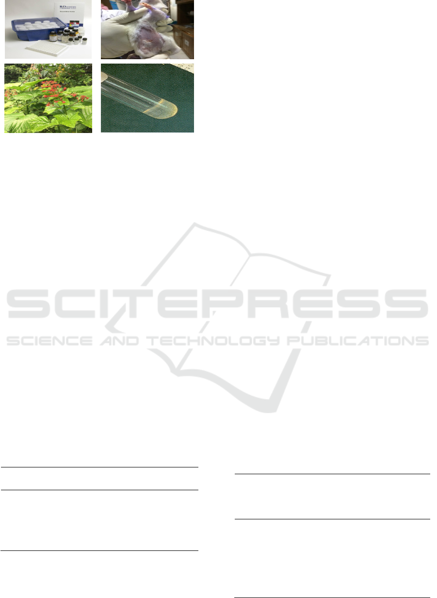
Figure 1. Research Tools and Materials
2.3.4 Data Analysis
Data analysis begins with test data
normality distribution. To compare TNF-α level in
the group after the induction of S. Aureus bacteria,
and after the intervention in the treatment group was
analyzed by using Paired T-Test. To compare
changes in TNF-α level between the treatment and
control group, statistical analysis used one way
Anova test. The significance limit used in this study
was 5% (p = 0.05).
3 RESULTS
Qualititative phytochemical test of bag flower
leaf extract (Clerodendrum Paniculatum L)
Table 1 shows the phytochemical test results of
secondary metabolite content in bag flower leaf
extract (Clerodendrum Paniculatum L), there was
secondary metabolite compounds include flavonoids
and tannins.
Table 1. Qualitatively analysis of phytochemical content
of sea leafy leaves (Scaevola taccada (Gaertn) Roxb.)
Extracts
Metabolit
Compunds
Analysis result
Alkaloid -
Flavonoid +
Steroid/triterpenoid -
Saponin -
Tanin +
From several studies that conducted
phytochemical testing of Clerodendrum
Paniculatum L plants, there were differences in
interpretation of the results of phytochemical
screening that researchers conducted on several
previous studies. Phytochemical screening results of
bag flower leaf ethanol extract in this study were
identified only 1 from 3 alkaloid solutions used
namely Mayer solution. The difference in
phytochemical screening results in this study can be
caused by several factors, namely the treatment of
the sample and the environmental conditions in
which the bag flower leaves grow (Pidugu and Arun,
2012; Andriani et al., 2017; Wang et al., 2018)
However, differences phytochemical
screening results conducted in this study with
previous studies did not affect the value of the study,
because the compound expected to exist in the
ethanol extract of bag flower leaves that had an
effect on anti-inflammatory activity was the
flavonoid compound. The tannin compound itself in
this study also influences anti-inflammatory activity
and can function as an antibacterial and the
compound does not differ from the results of
previous studies that bag flower leaf extract has
flavonoid compounds (Yadav et al., 2014; Wang et
al., 2018)
3.1 TNF Levels - after Bacterial
Induction and after Intervention
Table 2 shows that in negative control group there
were no differences in mean TNF-α levels on rats
that were not induced with S. Aureus bacteria with a
p value = 0.825. In the positive control group there
was no difference in the mean TNF-α levels after
bacterial induction with p = 0.894. In the treatment
group with a dose of bag flower leaf extract 150 mg
/ Kg BB there was a significant mean difference
between TNF-α levels after bacterial induction and
after administration of the intervention with a p
value = 0.003.
Table 2. Difference in mean TNF-α level before induction
of S. Aureus bacteria, after induction of S. Aureus bacteria
and after administration of Bag Flower Leaf Extract
Group
After
Induction
Average ±
SD
After
Treatment
Average ±
SD
P
Value
Control (-
)
251,3 ±
8,3*
249 ± 20,2*
P =
0,825
a
Control
(+)
305,5 ±
22,9
303,3 ±
35,6
P =
0,894
a
EDP 150
mg/Kg
BB
319 ±
11,1
247,0 ±
25,9
P =
0,003
a
The Effectiveness of Clerodendrum Paniculatum. L against TNF- in Rats Induced by S. Aureus
599
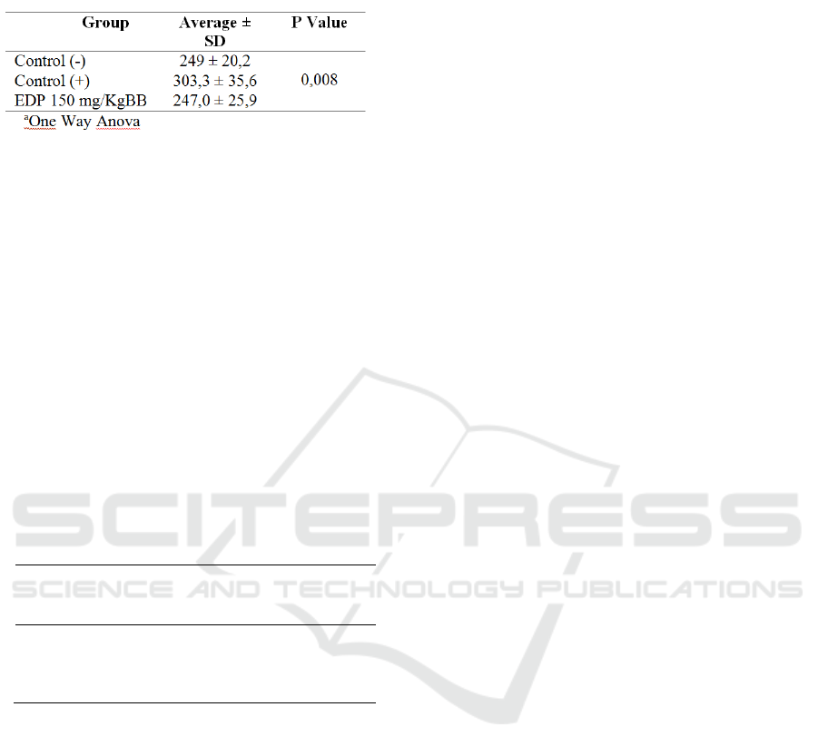
Table 3. Difference in mean TNF-α level after
administration of Bag Flower Leaf Extract
Table 3 shows that the statistical test results obtained
p value <0.008 so that there are significant
differences in average TNF-α levels in the negative
control group, positive control group, and EDP150
mg / Kg BB group after treatment. From the results
of continued tests (post hoc) in table 4 shows the
average comparison of TNF-α levels after giving
treatment in the negative control group with a
positive control group significantly different with a p
value = 0.022. In the negative control and EDP
treatment group 150 mg / kg BB there was no
significant difference in the average TNF-α level
after administration of the treatment with a p value>
0.05. In the positive control group and the EDP150
mg / Kg BB group, there was a significant difference
in the mean TNF-α levels after administration of
treatment with p = 0.016.
Table 4. Continued Test (Post Hoc) Differences in TNF-α
Levels after Giving Bag Flower Leaf Extracts
Group Control
(-)
Control
(+)
EDP150
mg/KgB
B
Control (-) - 0,022* 1,000
Control (+) - - 0,016*
EDP150
mg/KgBB
- -
-
* Significantly different groups
4 DISCUSSION
This study showed that there were
differences in the mean TNF-α level in rats between
groups that were not induced by S. Aureus bacteria
and those that were induced by S. Aureus. The
statistical results in this study showed that the
administration of bag flower leaf extract to rats at a
dose of 150 mg / Kg BB was able to reduce levels of
TNF-α in rat after being significantly induced by S.
aureus bacteria.
This study was in line with several previous
studies, namely mice infected with Staphylococus
aureus, and then there was an increase in TNF-α
production compared to mice that were not induced
by staphylococcus aureus. (Mufidah & Rifa'i., 2015;
Pereyra et al., 2017)
S. Aureus contains llipoteicoic acid which
is found on the surface of bacteria which is
recognized as toll-like receptors II (TLR II) which
will further stimulate IL-12 production so as to
stimulate INF- γ. This bacterium can also activate
the adaptive immune response through superantigen
induction. The interaction between superantigens
and cells can lead to greater stimulation of T cells
compared to other antigens. TNF-α cytokines and
IL-1 have immunostimulatory activity and work
synergistically with IFN-γ to enhance immune and
inflammatory reactions. However, if the levels of
cytokines are at high concentrations it can cause
epithelial cell damage and cause toxic shock (Liang
and Ji, 2007; Krakaeuer, 2011; Phuneerub et al.,
2015; Tong et al., 2015).
In the phytochemical test results on bag
flower leaf (Clerodendrum Paniculatum L), it is
found secondary metabolite compounds in the form
of flavonoids and tannins. The existence of
secondary metabolites is an important factor through
its mechanism of a bacterium. Flavonoids are the
largest group of phenol compounds. Flavonoid
compounds are good reducing compounds that
inhibit oxidation reactions both enzymes and non-
enzymes. Its mechanism as an antibacterial is to
form complexes with extracellular and dissolved
proteins and with microbial walls, flavonoids also
play a direct role by interfering with the function of
microorganism cells and inhibiting microbial cell
cycles, denaturating bacterial cell proteins and
damaging cell membranes that can result in lysis of
bacterial cells (Parubak, 2013; Nanda, Bora and
Tiwari, 2016; India Biodiversity Portal, 2017).
The work mechanism of tannin as an
antibacterial is to inhibit the reverse transcriptase
and DNA topoisomerase enzymes so that bacterial
cells cannot be formed. Tannin has antibacterial
activity related to its ability to inhibit and kill
bacterial growth by reacting toward cell membranes,
inactivation of essential enzymes in bacteria and
destruction of functions and genetic material and
interfering with the transport of proteins in the inner
layers of cells. Tannins also have targets on cell wall
polypeptides so that cell wall formation is less than
perfect. This causes the bacterial cell become lysis
due to osmotic and physical pressure so that the
bacterial cell will die. Tannin acts as an antibacterial
because it can form of protein complexes and
hydrovobic interactions, if the hydrogen bond
between tannin and protein enzymes contained in the
protein is likely to be denatured so that bacterial
ICHIMAT 2019 - International Conference on Health Informatics and Medical Application Technology
600

metabolism is disrupted (Daglia, 2012; Yadav et al.,
2014; Phuneerub et al., 2015).
Bag flower leaf extract has active compounds
namely flavonoids and tannins that can function as
anti-inflammatory mediators that can reduce the
release of proinflammatory cytokines in rats induced
by S. Aureus bacteria. From previous studies, bag
flower leaves that contain active compounds
flavonoids, tannins and steroids can reduce
inflammation that occurs in mice which is induced
carrageen (Hafiz, Rosidah and Silalahi, 2016a).
Other studies on the methanol extract of
mahogany with flavonoid active substances have
also been shown to reduce TNF-α levels in mice
induced by MLD-STZ. A similar study was carried
out by using purple leaf extract (Gratophyllum
pictum L.) with active flavonoid content, it has
proven to reduce TNF-α level and NO in mice which
is infected by S. Aureus bacteria(Suryani, Endang H
and Aulanni’am, 2013; Tjahjani, Kristina and
Endang Sri Lestari2, 2015; Bond, Morris and
Nassar, 2017)
In this study, the treatment group with a
dose of 150 mg / kg BW had a TNF-α value are
below TNF-α level in the negative control group or
it was not significantly different from the negative
control group where p value> 0.005 so that the
administration of bag flower leaf extract with a dose
of 150 mg / kg body weight can reduce TNF-α levels
in mice with normal conditions that are not induced
by S. Aureus bacteria.
The content of secondary metabolites found
in bag flower leaves (Clerodendrum Paniculatum L)
which is derived from bag flower leaf extract is
thought to act as an anti-inflammatory caused by S.
aureus bacteria. The main bioactive component of
phenols in flavonoids is quercetin. The mechanism
of bag flower leaf extract as an anti-inflammatory
can cause a decrease in TNF-α levels through
inhibition of Nuclear Factor kappa B (NF-kB). NF-
kB becomes active due to a stimulus from the
Reactive Oxygen Synthase (ROS) agent which
causes endothelial dysfunction, pathogen exposure,
DNA damage and physical stress. NF-κB has the
functions in controlling the genes expression of
cytokines and proinflammatory chemokins TNF-α,
IL-1β. The decrease in NF-kB activation is
influenced by inhibitory effect of monocytes on p56
Protein Tyrosin Kinase (PTK) enzyme, which causes
PTK to be inactive. Inactivated PTK causes NF-κB
transcription factors to remain bound to NF-κB
inhibitors so that it cannot trigger transcription and
translation of TNF-α proinflammatory cytokines
secreted by macrophages, thereby reducing TNF-α
levels (Nair, M.; Mahajan, 2006; Fassihi and Sabet,
2008; Rathee et al., 2009)
The dynamics of level changes in TNF-α
between positive control and treatment group
whereas in the treatment group after administration
of bag flower leaf extract, it is decreased TNF-α
levels after returning to the TNF-α level value equal
to the negative control group. It showed that the
content of compounds found in bag flower leaves
can reduce the inflammatory effect by suppressing
the release of TNF-α which is caused by S.Aureus
bacteria.
5 CONCLUSIONS
This research shows that bag flower leaf extract
contains secondary metabolite compounds including
flavonoids and tannins which can reduce TNF-α
level which are inflammatory mediators due to S.
Aureus bacteria. Bag flower leaf extract with a dose
of 150 mg / Kg BB is an effective dose to reduce
TNF-α level in rats induced by S. Aureus bacteria.
The administration of bag flower leaf extract can be
a solution in the treatment of complementary
therapies. It is necessary to evaluate the quantitative
test of bag flower leaf to determine the activity of
secondary metabolites and the percentage of
compound content in grams.
ACKNOWLEDGEMENT
This research was supported by Institut Kesehatan
Delihusada Delitua, Institut Kesehatan Medistra
Lubuk Pakam, Sembiring Hospital Foundation, and
Grand Med Hospital Foundation, Indonesia
REFERENCES
Abbas, A. K., Licthman, A. H. and Pillai, S. (2015)
Cellular and Molecular Immunology. Eighth Edi.
Philadelphia: Elsevier Saunders.
Acosta, A. C. et al. (2016) ‘Mastitis in ruminants in
Brazil’, Pesquisa Veterinaria Brasileira. doi:
10.1590/S0100-736X2016000700001.
Amir, L. H. et al. (2007) ‘A descriptive study of mastitis
in Australian breastfeeding women: incidence and
determinants’, BMC Public Health, 7(1), p. 62. doi:
10.1186/1471-2458-7-62.
Andriani, Y. et al. (2017) ‘Phytochemical analyses, anti-
bacterial and anti-biofilm activities of mangrove-
associated Hibiscus tiliaceus extracts and fractions
The Effectiveness of Clerodendrum Paniculatum. L against TNF- in Rats Induced by S. Aureus
601

against Pseudomonas aeruginosa’, Journal of
Sustainability Science and Management.
Berthold-Losleben, M. and Himmerich, H. (2008) ‘The
TNF-alpha system: functional aspects in depression,
narcolepsy and psychopharmacology.’, Current
neuropharmacology, 6(3), pp. 193–202. doi:
10.2174/157015908785777238.
Bond, D. M., Morris, J. M. and Nassar, N. (2017) ‘Study
protocol: evaluation of the probiotic Lactobacillus
Fermentum CECT5716 for the prevention of mastitis
in breastfeeding women: a randomised controlled
trial’, BMC Pregnancy and Childbirth. BMC
Pregnancy and Childbirth, 17(1), p. 148. doi:
10.1186/s12884-017-1330-8.
Chinchali, J. F. and Kaliwal, B. B. (2014) ‘Histopathology
of mammary gland in Staphylococcus aureus induced
mastitis in mice’, Asian Pacific Journal of Tropical
Disease, 4(S1). doi: 10.1016/S2222-1808(14)60463-1.
Cucumber, L. F., Virus, M. and Indonesia, D. (2009) ‘the
Poriferasta Compound-5,22E,25-Trien-3-O Β From’,
9(3), pp. 479–486.
Daglia, M. (2012) ‘Polyphenols as antimicrobial agents’,
Current Opinion in Biotechnology, pp. 174–181. doi:
10.1016/j.copbio.2011.08.007.
Das, S., Borah, M. and Ahmed, S. (2013) ‘Antibacterial
activity of the ethanolic extract of leaves of Citrus
maxima (Burm.) Merr. on escherichia coli and
pseudomonas aeruginosa’, Asian Journal of
Pharmaceutical and Clinical Research.
Fassihi, A. and Sabet, R. (2008) ‘QSAR study of p56 lck
protein tyrosine kinase inhibitory activity of flavonoid
derivatives using MLR and GA-PLS’, International
Journal of Molecular Sciences, 9(9), pp. 1876–1892.
doi: 10.3390/ijms9091876.
Florence, A. R., Joselin, J. and Jeeva, S. (2012) ‘Intra-
specific variation of bioactive principles in select
members of the genus Clerodendrum L’, Journal of
Chemical and Pharmaceutical Research, 4(11), pp.
4908–4914.
Green, B. N. et al. (2012) ‘Methicillin-resistant
Staphylococcus aureus: An overview for manual
therapists’, Journal of Chiropractic Medicine. Elsevier
B.V., 11(1), pp. 64–76. doi:
10.1016/j.jcm.2011.12.001.
Hafiz, I., Rosidah and Silalahi, J. (2016a) ‘Antioxidant
and anti-inflammatory activity of pagoda leaves
(clerodendrum paniculatum l.) ethanolic extract in
white male rats (Rattus novergicus)’, International
Journal of PharmTech Research, 9(5), pp. 165–170.
Hafiz, I., Rosidah and Silalahi, J. (2016b) ‘Antioxidant
and anti-inflammatory activity of pagoda leaves
(clerodendrum paniculatum l.) ethanolic extract in
white male rats (Rattus novergicus)’, International
Journal of PharmTech Research.
India Biodiversity Portal (2017) Clerodendrum
paniculatum L. Accepted Name. Available at:
http://indiabiodiversity.org/species/show/266140
(Accessed: 1 January 2017).
Indonesian Pediatric Society (2013) Mastitis: Pencegahan
dan Penanganan, 2013. Available at:
http://www.idai.or.id/artikel/klinik/asi/mastitis-
pencegahan-dan-penanganan.
Joseph, J., Bindhu, A. and Aleykutty, N. (2013) ‘In vitro
and in vivo antiinflammatory activity of
Clerodendrum paniculatum linn. leaves’, Indian
Journal of Pharmaceutical Sciences, p. 376. doi:
10.4103/0250-474X.117428.
Krakaeuer, T. (2011) ‘Comparative Potency of Green Tea
and Red Wine Polyphenols in Attenuating
Staphylococcal Superantigen Induced Immune
Responses’, American Journal Biomedical Sciences.
Lai, J. L. et al. (2017) ‘Indirubin Treatment of
Lipopolysaccharide-Induced Mastitis in a Mouse
Model and Activity in Mouse Mammary Epithelial
Cells’, Mediators of Inflammation, 2017. doi:
10.1155/2017/3082805.
Liang, X. and Ji, Y. (2007) ‘Involvement of alpha5beta1-
integrin and TNF-alpha in Staphylococcus aureus
alpha-toxin-induced death of epithelial cells.’, Cellular
microbiology, 9(7), pp. 1809–21. doi: 10.1111/j.1462-
5822.2007.00917.x.
Nair, M.; Mahajan, S. (2006) ‘The Flavonoid Quercetin
Inhibits Proinflammatory Cytokine ( Tumor Necrosis
Factor Alpha ) Gene Expression in Normal Peripheral
Blood Mononuclear Cells via Modulation of the NF- κ
β System’, Clinical and Vaccine mmunology, Vol
13.(3), p. 319.328. doi: 10.1128/CVI.13.3.319.
Nanda, G. C., Bora, M. and Tiwari, R. K. (2016) ‘Less
known medicinal plants of assam and odisha used for
treating diabetes with special reference to charaka’,
International Journal of Research in Ayurveda and
Pharmacy. doi: 10.7897/2277-4343.07249.
Notebaert, S. et al. (2008) ‘Inflammatory mediators in
Escherichia coli-induced mastitis in mice’,
Comparative Immunology, Microbiology and
Infectious Diseases, 31(6), pp. 551–565. doi:
10.1016/j.cimid.2007.10.004.
Parubak, S. A. (2013) ‘Senyawa Flavonoid yang Bersifat
Antibakteri dari Akway (Drimys becariana. Gibbs)’,
Chem. Prog., 6(1), pp. 34–37.
Phuneerub, P. et al. (2015) ‘In vitro anti-inflammatory,
mutagenic and antimutagenic activities of ethanolic
extract of Clerodendrum paniculatum root.’, Journal
of advanced pharmaceutical technology & research,
6(2), pp. 48–52. doi: 10.4103/2231-4040.154529.
Pidugu, S. and Arun, T. (2012) ‘Antibacterial activity and
phytochemical screening of mentha arvensis Linn.
against proteus mirabilis from urinary tract infected
patients’, International Journal of PharmTech
Research.
Poorter, H. et al. (2012) ‘Biomass allocation to leaves,
stems and roots: Meta-analyses of interspecific
variation and environmental control’, New Phytologist.
doi: 10.1111/j.1469-8137.2011.03952.x.
Rathee, P. et al. (2009) ‘Mechanism of Action of
Flavonoids as Anti-inflammatory Agents: A Review’,
Inflammation & Allergy-Drug Targets, 8(3), pp. 229–
235.
Sanchez, M. S., Ford, C. W. and Yancey, R. J. (2005)
‘Efficacy of tumor necrosis factor-alpha and
ICHIMAT 2019 - International Conference on Health Informatics and Medical Application Technology
602

antibiotics in therapy of experimental murine
staphylococcal mastitis.’, Journal of dairy science,
77(5), pp. 1259–66. doi: 10.3168/jds.S0022-
0302(94)77065-X.
Schwartz, K. et al. (2002) ‘Factors associated with
weaning in the first 3 months postpartum.’, The
Journal of Family Practice, 51(5), 439-444. The
Journal of family practice. 51. 439-44.
Suryani, N., Endang H, T. and Aulanni’am, A. (2013)
‘Pengaruh Ekstrak Metanol Biji Mahoni terhadap
Peningkatan Kadar Insulin, Penurunan Ekspresi TNF-
αdan Perbaikan Jaringan Pankreas Tikus Diabetes’,
Jurnal Kedokteran Brawijaya, 27(3), pp. 137–145.
doi: 10.21776/ub.jkb.2013.027.03.3.
Tjahjani, N. P., Kristina, T. N. and Endang Sri Lestari2
(2015) EFFECTIVENESS OF EXTRACT
Gratophyllum pictum (L.)LEAVES TO REDUCE
LEVEL OF TNF-α DAN NO.
Tong, S. Y. C. et al. (2015) ‘Staphylococcus aureus
infections: Epidemiology, pathophysiology, clinical
manifestations, and management’, Clinical
Microbiology Reviews, 28(3), pp. 603–661. doi:
10.1128/CMR.00134-14.
Wang, J. H. et al. (2018) ‘Traditional uses and
pharmacological properties of Clerodendrum
phytochemicals’, Journal of Traditional and
Complementary Medicine. doi:
10.1016/j.jtcme.2017.04.001.
Yadav, M. et al. (2014) ‘Innovare Academic Sciences
Preliminary Phytochemical Screening Of Six
Medicinal Plants Used In Traditional Medicine’,
International Journal of Pharmacy and
Pharmaceutical Sciences, 6(5), pp. 2–14.
The Effectiveness of Clerodendrum Paniculatum. L against TNF- in Rats Induced by S. Aureus
603
