
Sumbawa Horse Milk as a Prevention of Inflammatory Bowel Disease
(IBD) in Animal Model based on IL-6 Expression
and Gastric Histopathology
Nurina Titisari
1
*, Theresa Lidya Pramesti
2
, Tiara Widyaputri
3
, Rahadi Swastomo
4
1
Physyiology Departement of Veterinary Medicine, University of Brawijaya, PuncakDiengEksklusif, Malang, Indonesia
2
Student of Veterinary Medicine, University of Brawijaya, PuncakDiengEksklusif, Malang, Indonesia
3
Clinic Departement of Veterinary Medicine, University of Brawijaya, PuncakDiengEksklusif, Malang, Indonesia
4
Parasitology Departement of Veterinary Medicine, University of Brawijaya, PuncakDiengEksklusif, Malang, Indonesia
Keywords: Flowcytometri, Gastrointestinal, Inflamation, Rattusnorvegicus.
Abstract: Sumbawa horse milk contains bioactive components as exogenous antioxidants, anti-inflammatory
andantimicrobial which can prevent Inflammatory Bowel Disease. IBD is characterized by ongoing
inflammation of the digestive tract that could changed gastric histology and trigger pro-inflammatory
cytokines. The purpose of this study was to observe the preventive effects of Sumbawa horse milk on gastric
organs inflammation in IBD animal models. Twenty male rats, 8 weeks, 150-200 grams BW, divided into
five treatment groups; K- group (negative control), indometacin induced group; i.e K+ (positive group without
horse milk), P1(horse milk dose 0.5 mL/rat), P2 (horse milk dose1.0 mL/rat), and P3(horse milk dose
1.5mL/rat). Flowcytometry method was conducted to observe IL-6 expression, and then analyzed with one
way ANOVA followed by a tukey test (α 95%). While the gastric histopathological was analyzed
descriptively. The results showed that IL-6 expression were significantly lower (p <0.05) in P2 dan P3
compared with K+group. Whereas in the histological result, the p3 group was able to prevent
histopathological damage of the gastric organs compared to other treatment groups. The conclusion of the
study is Sumbawa horse milk able to prevent increased of IL-6 and gastric mucosal cell erosion, with the best
dose is 1.5 mL/rat.
1 INTRODUCTION
Inflammatory Bowel Disease (IBD) is a disease
characterized by inflammation of the digestive tract
associated with histopathological changes in the
digestive tract mucosa such as the stomach, intestine
and colon in the form of infiltration of inflammatory
cells in the lamina propria mucosa (Washabau, 2008).
Common symptoms of IBD are diarrhea and bleeding
in the digestive tract which is at high risk of causing
damage to the digestive tract (McFarland, 2008). IBD
is divided into two types, i.e ulcerative colitis
(chronic inflammation of the large intestine) and
chron's disease (chronic inflammation of the small
intestine) (Xavier and Podolsky, 2007). According to
Volkman's research (2017), there were 136 cases of
IBD (Inflammatory Bowel Disease) reported at the
Clinic Clinic for Small Animals, Faculty of
Veterinary Medicine, Freie Berlin, Germany
throughout 2017.
The causative factor of IBD is not known for
certain, but it is allegedly influenced by immune
regulation failure, exogenous factors, and the role of
intestinal flora (Thorenson et al. 2007). IBD also
occurs due to the use of non-steroidal anti-
inflammatory drugs (NSAIDs) such as indomethacin
(Podolsky, 2002). Indomethacin is an indole-acetic
acid derivate that is used as a treatment for arthritis.
In addition to its curative effects, indomethacin has
side effects which can inhibit COX-1 cyclooxygenase
which functions against prostaglandin synthesis and
mucous production to protect the small intestinal
mucosa from bacterial and viral infections (Takeuchi,
2003). Another side effect of indomethacin, it can
increase free radicals, which it can damage the
digestive organs marked by microflora reduction
(Strus et al., 2009).
Titisari, N., Pramesti, T., Widyaputri, T. and Swastomo, R.
Sumbawa Horse Milk as a Prevention of Inflammatory Bowel Disease (IBD) in Animal Model based on IL-6 Expression and Gastric Histopathology.
DOI: 10.5220/0009587801470151
In Proceedings of the 6th International Conference on Advanced Molecular Bioscience and Biomedical Engineering (ICAMBBE 2019) - Bio-Prospecting Natural Biological Compounds for
Seeds Vaccine and Drug Discovery, pages 147-151
ISBN: 978-989-758-483-1
Copyright
c
2020 by SCITEPRESS – Science and Technology Publications, Lda. All rights reserved
147

In this study we examined the ability of Sumbawa
horse milk to prevent IBD in animal model.
According to Saputro (2016), Sumbawa horse milk
has protein componeants namely lysozyme (BM 17
kDa) and lactoferrin (BM 75 kDa) which act as
indigenus proteins that function for antimicrobial
compounds and have bioactive compounds as
antioxidants which play an important role to protect
the body from conditions of oxidative stress due to
inflammatory processes. Elias (2008) states that the
bioactive compounds in Sumbawa horse milk have
the primary structure of the amino acid histidine
which has an imidazole group as a hydrogen giver, a
lipid-peroxyl radical scavenger, and a hydrifobic
potential as an exogenous antioxidant.
2 MATERIAL AND METHOD
2.1 Tools and Materials
Equipment include rats cages, masks, gloves,
micropipets, dissecting sets, glass objects, glass
cover, autoclaves, scales, sonde, filters, magnetic
stirrer, measuring cup, digital scale, incubator,
aluminum foil, blue tip, yellow tip, microtube, sample
pot, cover slip, fixation board, centrifugator,
waterbath.
Materials used in this study include 20 rats
(Rattusnovergicus), Wistar strain, male, 120-150
gram ofbody wieght, Sumbawa horse milk, corn oil,
10% formalin, HE staining (Hematoxyline Eosin),
aquades, xylol, PBS, liquid paraffin, IL-6 antibodies,
chromagen DAB (DiaminoBenzidine) dyes,
Biotinylated Rabbit Anti-Rat IgG Antibody
secondary antibodies.
2.2 Horse Milk Induction
Sumbawa horse milk was from horse farms located in
Dompu Regency, Sumbawa Island, NTB (West Nusa
Tenggara). Sumbawa horse milk was a fresh pure
milk with milking date on 31 December 2018.
Sumbawa horse milk is given as preventive once a
day with a volume of 0.5 mL/rat, 1 mL/rat, and 1.5
mL/rat for 7 days and then induced indomethacin
using gastric sonde on day 8 then continued giving
Sumbawa horse milk on day 9-14 (Reni et al., 2010).
2.3 Indometacin Induction
The IBD animal model in this study used a single
dose of NSAID induction in the form of
indomethacin. The induction dose of IBD with
indomethacin was 15mg / kgBB (Aulanni'am et al.,
2011). The administration of indomethacin in this
study was carried out orally using a gastric sonde.
Previously the indomethacin must be diluted with
corn oil. The dilution of indomethacin as much as 45
mg requires 4 mL of corn oil as the solvent (Bures,
2011).
2.4 Histopathological Preparation
Gastric organs that have been fixed with 10%
formalin then carried out the process of alcohol
dehydration using alcohol concentration levels of
70%, 80%, 90%, absolute alcohol I, absolute alcohol
II, then purified using xylol I and xylol II. The
paraffin process was carried out using paraffin I and
paraffin II. The preparation is put into a mold that
contains half volume paraffin and the sample is
placed vertically and horizontally so that the cross
section is attached to the base of the paraffin. After
starting to rigid, paraffin is added again until the mold
was full and left until the paraffin hardens. The
paraffin blocks are then cut thin 5 micrometers thick
using a microtome. The results of the ribbon-shaped
pieces are spread on warm water with a temperature
of 46 ° C and immediately removed which is useful
for stretching the pieces so they do not multiply or
eliminate the folds caused by cutting. The preparation
is then removed and placed on a glass object and dried
overnight in an incubator at 60 ° C so that staining of
Hematoxylin-Eosin (HE) (Febram, et al., 2010).
2.5 Interleukin 6 (IL 6) Expression
IL-6 expression measurements using the
Flowcytometry method. The initial stage was taken
the rat's gastric, washed and soaked in PBS then the
organs were crushed with the base of a new syringe
in 5 mL Phosphat Buffer Saline (PBS) Solution.
Filtered with wire and put into propylene 15 mL to a
certain volume. Homogenized by centrifuging with a
2500 rpm for 5 minutes at a temperature of 10ºC and
then discarded the supernatant. The pellet that remain
then resuspended with PBS 1 mL. Suspense results
are divided into several 1.5 mL microtubes according
to the needs of the type of coloring combination that
has been filled with PBS ± 0.5 mL, @ 50 μL. The
suspension is centrifuged at 2500 rpm for 5 minutes
at 10ºC then the supernatant is discharged from the
centrifugation until there is a pellet which is then
carried out intracellular staining to determine the
expression of IL-6 by adding a fixative solution of 50
μL, then incubated for 20 minutes at 4 ° C in a dark
room. The 500 μL permeability solution (1X) was
ICAMBBE 2019 - 6th ICAMBBE (International Conference on Advance Molecular Bioscience Biomedical Engineering) 2019
148
then homogenized with a centrifuge speed of 2500
rpm for 5 minutes at 10ºC then the supernatant was
removed. The pellet which was then added 50 μL
specific antibody solution, after that incubated it
for20 minutes at a 4°C in a dark room. After
incubation is complete, ± 400 μL PBS was added and
transferred to the flowcytometry cuvette for analysis.
2.6 Data Analysis
Flowcytometry test results were analyzed
quantitatively using BD CellquestProTM Software,
Microsoft Exel applications and statistical package
software for the social science (SPSS) for Windows
16 with One Way analysis of variance (ANOVA)
Test. The Tukey test was performed if there were
significant differences (α 95%). As for
histopathological features, descriptive analysis was
performed.
3 RESULT AND DISCUSSION
The test results showed that IL-6 expression of gastric
organ in negative control group (K-) was significantly
different with the positive control group (K+), group
P1 and group P2 but not with group P3 (Table 1). This
is due to the normal state of proinflammatory
cytokines was produced by the body to maintain
hemeostatic conditions in the immune system.
According to Erica et al. (2000), IL-6 is a
proinflammatory cytokine that is produced by
macrophages and is present in almost all tissues in the
body even in small amounts.
The positive control group (K+) showed
significantly different results from the negative
control group (K-), P2 group, and P3 group, but not
significantly different from P1 group. The results in
this study are in accordance with research conducted
by Riyansyah et al (2015), administration of
indomethacin at a dose of 15 mg/kgBW orally in rats
can cause irritation and damaged gastric mucosa,
which can trigger IL-6 expression as a pro cytokine
inflammation. Indomethacin inhibits COX-1
formation which destroys the performance of
mitochondria in the cell so it will interupted the
oxidative postforilation cycle. The disruption of
electron transfer on oxygen molecules can trigger
inflammation (Takeuchi et al., 2003). This can
triggers the formation of excess Reactive Oxygen
Species (ROS) that will stimulate the activation of
NF-kB which is a transcription factor in regulating
the expression of pro-inflammatory cytokine cells
such as IL-6 (Aulanni'am et al., 2012).
Table 1: Table of the average expression of interleukin 6
(IL-6) gastric organs of white rats.
Treatment
Average
IL-6 expression
(% gated) ± SD
K-
19,4925±4,33
a
K+
36,0475±1,535
c
P1
31,57±1,489
bc
P2
28,3525±2,464
b
Note: notations a, b, c show significant differences
between groups.
3.1 Histopathology of the Rat Gastric
Organ (Rattus Novergicus)
Gastric histology with hematoxylin eosin (HE)
staining (Figure 1). Based on on observation on the
gastric preparations of the negativ control group
(healthy mice) (Figure 1.A) showed that the mucosal
layer of tunica mucosa epithelial simplex with gastric
pit formation and gastric gland still arranged and
neatly lined with cylindrical simplex form.
Puspitasari (2008) state mucosal layers contain many
mitochondria and granules to produce pepsinogen
enzymes, while parietal cells are acidophilic because
these cells produce HCL of stomach acid. It also
appears a small number of neutrophill cells, which is
normal because to its role agains pathogenic agents.
Contrast result occur in the positive control group
(Figure 1.B), which showed a severe cell damage in
the tunica lamina propria layer of the gastric mucosa.
The epithelium erosion accompanied by infiltration
of inflammatory cells (neutrophils) when we
compared it with negative controls. Erosion that
occurs in the gastric mucosa suspected as a result of
free radicals from indomethacin induction, that can
stimulate leucocyte toward gastric mucosa cells.
Leucocyte wull produce H
2
O
2
to kill several types of
bacteria and fungi and for cell growth, but neither
attack specific targets. So it will also attack
polyunsaturated fatty acis from cell membranes, cell
organeles, or DNA, which can cause damage to
structure and function cell (Puspitasari, 2008).
The histopathological result showed no
significant difference between group P1 (Figure 1.C)
and K
+
(Figure 1.B). There were erosion in gastric
mucosal epithelial cells and inflammatory cell
infiltration still visible although its not as much as K
+
group. In group P2 (Figure 1.D) showed less erosion
of the epithelial cell and lower number of
inflammation cells when we compared with P1 group
Sumbawa Horse Milk as a Prevention of Inflammatory Bowel Disease (IBD) in Animal Model based on IL-6 Expression and Gastric
Histopathology
149
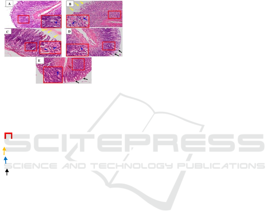
(Figure 1.C). In the last horse milk treatment, P3
(Figure 1.E) showed a better result compared to the
other treatment group. Epithelial cells erosion in
lamina propria tunica gastric mucosa was decreasing
and inflammation cells number was much lower when
we compared with P1 and P2 group.
Figure 1: Histopathology of rat gastric mucosa
(Rattusnorvegicus) (gastric preparations; cross sections;
HE staining. 100xenlargement).
Note: (a) Negative control rats (K-); (B) Positive control
rats (K+); (C) Group P1; (D) Group P2; (E) Group P3;
(400x magnification)
(epithelial erosion of the gastric mucosa)
(neutrophil cell infiltration)
(repair of epithelial cells)
4 CONCLUSIONS
Preventive administration of Sumbawa horse milk in
animal model of IBD induced by indomethacin were
able to prevent IL-6 expression and histopathological
changes in gastric organs with the best dose was 1.5
mL/rat. Further research needs to be done to prevent
Inflammatory Bowel Disease (IBD) using Sumbawa
horse milk on the entire digestive tract.
ACKNOWLEDGEMENTS
This research was funded by Veterinary Medicine
Faculty, University of Brawijaya through the
DPPSPP scheme 2019.
REFERENCES
Aulannia’am, Roosdiana A. & Rahmah, N.L. (2012). The
Potency of Sargassum duplicatum Bory Extract on
Inflammatory Bowel Disease (IBD) in Rattus
Norvegivus. Journal of Life Sciences 6: 144-154.
Aulanni’am, Roosdiana, A., Rahmah, N.L.,Tjaniadi P.,
Lesmana, M. & Subekti, D.(2003).Antimicrobial
Resistance of Bacterial Pathogens Associated
withDiarrheal Patiens in Indonesia. Am J Trop Med
Hyg. 68(6): 666-10. Bures, J, J. Pejchal, J.Kvetina, A.
Tichy, S.Rejchrt, M.
Kunes & Kopacova, M. (2011). Morphometric analysis of
the porcine gastrointestinal tract in a 10-day high-dose
indomethacin administration with or without probiotic
bacteria Escherichia coli Nissle 1917. Humanand
Experimental Toxicology. 30(12) 1955–1962.
Elias, R, J. & Decker E.A. (2008). Antiocidant Activity of
Proteins and Peptides. Food Sci Nutr. 48:430-441.
Erica, E. S.. Henn, A., Gurtoo, H.L., Wollman, R.M.,
Alderfer, J.L., Mihich. E., Ehrke M. J. (2001). A Novel
Tumor Necrosis Factoralpha Inhibitory Protein, TIP-
B1. International Journal of Immunopharmacology
22(12):1137-42
Febram, B., Wientarsih, I & Pontjo, B. (2010). Activity Of
Ambon Banana (Musa Paradisiaca Var. Sapientum)
Stem Extract In Ointment Formulation On The Wound.
Healing Process Of Mice Skin (Mus Musculus
Albinus).Majalah Obat Tradisional 15(3):121-137.
McFarland, L.V. (2008). State-of-the Art of Irritable
Bowel Syndrome and Inflammatory Bowel Disease
Reasearch in 2008.WorldJournalof Gasstroenterology.
14 (17): 2625 2629.
Puspitasari, D. A. (2008). Gambaran Histopatologi
LambungTikus Putih (Rattus novergicus) Akibat
PemberianAsam Asetil Salisilat. Institut Pertanian
Bogor.
Podolsky, D. K. (2002). Inflammatory Bowel Disease. N
Engl J Med. 347 (6): 417- 429.
Reni, S., Sumarno. & Widjajanto, E. (2011). Susu Kuda
Sumbawa Meningkatkan Respon Imun Seluler
Makrofag Peritoneal Mencit terhadap Salmonella
Typhimurium Jurusan Gizi Politeknik Kesehatan
Mataram. Jurnal Kedokteran Brawijaya, Vol. XXVI,
No. 1.
Riyansyah, Y., Lanny, M. & Ratu C. (2015). Uji aktifitas
Antiinflamasi Ekstrak Ethanol Daun Ubi Jalar Ungu
Terhadap Tikus Wistar Jantan. Prosiding Penelitian
SPESIA Unisba.Hal:630-636.
Saputro, M. (2016). Profil Protein, Aktivitas Antioksidan
dan Inhibitor ACE dari Susu Kuda dan Hidrolisisnya.
IPB: Bogor.
Strus, M., Gosiewski., Fyderek, K K. & Adamski, P.
(2009). A Role of Hydrogen Peroxide Producing
Commensal Bacteria Presentin Colon Of Adolescentes
with Inflammatory Bowel Disease in Prepetuation of
the Inflammatory Process. Journal of Physiology and
Pharmacology 60 (6):49-54.
Takeuchi, K., Tanaka, A., Ohno, R. & Yokota, A. (2003).
Role of COX Inhibition in Patogenesis of NSAID-
ICAMBBE 2019 - 6th ICAMBBE (International Conference on Advance Molecular Bioscience Biomedical Engineering) 2019
150

Iinduced Small Intestinal Damage. Research article.
Kyoto Pharmaceutical University. Kyoto. Healing: A
Review. Wound Repair and Regeneration. 6(12): 591-
599.
Tonnesen, M. G., Feng X. & Clark R. A. F. Angiogenesis
in Wound Healing. JID Symposium Proceedings. 2007;
5(1): 40-46.
Uniacke-Lowe, T. & Fox, P.F. (2010. Equine Milk
Proteins: Chemistry, Structure, and Nutritional
Signification. IntDairy J: 20:609-629
Volkmann M., Steiner, J.M., Fosgate, G.T., Zentek, J.,
Hartmann, S. & Kohn, B. (2017). Chronic Diarrhea in
Dogs- Rostropective Study in 136 Cases. JVet Intern
Med. Vol 3 (2): 121-126.
Washabau R.J. (2008). Summary of Findings and Reports
ofthe WSAVA Gastrointestinal Standardization Group.
The 33rd WSAVA Congress. Dublin. 20-24. 08.2008,
pp. 60-62.
Xavier, R.J. & Podolsky, D.K. (2007). Unravelling The
Patogenesis of Inflammatory Bowel Disease. Nature.
448 (7152): 427-434.
Sumbawa Horse Milk as a Prevention of Inflammatory Bowel Disease (IBD) in Animal Model based on IL-6 Expression and Gastric
Histopathology
151
