
Extract Gel of Chinese Petai Leaf (Leucaena leucocephala) for Acute
Wounds in Rats (Rattusnorvegicus)
Anis Ika Nur Rohmah
*
, Edi Purwanto and Vicky Nurdiansyah
Department of Nursing, Faculty of Health Science, University of Muhammadiyah Malang, Jalan. Bendungan Sutami
No. 188A, Malang, East Java, Indonesia
Keywords:
Chinese petai leaf, acute wounds, rats.
Abstract:
The many occurrences of acute injuries due to the surgical process or accident have a considerable risk of
complications if not given appropriately. Chinese Petaileaves contains saponins, tannins, and flavonoids
are useful for accelerating through the process of acute wound healing. In this study, extract Chinese Petai
leaf made in gel preparations to be practical, sterile, and become the latest innovation. The purpose of this
study was examined the extract gel Chinese Petai leaf effectively for the treatment of acute wounds to rats.
This study used true-experimental design with a randomized control group posttest design as an approach.
The samples in intervention group (n = 7) and control group (n = 6) used simple random sampling and for
data was analyzed using Man Whitney test. The results indicated that the first group (Extract Gel Chinese
Petai Leaf) and the second group (Normal Saline) had significant differences in the percentage of wound
closure on the eighth day with the score (p = 0.014). Difference in the percentage of wound closure was
seen starting early in the epithelialization phase of acute wounds and more clearly on the eighth day.
Extract gel of Chinese Petai leaf effectively accelerated the closure of wound acute stage II.
1 INTRODUCTION
Wounds come from trauma to the skin which is a
condition where the continuity of the body’s tissue
continuation can disrupt to the normal functioning of
the body (Damayanti, Pitriani, & Ardhiyanti, 2013).
According to the duration of healing, wounds are
divided into 2 types, namely acute and chronic
wounds. Acute wounds are wounds that heal
according to the physiology of the wound healing
process. Acute wounds can be caused by trauma
suddenly or planned as during the surgical process.
A chronic wound is a complication of an acute
wound, which is where acute wound care is not
appropriate (Wijaya, 2018).
Acute wounds have a high risk of complications
if they do not get proper wound care. Complications
of acute injuries can occur within 24 hours after the
trauma. Several kinds of complications can occur in
acute wounds, including bleeding, hematoma or
tissue edema, dehiscence or not joining the wound
edges, and infection (Wijaya, 2018). Infection In
acute wounds can occur in the inflammatory phase.
This can occur because of bleeding that is long
enough (10-20 minutes), so that the blood supply in
the inflammatory phase is greatly reduced and white
blood cells do not reach the site of the wound (Peate
& Glencross, 2015). Complications of acute wounds
can also lead to amputation caused by an infection in
the wound and sufferers who have risk factors for
diabetes mellitus (Supriyadi, 2017).
Several factors influence wound healing,
including: vascularization, age, and concomitant
diseases. Vascularization affects because the wound
requires a good circulatory condition. The speed of
cell repair takes place in line with the growth or
maturity of a person, but the aging process can
reduce the speed of cell repair. While other diseases,
such as anemia, diabetes mellitus and kidney
disorders that can slow wound healing (Damayanti
et al., 2013).
The national wound prevalence in Indonesia is
8.2%. Comparison of the results of Riskesdas 2007
with Riskesdas 2013 showed an increase in wound
prevalence from 7.5% to 8.2% (Trihono, 2013). The
three most common types of injuries suffered were
70.9% abrasions; sprained 27.5%, and torn wounds
23.2%. Whereas in 2018 Riskesdas there was an
increase in wound prevalence from 8.2% in 2013 to
9.2% in 2018 results (Riskesdas, 2018).
Rohmah, A., Purwanto, E. and Nurdiansyah, V.
Extract Gel of Chinese Petai Leaf (Leucaenaleucocephala) for Acute Wounds in Rats (Rattusnorvegicus).
DOI: 10.5220/0009125701370142
In Proceedings of the 2nd Health Science International Conference (HSIC 2019), pages 137-142
ISBN: 978-989-758-462-6
Copyright
c
2020 by SCITEPRESS – Science and Technology Publications, Lda. All rights reserved
137

Chinese Petai has a variety of chemical contents
both in the seeds or leaves. The chemical content of
old seeds in every 100 grams has 148 calories of
calories, 10.6 grams of protein, 0.5 grams of fat,
26.2 grams of charcoal hydrate, 155 grams of
calcium, 59 mg of phosphorus, 2.2 mg of iron ,
vitamin A 416 SI, vitamin B 1 0.23 mg, and vitamin
C 20 mg. In the results of a study found that Chinese
Petai leaf extract contains saponins, alkaloids,
tannins, and flavonoids (Widyantoro & Sugihartini,
2015). The saponin content contained in Chinese
Petai leaves can be antibacterial (Pratiwi, Soetjipto,
& Hartini, 2014). Flavanoid is a compound derived
from flavone compounds. Flavanoid is a protein
structure that plays a role in the process of wound
healing and can be a cleanser for wounds (Praja &
Oktarlina, 2016).
Chinese Petai leaves have a variety of chemical
contents, where the content can be used as an
ingredient in the treatment of new wounds and
swelling by pounding or chewing. Furthermore, the
results of leaves that have been smooth are
immediately affixed to the wound or swollen area
(Thomas, 2007). In another study showed that the
scouring of 30 grams of Chinese Petai leaves
provided significant healing results with the most
length of clean wound healing on day 5, while the
length of clean wound healing using povidone iodine
was 10% on the 8th day (Rahmawati, 2015).
The gel is a semi-solid preparation consisting of
organic and inorganic suspensions. Gel for topical
use can immediately melt if it comes into contact
with the skin and will form a layer. Absorption of
the skin in gel form is better than other ointment
preparations (Yanhendri & Yenny, 2012). Gel-
shaped ointments have finer consistency, are
generally liquid and contain little or no mucosa as a
base (Elmitra, 2017).
Treatment using Chinese leaves with crushed or
chewed cannot last long and must be made every
day. This can make people less interested in using it
and choosing a more practical way. Therefore, an
alternative is needed in the presentation of Chinese
Petaileaves so that it is easy to use. On this occasion,
the researchers wanted to know the effectiveness of
Chinese Petaileaf extract gel for the treatment of
incision wounds in rats. The presence of Chinese
Petaileaf extract gel is expected to simplify wound
care that can be used at any time, is more effective,
and is practical for storage as supplies.
2 METHODS
This study is an experimental study using a true
experimental research design and using a
randomized control group posttest design approach.
This study consisted of 2 groups, namely the
intervention group (n = 7) who were treated with
wound care using Chinese Petaileaf extracts, and the
control group (n = 6) who were given standard
treatment using 0.9% NaCl.
The sampling technique used is simple random
sampling. The method of taking the sample is by
drawing, so it is chosen randomly according to the
experimental design to place the rat in 2 groups
(Surahman, Rachmat, & Supardi, 2016).
White rat used in this study must meet the
following experimental animal requirements: (1)
White male rats (rattusnorvegicus), (2) 2-3 months
old, (3) White rat with normal body weight of 160
grams, (4) active (healthy) moves, and (5) have
never been used as experimental animals in other
studies.
In this study, ethical information was obtained
No.E.5.a / KEPK-UMM / II / 2019 from the UMM
Health Research Ethics Commission. This research
was conducted at the Pharmacology Laboratory of
the Faculty of Medicine, University of
Muhammadiyah Malang. When the research was
carried out on 05 to 12 April 2019.
The dose of Chinese Petaileaf extract gel was
0.006 g each time applying 1cm2 to the wound.
Measurement of gel dosage using micro scales that
can read the mass of objects under 1 gram and
adjusted to changes in wound area until the gel is
applied evenly. Giving Chinese Petaileaf extract gel
is done 2 times a day in the morning at 07.00 and in
the afternoon at 16.00 (Andrie & Sihombing, 2015).
Experimental animals adapted in rat cages were
given the same treatment for 7. During adaptation,
all try animals looked healthy, actively moving, and
nothing stressed. When intervening, animals try to
get food and drink according to their needs.
The data collection stage was carried out by
measuring the area of the wound on the 4th day and
the 8th day. Measuring the area of the wound using
image J software. The wound image is taken by
attaching mica paper over the wound, then draw by
following the edge of the wound using a marker and
after that, the picture is photographed and inserted
into the image J application.
The resulting wound area is then entered into the
formula for wound closure percentage. The formula
for measuring wound closure:
HSIC 2019 - The Health Science International Conference
138

Percentage of wound closure= (AO - AD)/AO
x100% (1).
Information:
AO = area of the wound on day 0.
AD = area of the wound on the day after
treatment (Avinash et al., 2016).
3 RESULTS AND DISCUSSION
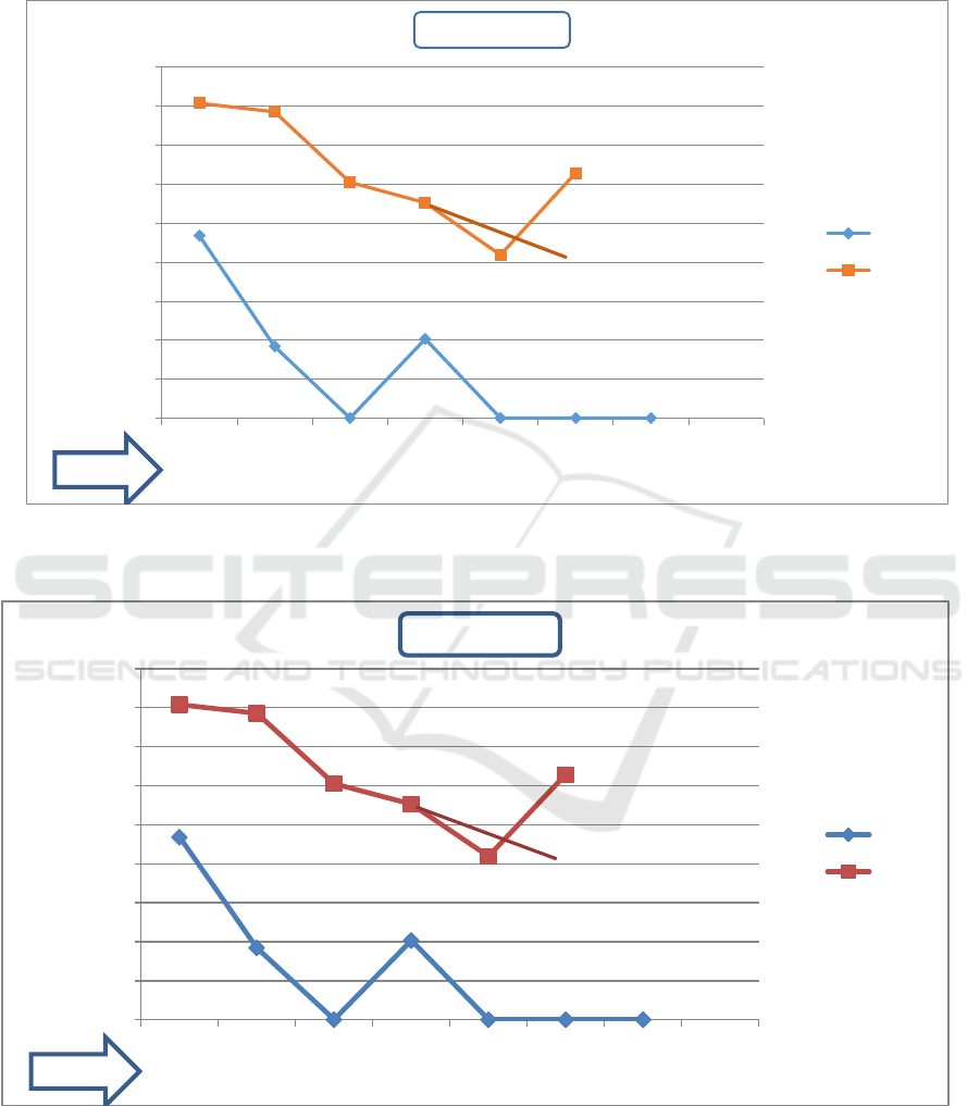
Based on the figure 1, the picture shows that all
white rat animals have been measured the
percentage of wound closure on day 4 in group 1
(intervention) with an average value of 14.6%, and
in group 2 (control) with a mean value an average of
9.7%. On the 8th day in group 1 with an average
value of wound closure was 81.2% and in group 2 it
was 62.9%. There was an increase in the percentage
of initial wound closure on day 4 by 14.6 in group 1
to 81.2% on day 8, whereas in group 2 the
percentage of wound closure initially on day 4 was
9.7% to 62.9% on the 8th day.
The results of the study on the percentage of
wound closure on day 4 obtained from the SPSS
output found that the significant value (p), in the
Man Whitney statistical test the significance value of
0.628 where the value is greater than the value of α
(0.05). The results of these calculations can be
concluded that there was no significant difference in
the percentage of wound closure in the intervention
group given Chinese Petai leaf extract gel and in the
control group given normal saline for acute wounds
in the rat on the 4th day.
The results of the study on the percentage of
wound closure on the 8th day obtained from the
SPSS output found that the significant value (p), in
the man whitney statistical test the significance
value of p = 0.014 where the value is smaller than
the value of α (0.05). The results of these
calculations can be concluded that there is a
significant difference in the percentage of wound
closure in the intervention group given Chinese
Petaileaf extract gel and in the control group given
normal saline for acute wounds in rats on the 8th
day.
Based on the results of research in the
intervention group that was given Chinese Petaileaf
extract gel showed that the wound healing process
went normal. None of the wound healing stages
were missed and there were no interruptions in each
process, which was proven in the intervention group
that there were no signs of wound infection. This
proves the theory revealed by Pratiwi (2014) and
Yuliarti (2009) that the saponin and tannin content
in Chinese Petai leaves that function as anti-bacterial
and anti-inflammatory properties can prevent
infection so that the inflammatory process is not
interrupted.
In the intervention group, the results of the study
showed the percentage of wound closure on day 4
with an average value of 14.6%, the condition of the
wound had begun to epithelialization phase, and no
signs of infection were found. Reassessment was
carried out on the 8th day which found that the
percentage of wounds had increased with an average
value of 81.2%, a good wound condition which was
proven that the scab (necrose) had been peeled off
on the 6th day so that a new layer of skin had been
seen.
In the control group, the results of the study were
the percentage of wound closure on day 4 with an
average value of 9.7%, the condition of the wound
had begun to epithelialization phase, and found signs
of infection in the form of edema. Reassessment was
conducted on the 8th day which found that the
percentage of wounds had increased with an average
value of 62.9%, found scabs (necrose) had only
begun to peel on the 8th day.
In the control group given normal saline showed
that the wound healing process proceeded normally,
even though on the 3rd and 4th day there were signs
of infection in the form of edema in some rats. That
is, not until there are severe complications that
become chronic wounds. Wounds in the control
group can heal according to the process of healing
acute wounds. In accordance with other studies
which state that irrigation wounds using normal
saline routinely can reduce the incidence of surgical
wound infections. Most liquids used in addition to
normal saline show inconsistencies as wound
irrigation fluid (Pieper et al., 2018).
During the study, the rat wound condition in the
intervention group healed faster, visible from the
polymerization phase faster than the control group
and visible signs of infection in the form of edema in
the wound area in 3 rats in the control group on days
3 and 4. In the intervention group, there was 6 rats
with a scab (necrose) covering the skin to peel off
earlier in the 6th day. Whereas in the control group
the new necrose peeled off on the 8th day.
Characteristics of scab (necrose) in the rat have a
clear color and when peeling the wound dries
immediately does not cause wound infection.
Extract Gel of Chinese Petai Leaf (Leucaenaleucocephala) for Acute Wounds in Rats (Rattusnorvegicus)
139
46.80%
18.40%
0%
20.30%
0% 0% 0
80.80%
78.60%
60.50%
55.30%
41.90%
62.90%
0.00%
10.00%
20.00%
30.00%
40.00%
50.00%
60.00%
70.00%
80.00%
90.00%
A B C D F G
Day 4
Day 8
Rat
Day 8
Figure 1: Change in percentage of wound closure on day 4.
Figure 2: Change in percentage of wound closure on day 8.
According to (Aponno et at., 2014) in (Fitriani,
2016) explains that the speed of the formation of a
scab indicates the speed of wound healing. This
indicates the occurrence of the growth of new cells
in the skin so that it helps accelerate the release of
46.80%
18.40%
0%
20.30%
0% 0% 0
80.80%
78.60%
60.50%
55.30%
41.90%
62.90%
0.00%
10.00%
20.00%
30.00%
40.00%
50.00%
60.00%
70.00%
80.00%
90.00%
A B C D F G
Day 4
Day 8
Rat
Day 8
HSIC 2019 - The Health Science International Conference
140

scabs and accelerate the contraction of the wound
edges.
The above results prove that Chinese Petaileaves
which have useful ingredients for the wound healing
process is proven to accelerate the polymerisation
process as seen from the percentage of wound
closure with an average value of 81.2% on the 8th
day. By the conceptual framework expected from
the content of flavonoids, saponins, and tannins
which can accelerate proliferation. These results are
consistent with Tsala, Nga, Thiery, Bienvenue, dan
Theophile (2015) research which explains that the
content of flavonoids, tannins, and saponins can be
antioxidants and accelerate the process of
epithelialization of incision wounds. Widyantoro
dan Sugihartini (2015) stated that in each extract of
Chinese Petaileaf contains saponin, alkaloid, tannin,
and flavonoid compounds. Saponins that are
abundant in the roots and leaves can provide many
benefits because it has anti-bacterial and antiviral
(Mardiana, 2012). Flavonoids, tannins, and saponins
can also be antibacterial and can accelerate the
contraction of wound edges (Begashaw et al., 2017).
Flavonoids also function as antioxidants and
protect collagen tissue (Yuliarti, 2009). This is in
line with other studies which explain that the
flavonoid content in hesperidin can accelerate the
wound healing process in the rat (Li et al., 2018).
Other studies on latex jatropha curcas leaves which
contain flavonoids, tannins and saponins are
effective in accelerating the wound healing process
(Balqis et al., 2018). In another study also explained
the effectiveness of flavonoids from butea
monosperms showed significant results in the
process of wound healing (Muralidhar et al., 2013).
Chinese Petaileaf extract gel which was used for
wound care during the research proved that after it
was applied it could immediately melt and fill the
wound area which could make the active ingredient
of Chinese Petaileaf absorbed into rat skin. Wounds
given by Chinese Petaileaf extract gel can also be
protected from contamination. Following the theory
(Elmitra, 2017) which explains that gels for topical
use can immediately melt when in contact with the
skin and will form a layer.
The results of this study which aims to determine
the percentage of wound closure in line with other
studies show that the scouring of 30 gram Chinese
Petaileaves provides significant healing results with
the most length of clean wound healing on day 5,
while the length of clean wound healing using 10%
providone iodine at 8th day (Rahmawati, 2015). The
results of this study indicate a higher percentage of
wound closure with an average value of 81.2% on
the 8th day when compared to other studies of the
combination of water-oil ointment cork fish extract
which obtained a 50% percentage of open wound
closure stadium II on the 8th day (Andrie and
Sihombing, 2015).
During the study, a limitation that could not be
avoided from the research was that there was one
mouse that died in the control group. As per the code
of ethics of animal studies, dead rats are buried. In
addition, necrosa on some rat peel prematurely due
to friction in places around the cage.
4 CONCLUSIONS
In this study, a significant difference in the
percentage of wound closure (p = 0.014) using
Chinese Petaileaf extract gel compared with normal
saline on the 8th day. In the intervention group, there
were no signs of infection and necrose was more
rapid peeling. This proves that Chinese Petaileaf
extract gel can accelerate the process of closing the
acute wound in the rat. Further research is needed to
get accurate results on human samples so that the
Chinese Petaileaf extract gel can be proven to
accelerate the process of wound closure in humans.
Future studies are expected to be able to control the
limitations of this study in the form of the influence
of friction around the place which results in peeling
off scabs prematurely.
REFERENCES
Andrie, M. and Sihombing, D. (2015) ‘Efektivitas Sediaan
Salep yang Mengandung Ekstrak Ikan Gabus ( Channa
striata ) pada Proses Penyembuhan Luka Akut
Stadium II Terbuka pada Tikus Jantan Galur Wistar
The Effectiveness of Snakehead ( Channa striata )
Extract- Containing Ointment on Healing P’, Pharm
Sci Res, pp. 88–101.
Balqis, U. et al. (2018) ‘Angiogenesis Activity of Jatropha
Curcas L. Latex in Cream Formulation on Wound
Healing in Mice’, Veterinary World, 11(9).
Begashaw, B. et al. (2017) ‘Methanol Leaves Extract
Hibicus Micranthus Linn Exhibited Antibacterial and
Wound Healing Activities’, Complementary and
Alternative Medicine, 17(337).
Elmitra (2017) Dasar Dasar Farmasetika dan Sediaan
Semi Solid. 1st edn. Yogyakarta: Deepublish.
Fitriani, N. (2016) Uji Aktivitas Gel Etil P-
Metoksisinamat Terhadap Penyembuhan Luka
Terbuka Pada Tikus Putih (Rattus norvegicus) Jantan
Galur Sprague Dawley.
Li, W. et al. (2018) ‘Hesperidin, A Plant flavanoid
Accelarated The Cutaneous Wound Healing in
Extract Gel of Chinese Petai Leaf (Leucaenaleucocephala) for Acute Wounds in Rats (Rattusnorvegicus)
141

Streptozotocin-Induced Diabetics Rats: Role of TGH-
B/SMADS and ANG-1/TIE-2 Signaling Pathways’,
EXCLI Journal, 17, pp. 399–419.
Mardiana, L. (2012) Daun Ajaib Tumpas Penyakit.
Jakarta: Penebar Swadaya.
Muralidhar, A. et al. (2013) ‘Wound healing activity of
flavonoid fraction isolated from the stem bark of
Butea monosperma ( Lam ) in albino wistar rats’, J.
Exp. Bio, 3(6), pp. 1–6.
Pieper, D. et al. (2018) ‘The Role of Saline Irrigation Prior
to Wound Closure in The Reduction of Surgical Site
Infection: Protocol for a Systematic Reviev and Meta
Analysis’, 7(158).
Pratiwi, M., Soetjipto, H. and Hartini, S. (2014) Isolasi
Saponin Daun Petai Cina ( Leucaena leucocephala
(Lam.) De Wit.) dan Aplikasinya Sebagai Pembusa
Alami Serta Antibakteri Dalam Shampo.
Rahmawati, I. (2015) ‘Perbedaan Efek Perwatan Luka
Menggunakan Gerusan Daun Petai Cina ( Leucaena
glauca , Benth ) dan Povidon Iodine 10 % Dalam
Mempercepat Penyembuhan Luka Bersih Pada
Marmut ( Cavia porcellus )’, Jurnal Wiyata, 2, pp. 73–
78.
Tsala, D. E. et al. (2015) ‘Evaluation of the Antioxidant
Activity and the Healing Action of the Etanol Extract
of Calotropis Procera Bark Against Surgical Wounds’,
Intercultural Ethnopharmacology, 4(1).
Widyantoro, O. B. and Sugihartini, N. (2015) ‘Uji Sifat
Fisik dan Aktivitas Ekstrak Daun Petai Cina (
Leucaena glauca , Benth ) Dalam Berbagai Tipe Basis
Salep Sebagai Obat Luka Bakar’, Media Farmasi, 12,
pp. 48–60.
Yuliarti, N. (2009) A to Z Food Supplement. 1st edn.
Edited by B. Rini. Yogyakarta: ANDI.
Andrie, M. and Sihombing, D. (2015) ‘Efektivitas Sediaan
Salep yang Mengandung Ekstrak Ikan Gabus ( Channa
striata ) pada Proses Penyembuhan Luka Akut
Stadium II Terbuka pada Tikus Jantan Galur Wistar
The Effectiveness of Snakehead ( Channa striata )
Extract- Containing Ointment on Healing P’, Pharm
Sci Res, pp. 88–101.
Balqis, U. et al. (2018) ‘Angiogenesis Activity of Jatropha
Curcas L. Latex in Cream Formulation on Wound
Healing in Mice’, Veterinary World, 11(9).
Begashaw, B. et al. (2017) ‘Methanol Leaves Extract
Hibicus Micranthus Linn Exhibited Antibacterial and
Wound Healing Activities’, Complementary and
Alternative Medicine, 17(337).
Elmitra (2017) Dasar Dasar Farmasetika dan Sediaan
Semi Solid. 1st edn. Yogyakarta: Deepublish.
Fitriani, N. (2016) Uji Aktivitas Gel Etil P-
Metoksisinamat Terhadap Penyembuhan Luka
Terbuka Pada Tikus Putih (Rattus norvegicus) Jantan
Galur Sprague Dawley.
Li, W. et al. (2018) ‘Hesperidin, A Plant flavanoid
Accelarated The Cutaneous Wound Healing in
Streptozotocin-Induced Diabetics Rats: Role of TGH-
B/SMADS and ANG-1/TIE-2 Signaling Pathways’,
EXCLI Journal, 17, pp. 399–419.
Mardiana, L. (2012) Daun Ajaib Tumpas Penyakit.
Jakarta: Penebar Swadaya.
Muralidhar, A. et al. (2013) ‘Wound healing activity of
flavonoid fraction isolated from the stem bark of
Butea monosperma ( Lam ) in albino wistar rats’, J.
Exp. Bio, 3(6), pp. 1–6.
Pieper, D. et al. (2018) ‘The Role of Saline Irrigation Prior
to Wound Closure in The Reduction of Surgical Site
Infection: Protocol for a Systematic Reviev and Meta
Analysis’, 7(158).
Pratiwi, M., Soetjipto, H. and Hartini, S. (2014) Isolasi
Saponin Daun Petai Cina ( Leucaena leucocephala
(Lam.) De Wit.) dan Aplikasinya Sebagai Pembusa
Alami Serta Antibakteri Dalam Shampo.
Rahmawati, I. (2015) ‘Perbedaan Efek Perwatan Luka
Menggunakan Gerusan Daun Petai Cina ( Leucaena
glauca , Benth ) dan Povidon Iodine 10 % Dalam
Mempercepat Penyembuhan Luka Bersih Pada
Marmut ( Cavia porcellus )’, Jurnal Wiyata, 2, pp. 73–
78.
Tsala, D. E. et al. (2015) ‘Evaluation of the Antioxidant
Activity and the Healing Action of the Etanol Extract
of Calotropis Procera Bark Against Surgical Wounds’,
Intercultural Ethnopharmacology, 4(1).
Widyantoro, O. B. and Sugihartini, N. (2015) ‘Uji Sifat
Fisik dan Aktivitas Ekstrak Daun Petai Cina (
Leucaena glauca , Benth ) Dalam Berbagai Tipe Basis
Salep Sebagai Obat Luka Bakar’, Media Farmasi, 12,
pp. 48–60.
Yuliarti, N. (2009) A to Z Food Supplement. 1st edn.
Edited by B. Rini. Yogyakarta: ANDI.
.
HSIC 2019 - The Health Science International Conference
142
