
Determination Method of Asbestos in Electrical and Electronic
Products by using a Scanning Electron Microscope with a X-Ray
Diffraction
Hongwei Wang
1, a
, Jun Liu
2,b
, Ziqiang Tao
1,c
, Yanling Fu
1,d
, Junfang Li
1,e
Haiqing Xiao
1,f
and
Hong Bai
1,g
1
Institute of Industrial and Consumer Product Safety, Chinese Academy of Inspection and Quarantine, China
2
Beijing Petrochemical Engineering Co., Ltd., Beijing 100107, China
f
haiqingx@caiq.gov.cn,
g
hbai@caiq.gov.cn
Keywords: Asbestos, Brake Pads, Scanning electron microscope, X-ray diffraction.
Abstract: Asbestos is a proven carcinogen. It can able to exist in fibrous form, and be suspended in atmosphere and
water for weeks or months, it constantly causing pollution on a large scale. Therefore, the technology of
testing for asbestos in products is particularly important, receiving great attention. In this paper, the method
of using a scanning electron microscope with a X-ray diffraction is used to identify the asbestos, and this
method is simple, reliable and suitable to be widely applied.
1 INTRODUCTION
Asbestos is a proven carcinogen, able to exist in
fibrous form, and be suspended in atmosphere and
water for weeks or months, constantly causing
pollution on a large scale. Long-term intake of
asbestos fiber or fibril at a certain amount can lead
to asbestosis, lung cancer, pleural endotheliomas,
gastrointestinal cancer, etc (Shi H Y, Mao L, 2009;
ISO 22262. 1: 2012). Therefore, testing for asbestos
in products is particularly import for ensuring human
health and living environment.
At present, conventional technologies of testing
for asbestos mainly involve count-concentration, X-
ray diffraction, microscopic, thermogravimetric,
infrared spectrum, and neutron activation methods
(Zhang M, Gao X L, et al, 2010; Zhenglong Chen,
Hongwei Hu, Shizhong Hou, et al, 2013; Hossein
Kakooei, et al, 2009). The components of asbestos
are structurally complicated, and thus are very
difficult to accurately identify by a single
technology, while the methods that can serve arbitral
purpose are over-dependant on expensive
instruments, failing to be widely used (Hossein
Kakooei, Masode Yunesian, Hossein Marioryad ,
Kamal Azam, 2009; Selikoff, I.J. et al, 1966)
Therefore, in this article, multiple methods are used
together to qualitatively identify asbestos. In this
way, not only can instruments are applied in a highly
universal manner, but results also accurate and
reliable, and as a result, this way is suitable to be
widely applied.
2 EXPERIMENT
2.1 Methods Overview
Scanning electron microscopes and X-ray
diffractometers are used for qualitative analyses to
determine whether asbestos is found in analytical
specimens. If failure to find totally 300 asbestos
fiber particles after three parallel measurements by
scanning electron microscopes, it will be determined
that no asbestos is found. If any asbestos is found, an
energy spectrum analysis will be conducted. If the
fibrous particle does not contain such elements as
magnesium, silicon, and oxygen after an energy
spectrum analysis, X-ray diffractometers will be
used for testing. If peaks characteristic of asbestos
diffraction are found after testing by X-ray
diffractometers, it will be determined that asbestos is
found. Otherwise, it will be determined that no
asbestos is found (as shown in Figure1).
Wang, H., Liu, J., Tao, Z., Fu, Y., Li, J., Xiao, H. and Bai, H.
Determination Method of Asbestos in Electrical and Electronic Products by using a Scanning Electron Microscope with a X-Ray Diffraction.
DOI: 10.5220/0008365302650269
In Proceedings of 5th International Conference on Vehicle, Mechanical and Electrical Engineering (ICVMEE 2019), pages 265-269
ISBN: 978-989-758-412-1
Copyright
c
2020 by SCITEPRESS – Science and Technology Publications, Lda. All rights reserved
265

Figure 1. Determination procedures with scanning
electron microscopes and X-ray diffractometers.
2.2 Pre-treatment of Specimens
Break up the specimen to appropriate size in an
appropriate way. Take about 5 g of the broken
pieces of the specimen, and put them into a crucible
immediately before placing it in a furnace.
Incinerate them for 1 h at 450°C±10°C, and remove
organic substances. Take out the crucible, and put it
into a dryer to cool until it reaches the room
temperature. Grind up the incinerated specimen in
an agate mortar, and sift them through a metallic
sieve, in which the openings are 425 μm - 500 μm in
diameter.
2.3 Preparation of Analytic Specimen
Take three appropriate portions of the specimen
above, and smear them evenly over the specimen
holder, on which electrically conductive glue is
applied, in the scanning electron microscope. Use an
aurilave to blow the particles off the surface of the
specimens.
2.4 The Method with A Scanning
Electron Microscope
Observe the analytic specimens under the scanning
electron microscope, move the field of view
magnified 500 to 50000 times, and do a count.
Observe 100 particles in the same area, and record
the number of fibrous particles in it. Repeat the
determination procedure three times, and do an
energy spectrum analysis if there is any asbestos
fiber among the total 300 particles observed in the
three parallel determinations, and continue to do the
energy spectrum analysis if the asbestos fiber
particles contain such elements as magnesium,
silicon, and oxygen. It will be determined that no
asbestos is observed, if no asbestos fiber particle is
found.
2.5 Energy Spectrum Analysis
If the results of the energy spectrum analysis
indicate that the analytic specimens of fibrous
particles contain such elements as magnesium,
silicon, and oxygen, the elements composing the
powdery substrate will need to be analyzed. If the
elements contained in the powdery substrate are
obviously inconsistent with fibers, it will be
determined that asbestos is observed. If the powdery
substrate contains such elements as silicon,
magnesium, and oxygen to a higher degree, the
determination procedure with the method of X-ray
diffraction will be gone through.
2.6 Determination with the Method of
X-Ray Diffraction
Put the analytic specimens on the specimen holder,
and press them until they become flat. Determine
whether the analytic specimen shows any diffraction
peak that is characteristic of asbestos, under the
analytic conditions for X-ray diffraction in
Appendix B. Repeat the test three times. If any
diffraction peak characteristic of asbestos is found
during any of the three tests, it will be determined
that asbestos is observed. If no diffraction peak
characteristic of asbestos is found, it will be
determined that no asbestos is observed.
ICVMEE 2019 - 5th International Conference on Vehicle, Mechanical and Electrical Engineering
266

3 RESULTS AND DISCUSSION
3.1 Results of Testing for the Standard
Substances in 6 Types of Asbestos
Figure2 is the picture showing the characteristic
features observed under a scanning electron
microscope of the standard substances in 6 types of
asbestos.
(a) The standard substances in chrysotile
(b) The standard substances in amosite
(c) The standard substances in crocidolite
(d) The standard substances in tremolite
(e) The standard substances in actinolite
(f) The standard substances in anthophylite
Figure 2. The scanning electron microscope images
for all types of asbestos.
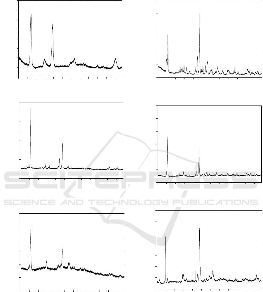
From Figure 3 we could get that the scanning
images of the X-ray diffraction peaks for all types of
asbestos.
Determination Method of Asbestos in Electrical and Electronic Products by using a Scanning Electron Microscope with a X-Ray Diffraction
267
5 1015202530354045505560
0
500
1000
1500
2000
2500
3000
3500
4000
Diffraction intensity( cps)
Angle(2theta. Cu Ka)
(a) Chrysotile
5 1015202530354045505560
0
2000
4000
6000
8000
10000
12000
14000
16000
Diffraction intensity( cps)
Angle( 2theta. Cu Ka)
(b) Amosite
5 1015202530354045505560
0
1000
2000
3000
4000
5000
6000
衍射强度( cps)
Angle( 2theta. Cu Ka)
(c) Crocidolite
5 1015202530354045505560
0
1000
2000
3000
4000
5000
6000
Diffraction intensity( cps)
Angle( 2theta. Cu Ka)
(d)Tremolite
5 1015202530354045505560
0
2000
4000
6000
8000
10000
12000
Diffraction intensity( cps)
Angle( 2theta. Cu Ka)
(e) Actinolite
5 1015202530354045505560
0
1000
2000
3000
4000
5000
6000
7000
Diffraction intensity( cps)
An
g
le( 2theta. Cu Ka)
(f) Anthophylite
Figure 3. The scanning images of X-ray diffraction
peaks for all types of asbestos.
ICVMEE 2019 - 5th International Conference on Vehicle, Mechanical and Electrical Engineering
268

Table 1. The data on the X-ray diffraction characteristics
of all types of asbestos
Chrysotile Crocidolite Amosite
2θ°
I
CPS
2θ°
I
CPS
2θ°
I
CPS
12.06 100 10.68 100 10.68 100
24.50 78 28.9 67 29.17 49
60.38 28 19.84 49 27.29 29
36.85 27 33.04 44 9.65 28
19.83 27 26.62 43 18.89 22
Anthophylite Tremolite Actinolite
2θ°
I
CPS
2θ°
I
CPS
2θ°
I
CPS
28.97 100 28.6 100 10.48 100
9.73 87 10.58 60 28.42 79
36.34 31 33.12 29 33.04 27
26.82 29 9.83 25 27.09 26
19.64 28 33.14 23 9.73 25
From Table 1 we could get that the data on the
X-ray diffraction characteristics of all types of
asbestos.
4 CONCLUSIONS
Based on the combination of SEM and XRD
methods, the method of testing for asbestos in
electronic and electric products has been worked
out. In this method, SEM is used to initially screen
the specimens. XRD is used to identify for the
second time the specimens with particles similar to
those of asbestos fibers, largely improving the
accuracy and reliability of the test.
ACKNOWLEDGEMENTS
The authors would like to acknowledge the financial
support provided for this research work by Fund:
National key R & D program of China
(2017YFF0210703) and (2017YFF0210002).
REFERENCES
Hossein Kakooei, Masode Yunesian, Hossein Marioryad ,
Kamal Azam, Assessment of airborne asbestos fiber
concentrations in urban area of Tehran, Iran, Air Qual
Atmos Health, 2009, 2:39.
ISO 22262. 1: 2012, Sampling and qualitative de-
termination of asbestos in commercial bulk materials.
Selikoff, I.J. et al., The occurrence of asbestosis among
insulation workers in the United States. Annals of the
New York Academy of Sciences., 132, pp139-155,
1966.
Shi H Y, Mao L. Research development of health hazards
and safe handling of asbestos. Shanghai Journal of
Preventive Medicine, 2009, 219(3): 125-128.
Zhang M, Gao X L, et al. Quantitative detection of trace
asbestos by X-ray diffraction system. Rock and
Mneral Analysis, 2010, 29 (3): 319-322.
Zhenglong Chen, Hongwei Hu, Shizhong Hou, et al.
Investigation of Several Common Methods for
Asbestos Detection. 2013.10: 89-94.
Determination Method of Asbestos in Electrical and Electronic Products by using a Scanning Electron Microscope with a X-Ray Diffraction
269
