
Active Contour Segmentation based on Histograms and
Dictionary Learning for Videocapsule Image Analysis
Gaetan Raynaud, Camille Simon-Chane
a
, Pierre Jacob and Aymeric Histace
b
ETIS UMR 8051, UPS, UCP, ENSEA, CNRS, 6 av. du Ponceau, 95014, Cergy, France
Keywords: Active Contour, Bag of Words, Small Bowel Videocapsule.
Abstract: This article deals with statistical region-based active contour segmentation using histograms and dictionary
learning. Following previous publication, the active contour segmentation using optimization alpha-diver-
gence family, leads to satisfying results. The method of the segmentation is based on histograms of the lumi-
nance of the pixels. To improve this method and to allow it to adapt to more types of images, we propose to
replace luminance histograms with histograms of features using a bag of features model. This approach will
be able to overcome the limitations of the luminance and give a better representation of the image. We will
present the approach to create the new representation of the image, first with associated histograms to show
its potential, using a local approach based on dictionary learning to compute the probability map of each pixel
of the image to belong to the targeted object. In a second step using histograms based on bag of features for
the representation of the image. We present experiments for the two methods on images extracted from small
bowel videocapsule acquisitions and for two types of targeted objects (angiodysplasia and ulcer). We show
that by replacing the luminance representation by a more complex one, we reach better performances for the
segmentation of the targeted objects.
1 INTRODUCTION
The purpose of segmentation is to extract homogene-
ous image regions representing objects. Active con-
tour segmentation methods, first introduced by (Kass
et al., 1988), consist of an iterative process that ap-
plies a velocity to a curve to fit the boundaries of the
targeted object. The forces are applied from the inside
and the outside of the curve. The most common ap-
proach is to use energies related to the segmentation
problem. The existing methods can be divided into
two categories: edge-based models (Kass et al.,
1988), Casselles et al., 1997), and region-based mod-
els (Chan and Vese, 2001). One of the most popular
edge-based models is the Geodesic active contours
(Casselles et al., 1997) which minimize the curve’s
length based on a function of the gradient of the im-
age. The region-based methods like (Chan and Vese,
2001) were introduced in order to overcome the limi-
tations of the edge-based methods. The region-based
methods use statistical descriptors like mean or vari-
a
https://orcid.org/0000-0002-4833-6190
b
https://orcid.org/0000-0002-3029-4412
ance, from the inside and the outside of the region,
which allows sturdiness to noise and better perfor-
mance with weak edges than the edge-based methods.
Another approach, inspired by the region-based
approach, is the statistical-region based active con-
tour. This last approach intents to improve the region-
based descriptors, like mean or variance of pixels,
that fail to segment regions in image that cannot be
easily discriminated by their first order statistics.
(Aubert et al., 2003) and (Lecellier et al., 2009) pro-
poses to use probability density function (PDF), de-
scribing both the inner (
) and outer (
) re-
gions, to evolve the curve. The strategy consists in a
minimization of the distance between the PDFs of in-
ner and outer regions and two reference PDFs de-
scribing the targeted object and the background of the
image. The usual way to do so consists in deriving the
related steering partial differential equation (PDE) of
the energy of the distance between the PDFs.
The key point of the method is to choose the dis-
tance function between two PDFs. Some of the most
used distance are the Kullback-Leibler divergence
(KL), the Hellinger distance or the divergence. In
this paper, we will use alpha-divergences as proposed
Raynaud, G., Simon-Chane, C., Jacob, P. and Histace, A.
Active Contour Segmentation based on Histograms and Dictionary Learning for Videocapsule Image Analysis.
DOI: 10.5220/0007694706090615
In Proceedings of the 14th International Joint Conference on Computer Vision, Imaging and Computer Graphics Theory and Applications (VISIGRAPP 2019), pages 609-615
ISBN: 978-989-758-354-4
Copyright
c
2019 by SCITEPRESS – Science and Technology Publications, Lda. All rights reserved
609

by (Meziou et al., 2011). The alpha-divergence be-
tween two PDFs
and
is given by Eq. (1):
(1)
with
=
(2)
In the case of the alpha-divergence between the
two PDFs of the inner and outer regions, the corre-
sponding energy J is given by Eq. (3):
(3)
where is the boundary between
and
,
is
the alpha-divergence between the two PDFs, the last
term is the regularization of the contour with being
a positive constant. The PDFs are calculated using
Parzen window approach.
Calculating the Euler derivative of (3), leads to
evolution equation (4) of :
(4)
With
(5)
In (4) and (5),
denotes the first order deriva-
tive of φ function with respect to the estimated PDFs,
is the Gaussian kernel (with standard-deviation σ)
used in estimation of the PDFs of the two regions, I(x)
is the statistical function representing the segmented
image at the pixel x, and N the inward local normal
vector of the moving curve Γ.
In this article, the optimization of the φ function
is not the main purpose; a method to do so is pre-
sented in (Meziou et al., 2014). As a consequence, we
use the α parameter as a constant of
, leading the al-
pha-divergence to correspond to:
In fact, we propose a method to improve the sta-
tistical-region based active contour used in (Meziou
et al., 2014) in which the histogram used for the sta-
tistical representation, only takes into account the sta-
tistical luminance distribution of the data in the im-
age. This paper proposes to replace the luminance by
more complex representation and to use bags of fea-
tures histograms as statistical representation.
The paper is organized as follow: In section 2, we
begin by introducing the bags of features method,
how to create them, and show some results. Then sec-
tion 3 presents how we can use this approach to im-
prove the statistical-region based method and the re-
sults on medicals images related to gastrointestinal
image extracted from small-bowel videocapsules ac-
quisition. Finally, section 4 concludes the paper.
2 LOCAL PROBABILITY USING
DICTIONARY LEARNING
2.1 Method
In this section, we want to test if a bag-of-features ap-
proach can be a promising idea and if it can be fruit-
fully used in the context of histogram-based ap-
proach.
The alpha-divergence method previously intro-
duced is based on histograms analysis to perform the
segmentation. however as mentioned before the lumi-
nance histogram can be not discriminating enough for
complex segmentation task where usual hypothesis
(Gaussian distribution) are not fulfilled. Thus, it could
be interesting to look for another way to represent the
statistical information of the image; the closest
method of the classic luminance histograms being the
bag of words, we investigated this approach. To rep-
resent an image as a bag of words model, we need
first to define the words that will form the dictionary,
that is to say, the patterns that will embed the most
representative statistical properties of the objects to
segment in a further step. A good descriptor for those
words is the Scale-invariant feature transform (SIFT)
descriptor for instance. These descriptors will allow
to get the interest (or most saillant) points inside the
image. Considering those extracted interest points,
we can use the SIFT descriptor, to describe interest
areas around them that will be used for the bag of fea-
tures dictionary.
GIANA 2019 - Special Session on GastroIntestinal Image Analysis
610
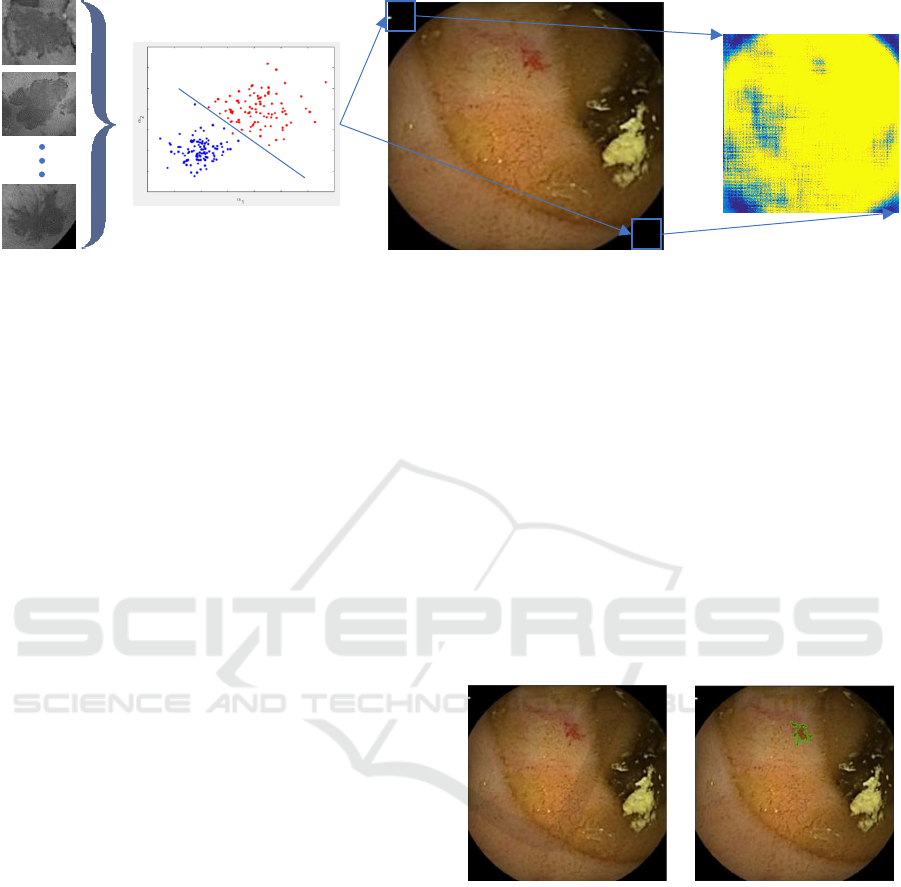
Figure 1: First method consists as a k-means clustering of all the SIFT descriptors extracted from patches containing the type
of pathology which are targeted (learning step). In a second step, the learned SVM classifier is computed on the overall test
image (sliding window strategy) whih is converted into a probability map using the SVM classification score obtained.
The next step consists in creating the dictionary it-
self using the all set of SIFT descriptors obtained from
the images. In that purpose, we use the classic k-
means clustering over the descriptors to perform the
classification and obtain the most representative fea-
tures of the images, which will be the words of the
dictionary.
In this section, we want first to evaluate if diction-
ary learning and bag of features are of related interest
for image segmentation. To do so, an interesting idea
is to have a classifier that can tell if a histogram of
features transposed into the bag of features basis may
properly describe an image which belongs to the class
of object we want to segment. To achieve so, a two-
classes Support Vector Machine (SVM) classifier is a
good option because it will provide the class an image
belongs to, the targeted object or not, and can gives a
score of this classification based on the loss function.
This score will be used to have the probability of the
image to be part of the targeted object. The all pro-
cessing scheme leading to the energy map that will be
used for active contour segmentation is shown in
Fig.1.
At last we associate the heat map with the gray
scale image to generate a weighted gray scale image
and do the active contour segmentation on the new im-
age coming from this association using the maximiza-
tion of alpha-divergence, detailed in (Meziou et al.,
2012).
2.2 Experiments
In this paper, we worked on small-bowel videocapsule
images with a focus on two types of lesions: vascular
lesions (angiodysplasia) and inflammatory lesions (in-
cluding ulcers). The image set we have contains 600
images of angiodysplasia and 450 images of ulcer. It
is a part of the CAD-CAP database (Computer-As-
sisted Detection in Capsule) presented in (Leenhardt
et al., 2019). All the images have an associated mask,
which is the ground truth manually segmented by phy-
sicians and that is used to generate a set of patches
containing the pathology (Fig. 2). An equivalent set of
negative patches is randomly created by taking square
area which does not overlap the ground truth. For all
the methods presented in this paper, we used 80% of
the patches set as a learning base and kept 20% for the
validation process, that is to say to evaluate if the dic-
tionary and SVM classifier leads to a good classifica-
tion of the testing database. The active contour seg-
mentation process using the learned dictionary is eval-
uated on two specific images that were not used in the
learning process nor in the testing.
Figure 2: Left: Example of an angiodysplasia image used for
testing. Right: The image with the associated mask.
In Fig. 3 and Fig. 4 are shown segmentation results
obtained with the classic histogram-based approach of
(Meziou et al., 2014). For this segmentation, we
choose an initialization of the active contour as a circle
inside the targeted object. The segmentation process is
designed to work with a detector that can certify the
presence of the targeted object and gives an approxi-
mate location of it, like the detector proposed in (An-
germann et al., 2016). So, the location returned will be
used as the center of the initialization circle for the
segmentation process.
SV
M
Active Contour Segmentation based on Histograms and Dictionary Learning for Videocapsule Image Analysis
611
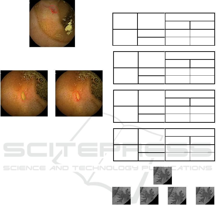
Figure 3: Segmentation done by the alpha-divergence
method based on luminance alone. Red is the result ad green
the ground truth segmentation.
Figure 4: Left: The ulcer image used with the associated seg-
mentation (ground truth). Right: The segmentation done by
the alpha-divergence method based on luminance alone.
The segmentation done using the alpha-divergence
based on luminance alone, that is to say, the classic
approach, doesn’t lead to a satisfying result (80% of
misclassified pixels) on this image, and does not
converge (the process was manually stopped).
Condidering now the first approach proposed in
this paper, to constitute the bag of features we need
first to define the size of the dictionary (number of
clusters for the k-means clustering), if it is obvious to
have 256 clusters when using the luminance of pixels
like for the alpha-divergence approach, it is not that
simpleto determine the optimal size of the considered
bag of features. To decide which size would lead to
the best representation of the images, we tried several
ones and evaluated the SVM classifier trained with
the different dictionaries. The results are computed in
Table 1 under the form of confusion matrices.
From those results one can notice that all
dictionary sizes give high scores for the classification
task. Nevertheless we chose the 4000-cluster one as it
has the best results in terms of performance.
We propose to try our first method with two
different bags of features, using two different
dictionaries: The first one is based on chunk of gray
scale images using only one patch for a given area
presenting with a pathology. For the second, we
augmented the size of the patches set by adding
chunks of images but with a center shifted by 10%, for
each direction, so we have five more patches in
Table 1: Result of the evaluation of the SVM classifier with
several dictionary sizes.
Number of
clusters
500
Predicted
Target
Background
Known
Target
0.91
0.09
Background
0.10
0.90
Number of
clusters
1000
Predicted
Target
Background
Known
Target
0.93
0.07
Background
0.09
0.91
Number of
clusters
2000
Predicted
Target
Background
Known
Target
0.94
0.06
Background
0.06
0.94
Number of
clusters
4000
Predicted
Target
Background
Known
Target
0.97
0.03
Background
0.06
0.94
Figure 5: Example of chunk of images, top row is the chunk
used for the first method, and bottom row is the four chunks
added for the second method (from left to right shifts are
left, right, up, down).
the positive set (Fig. 5). We also added the same
amount of background images (negative examples) to
keep a well balanced image set.
Considering the same initialization scenario as
previously introduced for the classic luminance-histo-
gram approach, the first method leads to a satisfying
result with less than 30% of misclassified pixels (Fig.
6). But seams less robust that the second method that
give stronger results with high probability on the tar-
get area and give less probability in the corner of the
image where all the pixels are black (Fig. 7). This is
from the fact that the second method has more
GIANA 2019 - Special Session on GastroIntestinal Image Analysis
612
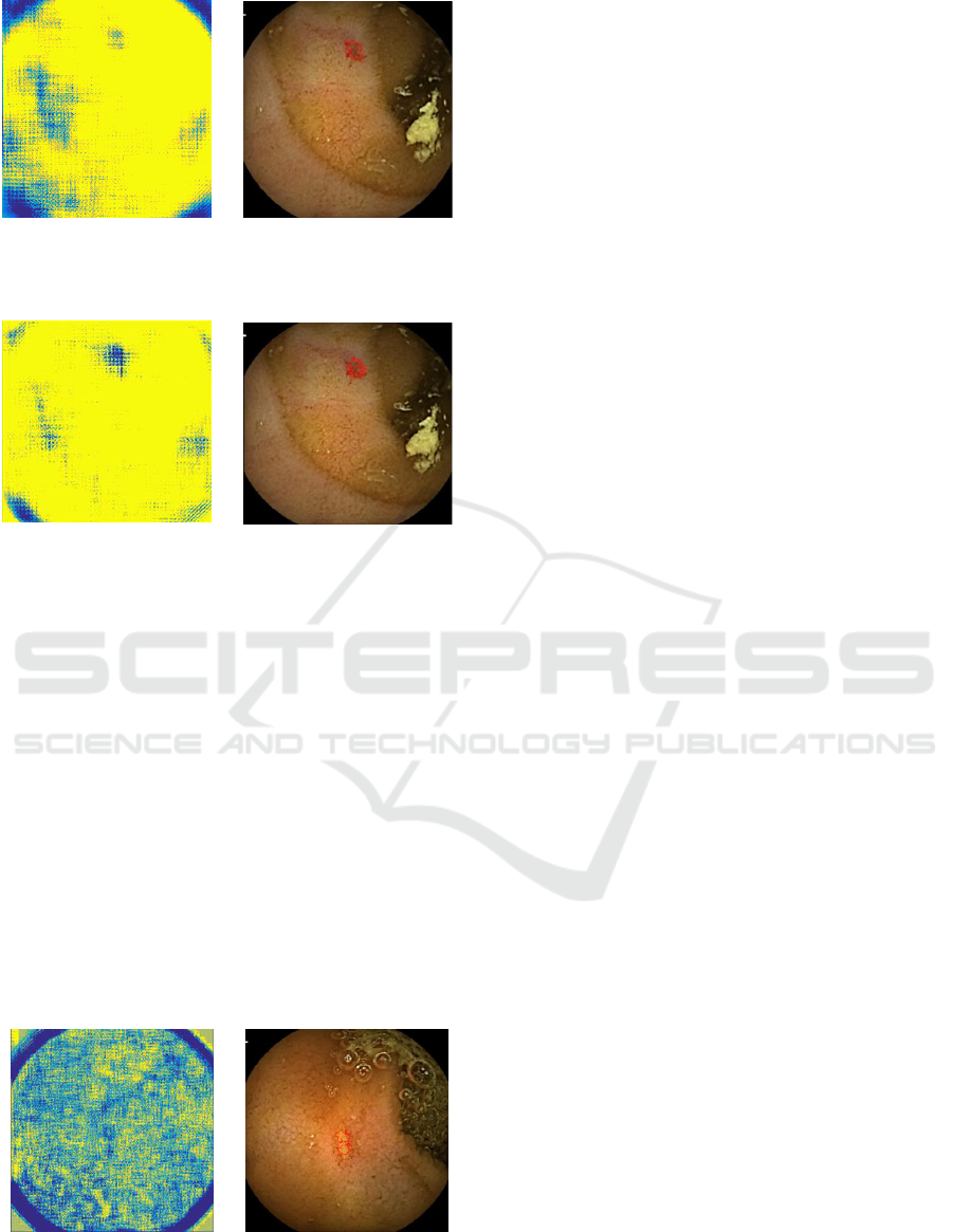
Figure 6: Example of the local probability method, using the
first learned dictionary. Left: The probability map. Right:
Obtained result at convergence.
Figure 7: Example of the local probability method, using the
second learned dictionary. Left: The probability map. Right:
The result.
background features, so it can better recognize them.
Additionally, a key point of this method is in the fact
that segmentation process has a convergence state,
which was not the case with the only luminance based
method.
We also challenged our method on a different tar-
geted object, this time we want to segment an ulcer
using the second dictionary method with more fea-
tures extracted from every image from the data set.
This example means to prove the robustness of the
proposed method. To use the method with another
type of targeted object, it is necessary only to change
the bag of features dictionary with one providing a
good representation of the targeted object. We can see
that even if as the probability map seams chaotic, the
result of the segmentation is promising (Fig. 8).
Figure 8: Example of the local probability method on an ul-
cer, using the second dictionary method. Left: The probabil-
ity map. Right: The result.
From those results, we know that a method based
on bag of features can provide satisfying results. But
this method comes with a strong drawback in terms of
the computation time: a probability map, in our exam-
ples, takes more than 1 hour to be computed for a
500x500 image (on a quad-core 3.3GHz), which is not
compatible with a reasonable use of the method.
Moreover, if it demonstrates that Bags of Features
can be efficiently used for patch classification, the
segmentation process does not use the histogram di-
rectly which is not fully satisfying considering the
work proposed in (Meziou et al., 2014).
3 ACTIVE CONTOUR
OPTIMIZTION USING
HISTOGRAMS OF BAG OF
FEATURES
3.1 Method
Now that it has been demonstrated that a bag of fea-
tures can be valuably used in the context of statistical
region-based active contour segmentation, we can use
them as the base histograms for the alpha-divergence
method to improve the statistical representation of the
image by replacing the histogram of luminance of the
pixels by a histogram of our new bag of features rep-
resentation of the image.
Here what we want to achieve is to create two refer-
ences histograms, a histogram that represents the tar-
geted object and another histogram representing what
is not the targeted object (background). Those
histograms need to be on the same bean-base, so we
decided to use the same database we used previously
(second dictionary with more features) as we know
this bag of features is a good representation for the
targeted object and the background.
As introduced previously, the objective is now to
use the alpha-divergence approach in a competition
scenario, that is to say that the distance between the
reference histogram of the targeted object, and the his-
togram of the inner region of the segmentation and at
the same time the distance between the reference his-
togram for the background and the one of the outer
region will be minimized.
This new definition of the statistical representation
of the inner and outer regions leads Eq. (6) for the en-
ergy :
Active Contour Segmentation based on Histograms and Dictionary Learning for Videocapsule Image Analysis
613
(6)
To compute the histograms, we will consider all
the images that we manually segmented based on
ground truth masks, and we represent them all in the
dictionary base. If we take all the object patches and
represent them in the dictionary base, we have a refer-
ence histogram which represent the occurrences of all
the SIFT descriptors for the targeted object. For the
background, we do the same but with patches that are
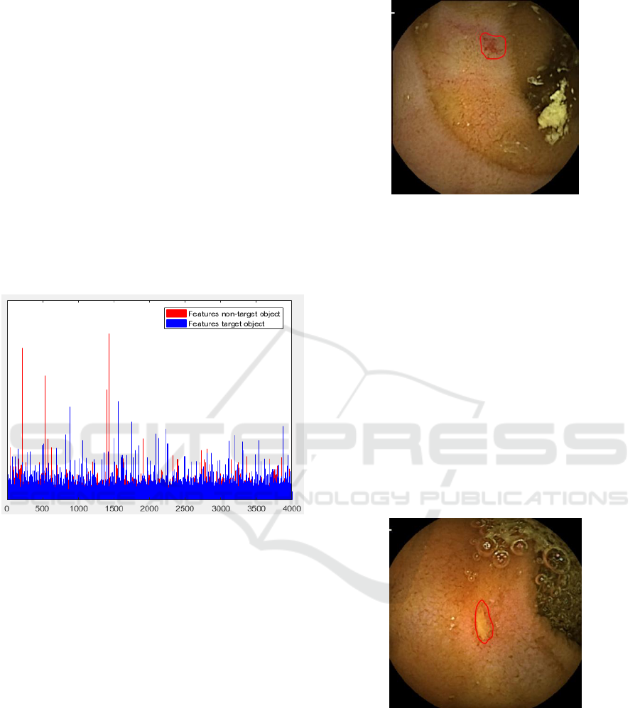
not part of the targeted object. Fig. 9 shows the two
reference histograms that are going to be used for the
alpha-divergence active contour segmentation ap-
proach.
Figure 9: Example of histogram of the features for the target
object (blue) and background (red).
Here we can see that the two histograms are differ-
ent, with for instance the highly-represented features
of the background different from the ones for the tar-
geted object.
3.2 Experiments
To implement the use of the histograms of Fig. 9, at
each iteration of the evolution process of the active
curve, we compute the new histograms for the inner
and outer regions and minimize the distance with the
references (see Eq. (6)).
Fig. 10 shows segmentation results obtained on the
same previously introduced image containing an an-
giodysplasia. With this new base histograms for the
alpha-divergence method, the results are better than
with luminance histograms. That method segment
99% of the targeted pixels, and has 45% of misclassi-
fied pixels, which is far less than the 80% of the
Figure 10: Example of segmentation done with the alpha-
divergence method based on bag of features.
alpha-divergence based on luminance alone, and the
results are the same on both types of images. A key
point here is the fact that this segmentation converges,
unlike the one based on luminance.
The minimization results in a less fit segmentation
on the object than the first method because the refer-
ence histograms are representations of a “BoF” region
that contains the targeted object, so it is not as precise
as a pixel-by-pixel computation. Nevertheless, it does
not require as much time as the first method to com-
pute.
Fig. 11 shows results obtained on the image con-
taining an inflammatory lesion. Obtained result is
quite satisfying and in this case, the final segmentation
does not suffer as much as in the previous case of the
“mean” effect related to the BoF representation.
Figure 11: Example of segmentation done with the alpha-
divergence method based on bag of features.
4 CONCLUSION
In this paper, we proposed a method to improve a sta-
tistical-based active contour segmentation using the
alpha-divergence family to assess the similarity be-
tween two images. The method uses a bag of features
GIANA 2019 - Special Session on GastroIntestinal Image Analysis
614

and SIFT descriptors as base for the representation of
the images. And use references histograms for the tar-
geted object and background of the image, computed
from a learned dataset. We try to minimize the dis-
tance between the reference histogram of the targeted
object and the histogram of the inner region of the seg-
mentation and at the same time the distance between
the reference histogram of the background of the im-
age and the histogram of the outer region. This ap-
proach provides a good combination of the statistical
properties of the whole image. We presented an appli-
cation of this method on two types of medicals images
leading to better results than the luminance base.
As a future work, several approaches can be added
to the method, the first one would be to use the opti-
mization of the alpha parameter of the alpha-diver-
gence (Meziou et al., 2014). Another approach can
consist in changing the minimization between the his-
togram of the region and the reference by a maximiza-
tion of the distance between the histograms of the two
regions. It is also possible to investigate further in the
statistical representation of the image using more
complex representations, as deep features computed
from a convolutional neural network.
REFERENCES
Meziou, L., Histace, A., Precioso, P., Matuszewski, B., and
Murphy, M., 2011, Confocal Microscopy Segmentation
Using Active Contour Based on Alpha-Divergence,
IEEE International Conference on Image Processing
(ICIP), pp. 3138-3141.
Kass, M., Witkin, A, and Terzopoulos, D., 1988, Snakes:
Active contour models, International Journal of Com-
puter Vision, vol. 1, no. 4, pp. 321-331.
Caselles, V., Kimmel, R., and Sapiro, G., 1997, Geodesic
active contours, International Journal of Computer Vi-
sion, vol. 22, no. 1, pp. 61-79.
Chan, T.F., and Vese, L. A., 2001, Active contours without
edges, IEEE Transactions on Image Processing, vol. 10,
pp. 266-277.
Aubert, G., Barlaud, M., Faugeras, O. and Jehan-Besson,
S., 2003, Image segmentation using active contours:
Calculus of variations or shape gradients?, SIAM Jour-
nal of Applied Mathematics, vol. 63, pp. 2128-2154.
Meziou, L., Histace, A., Precioso, F., 2014, Statistical re-
gion-based active contour using optimization of alpha-
divergence family for image segmentation, IEEE Inter-
national Conference on Image Processing (ICIP), pp.
6066-6070.
Angermann, Q., Histace, A., Romain, O., 2016, Active
Learning For Real Time Detection Of Polyps In Vide-
ocolonoscopy, Medical Image Understanding and Anal-
ysis Conference, pp. 182-187.
Meziou, L., Histace, A., Precioso, F., Matuszewski, B. and
Carreiras, F., 2012, Fractional Entropy Based Active
Contour Segmentation of Cell Nuclei in Actin-Tagged
Confocal Microscopy Images, Medical Image Under-
standing and Analysis Conference, pp. 117-123.
Lecellier, F., Jehan-Besson, S., Fadili, J., Aubert, G. and
Revenu, M., 2009, Optimization of divergences within
the exponential family for image segmentation, Interna-
tional Conference on Scale Space and Variational Meth-
ods in Computer Vision, Berlin, Heidelberg, 2009, pp.
137–149, Springer-Verlag.
Leenhardt, R., Vasseur, P. Li, C., Rahmi, G., Cholet, F.,
Saurin, J.-C. …, Histace, A. and Dray, X., 2019, A neu-
ral network algorithm for detection of GI angiectasia
during small-bowel capsule endoscopy, Gastrointesti-
nal Endoscopy,
Active Contour Segmentation based on Histograms and Dictionary Learning for Videocapsule Image Analysis
615
