
Segmentation of Shoulder MRI Data for Musculoskeletal Model
Adaptation
Tom
´
a
ˇ
s Ryba and Zden
ˇ
ek Kr
ˇ
noul
Department of Cybernetics, University of West Bohemia, Univerzitni 8, Pilsen, Czech Republic
Keywords:
Image Segmentation, MRI, Medical Imaging, Deltoid Muscle, Humerus, Clavicle, Scapula.
Abstract:
Applying image processing techniques to medical images has already brought many useful applications. This
work is focused on using these methods in the process of adapting a musculoskeletal model of the shoulder
joint. Comparing the model of healthy individuals and the patient with joint damage leads to a subject-
specific convalescence treatment. This work describes the procedure for segmentation of magnetic resonance
imaging (MRI) of the shoulder joint. Firstly three bones inside the shoulder area: humerus, clavicle, scapula
are identified and thereby it provides initial reference objects. A major step is the segmentation of the deltoid
muscle needed for the subsequent adaptation of the musculoskeletal model. This step is challenging in terms of
image processing due to the closeness of soft tissues, which are almost identical in intensity and the boundaries
between them are often barely visible. The approach to resolving this problem is described and possible
improvements and future work are described.
1 INTRODUCTION
Automated segmentation of medical image data is
still an unresolved task. There are computed to-
mography (CT) data that are characteristic of their
high image quality. At work (Kodym and
ˇ
Span
ˇ
el,
2018), the authors deal with a semi-automatic seg-
mentation method for general use. However, existing
applications or algorithms for segmentation of mag-
netic resonance imaging (MRI) data are always semi-
automatic and require some user intervention to set
initial parameter values that are often specific to the
task only. Therefore, for MRI data, we can find Vas-
cular modeling toolkits (2016) or voice tract segmen-
tation capturing the articulation of healthy subjects
(Engwall and Badin, 1999), (Ojalammi and Malinen,
2017), etc.
There are also applications for MRI segmenta-
tion usable for general task but require very time-
consuming user inputs to define initial configurations.
These parameter values are often different for dif-
ferent anatomies, can not always be derived directly
from the data, and the user usually has to proceed
based on an attempt of error. Recently, there are
very successful general segmentation methods of the
supervised machine learning based on convolutional
neural networks (Xue et al., 2018), (Liu et al., 2018).
This solution, however, leads to an enormous amount
of manual work during the preparation of the large
training data needed as reference for their learning.
On the other hand, in (Ojalammi and Malinen, 2017),
the aim is to process extensive MRI data sets of upper
respiratory and oral routes with minimal user interfer-
ence.
The examination of MRI is useful to improve the
current understanding of the human body’s relation-
ships. The MRI imaging technique is an attractive
alternative to CT because it has no-ionizing radia-
tion. This aspect is particularly important for the pro-
cessing of healthy subjects. We are considering the
healthy subjects for an identification of the muscu-
loskelatal model ((Havelkov
´
a et al., 2017)). On the
other hand, the disadvantage of MRI against CT, is the
worse spatial resolution given by the low raw voxel
data quality. This is caused by the imaging princi-
ple including motion artifacts due to a scanning time,
which can exceed 10 seconds for one high resolution
stationary 3D image.
The aim of this work is segmentation and sub-
sequent 3D reconstruction of the deltoid muscle of
the shoulder complex from MRI volume data. The
task is to separate the muscle from the background.
The background consist of other muscles and bones,
moreover some of them anatomically attached to the
segmented muscle. The 3D reconstruction transforms
segmented deltoid muscule to 3D surface model that
Ryba, T. and Kr
ˇ
noul, Z.
Segmentation of Shoulder MRI Data for Musculoskeletal Model Adaptation.
DOI: 10.5220/0007580701550160
In Proceedings of the 12th International Joint Conference on Biomedical Engineering Systems and Technologies (BIOSTEC 2019), pages 155-160
ISBN: 978-989-758-353-7
Copyright
c
2019 by SCITEPRESS – Science and Technology Publications, Lda. All rights reserved
155

is the input to musculoskeletal simulation of the
shoulder complex.
We try to perform the segmentation with minimal
user interference. Our work is directed to subsequent
research needing processing of extensive data of both
healthy individuals and patients with a shoulder com-
plex disorder. Therefore the application is not specifi-
cally designed for doctors to examine the patient, nor
is it an alternative to the software and user interface
provided by the MRI scanner manufacturer.
2 RELATED WORK
The hard tissue segmentation is a well-managed
technique in terms of their good visibility, in both
computed tomography (CT) and magnetic resonance
imaging (MRI). It is often used for diagnostic pur-
poses, such as assistance in preoperative planning
or initiation of downstream segmentation techniques
of other non-hard tissues. Precise and, at the same
time, fully automatic segmentation of hard tissue is
achieved when the patient’s anatomy does not devi-
ate significantly from the standard. The results are
currently also used for example for 3D implant print-
ing (Tetsworth et al., 2017) or teaching aids (Ji
ˇ
r
´
ık
et al., 2014).
The different non-hard tissues have low contrast
borders with each other in both CT and MRI data and
make its automatic segmentation very difficult also
due to anatomical variability or various pathologies.
In this case, the semi-automatic approaches are cur-
rently successful. The user searches for specific al-
gorithm parameter values to achieve the required seg-
mentation result accuracy. Although some applica-
tions do not require very anatomically precise surface
models, for the precise segmentation, it must to be
done by clinical professionals processing volumetric
data one slice after another. This is very challenging
due to tedious processing of a large number of slices
in volume data.
The segmentation of the hard tissue is sufficient by
adaptive thresholding techniques (Rathnayaka et al.,
2011) or area growth algorithm (Xi et al., 2014). The
segmentation failure is where the boundaries of an
object pass through areas of the low contrast. To
deal with this problem, there are approaches based
on models of active contours, (Pinheiro and Alves,
2015), optimizing the smoothness and a continuity
criteria.
Moreover, the methods of active or statistical
shapes (He et al., 2016) and (Yokota et al., 2013) as-
sume prior shape of a segmented object. However,
this must be obtained by learning from previous (often
manual) ideal segmentation of all potentially possi-
ble shapes. This can be a problem with unpredictable
anatomical pathologies. On the other hand, the user
input during their application degrades only on the
pre-positioning of the model in reference pose as near
as possible to the location (Virz
`
ı et al., 2017) or, in the
case of a fully automated method, this step is com-
pletely eliminated (Antong et al., 2010).
As another form of segmentation, the image reg-
istration can be considered (Hajnal and Hill, 2001).
This is not the pure segmentation technique because
needs a reference image in addition. It determines
the geometric relationship between each point of the
reference image and the processed image as a cost-
optimization function. However there are unregulated
registration algorithms for comparing deform-able or-
gans such as the brain, liver or lung (Rohlfing and
Maurer, 2003), (Ino et al., 2005). The failure of regis-
tration caused by dropping search algorithm into local
optima is prevented through generating a large train-
ing set for the deep-learning image registration (Ito
and Ino, 2018).
More recently, graph-based methods provide bi-
nary segmentation as the search for a global optima
separating object from the background. These meth-
ods are reliable if the user again provides a sufficient
amount of accurate user inputs. For MRI or CT, it
has a form of seeds labeled in many slices of vol-
umetric images. Furthermore automation reducing
amount of manual inputs benefits from a combina-
tion of graph-cut techniques with prior information
provided by some edge detection method (Keuster-
mans et al., 2012), (Kr
ˇ
cah et al., 2011) or a classifica-
tion technique based e.g. on random decision forests
(RDF) (Kodym and
ˇ
Span
ˇ
el, 2018). The last men-
tioned method searches for optimal binary segmenta-
tion of volumetric data with respect to the probability
field obtained from RDF classifiers online trained on
only a few expertly annotated sections.
In general, the convolutional networks are ma-
chine powerful learning techniques and are currently
a successful segmentation technique for the medical
imaging data (Ghosal and Ray, 2017), (Prasoon et al.,
2013). They overcome previously popular segmen-
tation techniques based on RDF classification (Loh,
2011), which uses random subsets of available train-
ing data to build a set of binary decision trees. In
the context, these data-driven and supervised tech-
niques need training data that is very varied due to
a wide range of imaging techniques used in medicine.
There are methods increasing accuracy and robust-
ness by generating thousands of synthetic training
data from only a few input original images (Ito and
Ino, 2018) and-or often combining with an augmen-
BIOIMAGING 2019 - 6th International Conference on Bioimaging
156

tation method (Milletari et al., 2016), (Ronneberger
et al., 2015).
3 METHODS
3.1 Image Preprocessing
A common problem in image processing is the image
noise. In order to deal with it, several options may
be used. The commonly used method is the Gaussian
filtering. Unfortunately, an obvious disadvantage of
this approach is blurring of image edges. As already
stated main goal of this work is to segment one cer-
tain muscle in the shoulder complex. The boundary
between this muscle and background is barely visi-
ble. Therefore, it’s not a reasonable approach to blur
them even more.
To reduce the image noise and leave the edges
untouched at the same time an edge preserving filter
must be used. In this work the bilateral filter (Tomasi
and Manduchi, 1998) is used. The weights in this fil-
ter correspond not only to distance as in the Gaussian
filter but to the intensity difference of pixels as well.
This way, only pixels that are geometrically close and
have similar intensity are taken into account during
filtering. And because edges are defined as pixels
with high intensity gradient, they’re usually filtered
very slightly.
Comparison of the Gaussian filter and the bilateral
filter is shown in Figure 1.
3.2 Image Segmentation
For the purpose of image segmentation, a semiauto-
matic tool was developed. It works on an established
system of placing seed points representing the object
of interest and another group of points representing
the background. For the algorithm processing the data
with defined seed points, we tested three commonly
used methods - Graph Cut (GC) (Boykov and Jolly,
2000), Random Walker (RW) (Grady, 2006) and Wa-
tershed (WS) (Dobrin et al., 1994).
The GC method is based on creating intensity
models for each object. These models are calcu-
lated from given seed points. Using the GC often
leads to overtrained models. This is due to very
similar object densities when segmenting one spe-
cific muscle. In this case, points with similar den-
sity are marked as two different objects. Moreover, to
mark the background object properly the correspond-
ing model needs to describe image parts that signifi-
cantly differ in intensity. The resulting model is not
descriptive enough and the algorithm gets confused.
Given the set of seed points, the RW algorithm
finds the closest path to one of the seed point for each
unlabeled pixel. There are no direct connections to an
intensity model like in the GC algorithm. Therefore,
the closest path depends strongly on the seed position,
which often yields to a significantly higher amount
of needed seed points that spreads on all parts of the
object. The needed interactivity was overwhelming
especially when segmenting the muscle.
Using the simple WS algorithm provides us with
the best results regarding the precision of segmen-
tation and the amount of needed interaction. An-
other important advantage of this approach is its ef-
ficiency and computation speed. Using the WS al-
gorithm makes the whole segmentation process faster
and more fluent.
To help the operator with orientation it’s possible
to switch between different views - coronal, sagittal
and axial. This way, it is possible to change the views
during the segmentation process to define the seed
points more accurately.
3.2.1 Extracting the Bones
For easier orientation and further data processing,
three bones are segmented: humerus, scapula, and
clavicle. The Humerus is a long bone of the arm that
forms the shoulder joint on the one end and the el-
bow joint on the second end. The Scapula or shoulder
blade is a triangular bone that lies on the upper back.
The Clavicle is an anterior bone of the shoulder. Its
main function is to support the shulder.
In the MRI the bones are well separated due to
their high intensity. The contrast between a bone and
near soft tissues is significant. Therefore, the needed
amount of interactivity is much lower comparing to
the muscle segmentation.
Nevertheless, to use a fully autonomous approach,
e.g. thresholding, is not recommended. Despite the
bones, the’re different objects with the similar inten-
sity that would be segmented as well. Using such an
approach often yields to results where for example the
clavicle and the skin are connected into one big ob-
ject.
On the other hand, the WS algorithm is perfectly
suited for this task. The final segmentation of the
humerus is shown in the second image in Figure 2.
3.2.2 Extracting the Deltoid
Segmentation of one specific muscle is a much more
challenging task. The reason for this is the intensity
similarity of the soft tissues and an unclear boundary
between individual muscles. The risk of overtraining
a segmentation algorithm based on intensity models is
Segmentation of Shoulder MRI Data for Musculoskeletal Model Adaptation
157
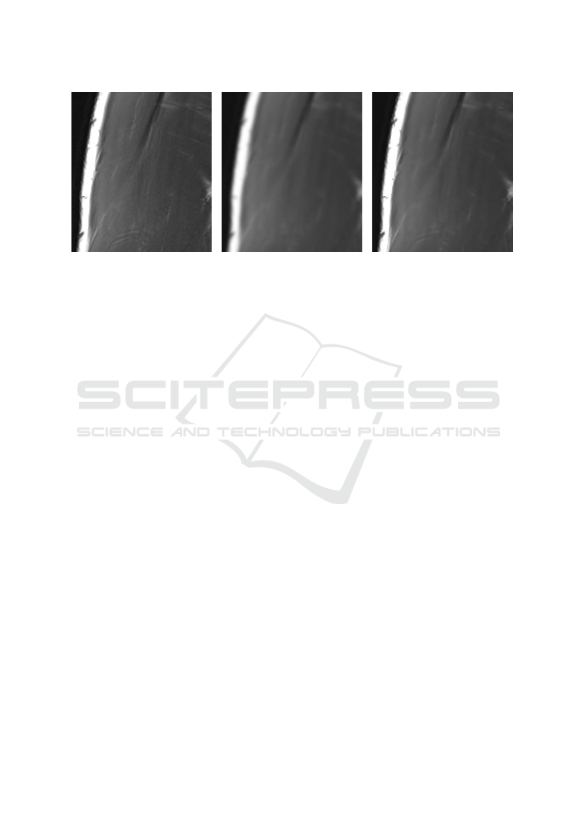
Figure 1: Input image (left) filtered by Gaussian (middle) and by a bilateral filter (right).
significantly higher than in the bones extraction case
and the amount of interactivity increases.
Moreover, this task is much more focus demand-
ing than the bone segmentation. A certain amount of
anatomy knowledge is also needed, especially in parts
with an unclear boundary between individual mus-
cles. Segmentation of bones prior to the muscle seg-
mentation is recommended for a better orientation.
The WS algorithm meets its limits here but still
provides reasonable results. Therefore, the segmenta-
tion refinement that is described in the next section is
very important in these cases.
3.3 Segmentation Refinement
The segmentation obtained by the WS algorithm is
sometimes very coarse and inaccurate. The reason
for that is the algorithm’s sensitivity to the noise and
unclear object boundaries. Despite the preprocessing
and proper noise filtration, the filtered data are still not
perfect. Is it, therefore, appropriate to use a segmenta-
tion method that will start at the coarse segmentation
and will refine the result to better match the reality.
An information that could be used in this step is
that most of the objects in the human body tend to be
compact with no sudden changes in shape. A perfect
approach that respects such an information is the ac-
tive contours approach (Pinheiro and Alves, 2015).
Methods based on active contours need to be ini-
tialized by an initial curve. This initial curve should
be as close to the desired result as possible, which
minimizes the possibility of getting stuck in a local
energy minimum. In this work, this initial curve cor-
responds to the boundary of the coarse segmentation
achieved from the image segmentation. During the
iterative process the curve evolves and due to the cal-
culation tends to be smooth and compact.
The last step in the postprocessing procedure is
the morphological filtering. This way the boundary
of the segmentation is smoothed even more, which
yields more reliable results.
4 RESULTS
In this work, we used the MRI of a 30 years old
healthy male subject. The final segmentation of the
humerus and the deltoid muscle is shown in Figure 2.
The main result of this work is the software for
MRI processing. The developed software is used for
segmentation of specific bones and soft tissues in the
shoulder.
The segmentation is done with an interactive ver-
sion of the watershed algorithm. Using this approach
yields to fast responses of the algorithm as the user
draws seed points over the input data. This coarse
segmentation is then refined using an active contours
method.
As more data are processed the software will learn
and the future processing should be faster.
5 CONCLUSIONS AND FUTURE
WORK
To develop a fully autonomous segmentation pro-
cess is a very challenging task especially regarding
muscle segmentation. We faced this challenge using
an interactive method based on the watershed algo-
rithm. Probably the biggest disadvantage of this ap-
proach is the amount of needed interactivity in some
cases. This interactivity could be reduced using sev-
eral ways.
For example, a thresholding algorithm could be
used for segmentation of the bones. As already men-
BIOIMAGING 2019 - 6th International Conference on Bioimaging
158

Figure 2: Segmentation of bones (the humerus on the first two images) and segmentation of soft tissues (deltoid muscle on
the last two images).
tioned, using such an approach could yield to a big
object consisting of a bone and other objects. We be-
lieve that with a proper postprocessing this problem
could be solved. Therefore, at least some steps could
be automized in the future work.
At this moment, the segmentation process is cal-
culated over the whole image. To improve the pro-
cessing speed defining a region of interest could be
implemented.
Another goal of our work is to create a statisti-
cal representation of the position of the deltoid mus-
cle. This means that for each point we would like to
calculate a probability that this point belongs to the
deltoid muscle. Segmented humerus, scapula, and
clavicle will be taken as the reference objects. Cre-
ating such an atlas could autonomously propose seed
points defining the deltoid thus decreasing the needed
amount if interactivity.
The problem of this approach is the uniqueness
and difference between subjects - factors such as
height, weight, musculature, sex etc. plays a signif-
icant role regarding the position and shape of the cor-
responding deltoid muscle. Having a large number of
data containing all of these factors, it could be possi-
ble to create more statistical atlases and then use the
one that best fits the given subject.
ACKNOWLEDGEMENTS
This research was supported by the project ”38 Vir-
tual human body model for prevention, therapy and
rehabilitation of shoulder diseases” realized within
the frame of the Program INTERREG V-A: Cross-
border Cooperation between the Czech Republic and
the Federal State of Germany Bavaria, Aim European
Cross-border Cooperation 2014 - 2020. The realiza-
tion is supported by financial means of the European
Regional Development Fund and the state budget of
the Czech Republic.
REFERENCES
Antong, C., Deeley, M. A., Niermann, K. J., Moretti, L.,
Dawant, B. M., Department of Radiation Oncology,
Vanderbilt-Ingram Cancer Center, . n. A. S. N. T. .,
of Electrical Engineering, D., and Computer Science,
Vanderbilt University, N.-T. . (2010). Combining reg-
istration and active shape models for the automatic
segmentation of the lymph node regions in head and
neck CT images. Medical Physics, 37(12).
Boykov, Y. and Jolly, M.-P. (2000). Interactive organ seg-
mentation using graph cuts. In International con-
ference on medical image computing and computer-
assisted intervention, pages 276–286. Springer.
Dobrin, B. P., Viero, T. J., and Gabbouj, M. (1994). Fast wa-
tershed algorithms: analysis and extensions. In Non-
linear Image Processing V, volume 2180, pages 209–
221. International Society for Optics and Photonics.
Engwall, O. and Badin, P. (1999). Collecting and analysing
two- and three-dimensional MRI data for swedish.
Quarterly Progress and Status Report - Royal Insti-
tute of Technology, Department of Speech, Music and
Hearing, 3.
Ghosal, S. and Ray, N. (2017). Deep deformable registra-
tion: Enhancing accuracy by fully convolutional neu-
ral net. Pattern Recognition Letters, 94:81 – 86.
Grady, L. (2006). Random walks for image segmentation.
IEEE transactions on pattern analysis and machine
intelligence, 28(11):1768–1783.
Hajnal, J. and Hill, D. (2001). Medical Image Registration.
Biomedical Engineering. Taylor & Francis.
Havelkov
´
a, L.,
ˇ
Spi
ˇ
cka, J., and Hyn
ˇ
c
´
ık, L. (2017). The new
torus obstacle method used for musculoskeletal mod-
eling. In 23st CONGRESS OF EUROPEAN SOCIETY
OF BIOMECHANICS.
He, B., Huang, C., Zhou, S., Hu, Q., and Jia, F. (2016). Fast
automatic 3D liver segmentation based on a three-
Segmentation of Shoulder MRI Data for Musculoskeletal Model Adaptation
159

level adaboost-guided active shape model. Medical
Physics, 43(5).
Ino, F., Ooyama, K., and Hagihara, K. (2005). A data dis-
tributed parallel algorithm for nonrigid image regis-
tration. Parallel Computing, 31:19–43.
Ito, M. and Ino, F. (2018). An automated method for
generating training sets for deep learning based im-
age registration. In Proceedings of the 11th Inter-
national Joint Conference on Biomedical Engineering
Systems and Technologies (BIOSTEC 2018) - Volume
2: BIOIMAGING, Funchal, Madeira, Portugal, Jan-
uary 19-21, 2018., pages 140–147.
Ji
ˇ
r
´
ık, M., Ryba, T., Svobodov
´
a, M., M
´
ırka, H., and
Li
ˇ
ska, V. (2014). LISA - liver surgery ana-
lyzer software development. In Proceedings of the
11th World Congress on Computational Mechanics
(WCCM 2014), Barcelona.
Keustermans, J., Vandermeulen, D., and Suetens, P. (2012).
Integrating statistical shape models into a graph cut
framework for tooth segmentation. In Wang, F., Shen,
D., Yan, P., and Suzuki, K., editors, Machine Learn-
ing in Medical Imaging, pages 242–249, Berlin, Hei-
delberg. Springer Berlin Heidelberg.
Kodym, O. and
ˇ
Span
ˇ
el, M. (2018). Semi-automatic CT im-
age segmentation using random forests learned from
partial annotations. In Proceedings of the 11th In-
ternational Joint Conference on Biomedical Engineer-
ing Systems and Technologies - Volume 2: BIOIMAG-
ING,, pages 124–131. INSTICC, SciTePress.
Kr
ˇ
cah, M., Sz
´
ekely, G., and Blanc, R. (2011). Fully auto-
matic and fast segmentation of the femur bone from
3D-CT images with no shape prior. In 2011 IEEE In-
ternational Symposium on Biomedical Imaging: From
Nano to Macro, pages 2087–2090.
Liu, F., Zhou, Z., Jang, H., Samsonov, A., Zhao, G., and
Kijowski, R. (2018). Deep convolutional neural net-
work and 3d deformable approach for tissue segmen-
tation in musculoskeletal magnetic resonance imag-
ing. Magnetic resonance in medicine, 79(4):2379–
2391.
Loh, W.-Y. (2011). Classification and regression trees.
Wiley Interdisciplinary Reviews: Data Mining and
Knowledge Discovery, 1(1):14–23.
Milletari, F., Navab, N., and Ahmadi, S.-A. (2016). V-
net: Fully convolutional neural networks for volumet-
ric medical image segmentation. 2016 Fourth Inter-
national Conference on 3D Vision (3DV), pages 565–
571.
Ojalammi, A. and Malinen, J. (2017). Automated segmen-
tation of upper airways from MRI: Vocal tract geom-
etry extraction. In Proceedings of the 10th Interna-
tional Joint Conference on Biomedical Engineering
Systems and Technologies, volume 2, pages 77–84.
SciTePress.
Pinheiro, M. and Alves, J. L. (2015). A new level-set-
based protocol for accurate bone segmentation from
CT imaging. IEEE Access, 3:1894–1906.
Prasoon, A., Petersen, K., Igel, C., Lauze, F., Dam, E., and
Nielsen, M. (2013). Deep feature learning for knee
cartilage segmentation using a triplanar convolutional
neural network. In International conference on medi-
cal image computing and computer-assisted interven-
tion, pages 246–253. Springer.
Rathnayaka, K., Sahama, T., Schuetz, M. A., and Schmutz,
B. (2011). Effects of CT image segmentation meth-
ods on the accuracy of long bone 3D reconstructions.
Medical engineering & physics, 33(2):226–233.
Rohlfing, T. and Maurer, C. R. (2003). Nonrigid im-
age registration in shared-memory multiprocessor en-
vironments with application to brains, breasts, and
bees. IEEE Transactions on Information Technology
in Biomedicine, 7(1):16–25.
Ronneberger, O., Fischer, P., and Brox, T. (2015). U-net:
Convolutional networks for biomedical image seg-
mentation. In International Conference on Medical
image computing and computer-assisted intervention,
pages 234–241. Springer.
Tetsworth, K. D., Block, S., and Glatt, V. (2017). Putting
3D modelling and 3D printing into practice: virtual
surgery and preoperative planning to reconstruct com-
plex post-traumatic skeletal deformities and defects.
In SICOT-J.
Tomasi, C. and Manduchi, R. (1998). Bilateral filtering for
gray and color images. In Computer Vision, 1998.
Sixth International Conference on, pages 839–846.
IEEE.
Virz
`
ı, A., Marret, J. ., Muller, C. O., Berteloot, L., Boddaert,
N., Sarnacki, S., and Bloch, I. (2017). A new method
based on template registration and deformable models
for pelvic bones semi-automatic segmentation in pe-
diatric MRI. In 2017 IEEE 14th International Sympo-
sium on Biomedical Imaging (ISBI 2017), pages 323–
326.
Xi, T., Schreurs, R., Heerink, W. J., Berg
´
e, S. J.,
and Maal, T. J. J. (2014). A novel region-
growing based semi-automatic segmentation protocol
for three-dimensional condylar reconstruction using
cone beam computed tomography (CBCT). In PloS
one.
Xue, Y., Xu, T., Zhang, H., Long, L. R., and Huang, X.
(2018). Segan: Adversarial network with multi-scale
l 1 loss for medical image segmentation. Neuroinfor-
matics, pages 1–10.
Yokota, F., Okada, T., Takao, M., Sugano, N., Tada, Y.,
Tomiyama, N., and Sato, Y. (2013). Automated CT
segmentation of diseased hip using hierarchical and
conditional statistical shape models. In Mori, K.,
Sakuma, I., Sato, Y., Barillot, C., and Navab, N.,
editors, Medical Image Computing and Computer-
Assisted Intervention – MICCAI 2013, pages 190–
197, Berlin, Heidelberg. Springer Berlin Heidelberg.
BIOIMAGING 2019 - 6th International Conference on Bioimaging
160
