
Detection of Enterohemorrhagic Escherichia coli (EHEC) in
Consumption Water Source using Multiplex PCR Method
Khoirun Nihayati
1
, Yuanita Rachmawati
1*)
, Saiku Rokhim
1
, Linda Prasetyaning
2
1
Science & Technology Faculty, Universitas Islam Negeri Sunan Ampel, Surabaya, Indonesia
2
Psychology & Health Faculty, Universitas Islam Negeri Sunan Ampel, Surabaya, Indonesia
Keywords : Escherichia coli O157: H7, water consumption source, Multiplex PCR.
Abstract :
Escherichia coli is microorganism that often causes diarrhea. One of the most pathogenic E. coli bacteria for
humans is Escherichia coli O157: H7. These bacteria are included in E. coli Enterohemorrhagic (EHEC)
bacteria which produce shiga like toxin. Shiga like toxin causes Hemorrhagic Colitis (HC) and Hemolytic
Ureamic Syndrome (HUS). Water is a basic need for living things. Water that is suitable for use as a source
of consumption must fullfill certain chemical, physical and biological requirements. Biologically, the source
of consumption water should not contain E. coli. The presence of these bacteria indicates fecal contamination.
Water, especially consumption water, is a potential source for the spread of microorganisms that cause
infection, including E. coli O157: H7 bacteria. The purpose of this research is to detect Escherichia coli O157:
H7 bacteria in consumption water source sample using multiplex PCR method. The primers used in this study
were fliCh7 (625 bp), rfbE (296 bp), stx1 (210 bp), stx2 (484 bp), eaeA (397 bp), and hly (166 bp) genes. The
process of bacterial isolation was carried out through several stages, namely presumptive test, confirmed test,
and completed test. Isolation of bacterial DNA was carried out using boiling cell method. Amplification
process was carried out in conditions of 94˚C-2 minutes, followed by 35 cycles of 94˚C-20 seconds, 63˚C-1
minute annealing, and 72˚C-1 extension minutes, and post-extension 72˚C-10 minutes. Next, 2% agarose gel
electerophoresis was done at 50 volts for 65 minutes. The results showed that from the total 7 samples tested,
all are positive samples of coliform bacteria, 6 are positive samples of Escherichia coli bacteria, and 3 are
positive samples of Escherichia coli O157: H7 bacteria with virulent expression of different genes including
stx1 and eaeA genes. An analysis of all irrigation systems is needed to determine the entry of coliform
contamination, E. coli and E. coli O157: H7.
1. INTRODUCTION
Escherichia coli is a normal flora microorganism in
the human digestive tract and warm-blooded animals
that are facultative anaerobic (Drasar and Hill, 1974).
Based on its pathogenicity, E. coli is divided into 6
groups: Enteropathogenic E. coli (EPEC),
Enterotoxigenic E. coli (ETEC), Enterohemorrhagic
E. coli (EHEC), Enteroaggregative E. coli (EAEC),
Enteroinvasive E. coli (EIEC), and Diffusely
Sdherent E. coli (DAEC) (Nataro and Kaper, 1998).
Escherichia coli O157: H7 is part of the
Enterohemorrhagic E. coli group (EHEC) which is
the most pathogenic of other types due to its ability to
produce shiga like toxin which can cause Thrombotic
Thrombocytopenic Purpura (TTP), Hemorrhagic
Colitis, and Hemolytic Ureamic Syndrome (HUS)
(Law, 2000)
Water that is suitable for use as a source of
consumption must meet the requirements chemically,
physically, and biologically. Chemically,
consumption water must not contain toxic chemicals.
Physically, water should be odorless, tasteless and
colorless. Meanwhile, biologically, consumption
water should not contain E. coli bacteria. The
presence of these bacteria indicates that the water has
been contaminated by feces (Mubarak and Chayatin,
2009). Escherichia coli O157:H7 infections are
mostly caused by food or milk originating from
livestock, including fresh fruit, vegetables and water.
In America, the first incident reported
Escherichia coli O157:H7 infection after drinking
contaminated water occurred in 1989 in Missouri
Village. Food and water contamination has been
identified as a potential source of the spread of
pathogenicity of EHEC O157 in humans (Tutenel et
al., 2003). The Ministry of Health of the Republic of
Indonesia has urged the public to be aware of diseases
caused by E. coli bacteria. Because, according to the
Ministry of Health data, the outbreak of this disease
108
Nihayati, K., Rachmawati, Y., Rokhim, S. and Widayanti, L.
Detection of Enterohemorrhagic Escherichia coli (EHEC) in Consumption Water Source using Multiplex PCR Method.
DOI: 10.5220/0008907000002481
In Proceedings of the Built Environment, Science and Technology International Conference (BEST ICON 2018), pages 108-112
ISBN: 978-989-758-414-5
Copyright
c
2022 by SCITEPRESS – Science and Technology Publications, Lda. All rights reserved

actually began to occur in Germany in mid-May
2011. Until June 2, 2011, Germany found 520 cases
of haemolytic uraemic syndrome (HUS) with 11
deaths. There were 1,213 cases of
enterohaemorrhagic Escherichia coli (EHEC); 6 of
them died. In Germany, there are 1,733 cases and 17
deaths (Zakki, 2015). Cases of E. coli O157:H7 cases
have been widely reported in developed countries
such as the United States and Japan, but there are few
data for cases that occur in Indonesia. This is due to
the unavailability of selective media for the isolation
of these pathogenic bacteria (Aziz et al., 2009). Hill
and Jinneman (2000) explained that, for the purpose
of epidemiological studies, a zoonotic agent should
be used in the application of genetic techniques
because the data produced have very high accuracy.
One of the molecular methods that can be used is the
multiplex PCR method. This method uses a variety of
primers to produce amplicons of various sizes
specific to different DNA sequences in one running-
test.
Multiplex Polymerase Chain Reaction is a type
of PCR technique that uses several primary pairs in
one reaction to produce amplicons from various target
genes with different DNA sizes. The use of molecular
techniques is done because it is specific to the target
genes to be detected. Besides that, the time used is
shorter compared to conventional testing. Therefore,
the purpose of this study is to detect the presence of
Escherichia coli O157: H7 bacteria in consumption
water sources samples using Multiplex PCR method.
2. RESEARCH METHODS
2.1. Tools and materials
The tools used in this study are Laminar Air Flow
(LAF), glass beaker, hot plate, ose needle, measuring
pipette, 10 µl micropipette, 20 μl, 200 μl, and 1000
μl, analytic balance sheet, centrifuge, autoclave,
incubator, water heater, thermocycler (Labnet),
spectrophotometer (Bio-Drop), electrophoresis
devices (Mupid-Exu), and gel documentation tool
(Enduro GDS-1302 Labnet).
The materials used in this study include 7
samples of consumption water, Lactose Broth (LB)
(Merck) media, Eosin Methylen Blue (EMB)
selective media (Merck), Nutrient Agar (NA) media (
Merck), sterile aquadest, Go Taq® Green mastermix,
six pairs of forward and reverse primary for multiplex
PCR detection including fliCh7 (flagellar antigen),
rbf E (antigen O157), stx 1 (shiga toxin 1), stx 2 (
shiga toxin 2), eaeA (intimin), and hly (haemolysin)
(Macrogen) (Table 1), agarose (Promega), DNA
scavengers Diamond Nucleic Acid Dye (Promega),
TAE Buffer solutions (Promega), loading dye
(Promega ), DNA Ladder 100 bp (Promega), isolates
of Escherichia coli O157: H7 bacteria as positive
controls obtained from the Veterinary Public Health
Laboratory of the Faculty of Veterinary Medicine,
Universitas Gadjah Mada of Yogyakarta, and isolates
Candida albicans as a negative control.
2.2. Sampling Method
The sampling process was carried out using purposive
sampling method. Total samples used in this study are
7 samples including tap water and ice cubes.
2.3. Presumptive Test
1 ml of water sample was inoculated into 9 ml Lactose
Broth (LB) media which was sterilized using
autoclave at 121
o
C for 15 minutes. Then, it was
incubated at 37° C for 24-48 hours. This process was
aimed at detecting the presence or absence of
coliform bacteria in a sample. Positive results of
coliform are indicated by changes in the color of the
media from cloudy to clear and gas appears on the
durham tube.
2.4. Confirmed Test
The testing process was carried out using Eosyn
Methylen Blue Agar (EMBA) media which is a
selective medium of E. coli bacteria. Positive results
on LB media were then inoculated on EMBA media
using needle ose, then incubated at 37° C for 24-48
hours. The positive results of E. coli bacteria are
marked by the appearance of a metallic green colony.
2.5. Completed Test
Purposed to multiply and purify E. coli bacteria
obtained from positive results in the confirmed test.
Performed using Nutrient Agar (NA) media which is
a universal medium for bacterial growth.
2.6. DNA Extraction
Isolation of bacterial DNA was carried out using
boiling cell method (BPOM Work Instructions,
2008). DNA isolates from NA media were taken as 2-
8 colonies using ose needles, then suspended with 500
µl sterile aquadest and distorted. The suspension
results were then roasted at a temperature of ± 100
o
C
for 15 minutes, then put into the freezer at ± -4
o
C for
Detection of Enterohemorrhagic Escherichia coli (EHEC) in Consumption Water Source using Multiplex PCR Method
109
3 minutes. After that, the suspension was centrifuged
at 12.000 rpm for 5 minutes. supernatant was taken as
much as 400 μl as a result of DNA isolates. The
results of DNA isolates were then measured in terms
of concentration and purity using a
spectrophotometer (Bio-Drop).
2.7. DNA Amplification
Amplification was done using the following 6
primary pairs as seen in Table 1. PCR reaction was
carried out on a total volume of 40 μl in each tube
consisting of 15 μl PCR Mastermix Solution, 9 µl
DNA Template, 12 primary pairs of 0.5 Primary
Primer and reverse µl, and 10 µl Free Water
Nuclease. Amplification was carried out using
Thermocycler with initial denaturation at 94˚C for 2
minutes, followed by 35 cycles of denaturation at
94˚C for 20 seconds, annealing at 63˚C for 1 minute,
and extension for 1 minute at 72˚ C, with the final
extension for 10 minutes at 72˚C ending with
maintenance at 4˚C.
2.8. Electrophoresis
Electrophoresis was carried out using 2% agarose gel
at 50 volts for 55 minutes
3. RESULTS AND DISCUSSION
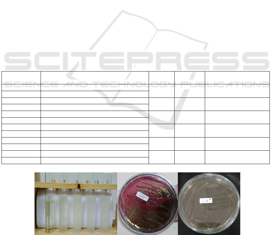
The presumtive test results showed that of the total 7
samples tested; all of them were positive for coliform
bacteria, characterized by acid formation, indicated
by the color change of the media from clear to cloudy,
and gas appearing on the durham tube. This is due to
the ability of coliform group bacteria to ferment
lactose found in the growth media. Coliform bacteria
is one indicator of water quality. Coliform is found in
nature like soil, but consumption water is not a natural
environment for coliform bacteria (Sengupta and
Saha, 2013). Meanwhile, the results of the
connfirmed test showed that of the 7 samples tested,
6 samples were positive for E. coli bacteria and 1 was
negative. Positive results are indicated by the
appearance of a metallic green colony on EMBA
media while on the NA media, the colonies appeared
white as milk. The results of DNA isolates in this
study have a good value of purity and concentration,
it can be seen in the following table 2.
Table 1: Multiplex PCR Primer used in Amplification
Primers Sequences (5’-3’)
Target
gene
Amplicon
size (bp)
Reference
FLICH7-F GCGCTGTCGAGTTCTATCGAGC
fliCh7 625
Sarimehmetoglu et al., 2009
FLICH7-R CAACGGTGACTTTATCGCCATTCC
rfb E-F CAGGTGAAGGTGGAATGGTTGTC
rfb E 296
Bertrand et al., 2007
rfb E-R TTAGAATTGAGACCATCCAATAAG
SLT1-F TGTAACTGGAAAGGTGGAGTATACA
stx1 210
Sarimehmetoglu et al., 2009
SLT1-R GCTATTCTGAGTCAACGAAAAATAAC
SLT1 1-F GTTTTTCTTCGGTATCCTATTCC
stx2 484
Sarimehmetoglu et al., 2009
SLT1 1-R GATGCATCTCTGGTCATTGTATTAC
AE22 ATTACCATCCACACAGACGGT
eaeA 397
Sarimehmetoglu et al., 2009
AE20-2 ACAGCGTGGTTGGATCAACCT
MFS1-F ACGATGTGGTTTATTCTGGA
hly 166
Sarimehmetoglu et al., 2009
MFS1-R CTTCACGTCACCATACATAT
a b c
Figure 1. a. Presumptive Test result; b. Confirmed Test result; c. Completed Test result.
BEST ICON 2018 - Built Environment, Science and Technology International Conference 2018
110
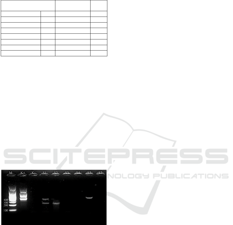
Table 2. DNA Concentration and Purity
High purity DNA and free contaminants are
needed in molecular technology. The presence of
contaminants can inhibit the molecular testing
process. Generally contaminants found in DNA
isolates are in the form of enzymes, proteins, and
lipids (Padhye et al., 1997). DNA isolates are said
to be pure if the ratio value in Å260/280 is between
1.8 and 2.0. If the ratio value at Å260/280 is less
than 1.8, then DNA isolates are contaminated with
phenol or too much solvent is used, and the DNA
taken is too little. On the other hand, if the ratio
value at Å260/280 is more than 2.0, then DNA
isolates contain protein membrane contaminants or
other compounds (Sambrook and Ruslle, 2001).
Agarose gel electrophoresis results and
visualization using gel documentation showed the
following results:
Figure 2. Multiplex PCR results in sample S1-S7
M = Marker (DNA Ladder 100 bp); C+ = Positive
Control used E. coli O157:H7; C- = Negative
Control
The picture above shows that from the 6
samples tested, 3 positive samples expressed the
virulent gene of E. coli O157: H7 bacteria. Positive
samples include the S1 sample (ice cube 1) DNA
bands appear in the area of 210 bp and 397 bp which
is an area of amplification products from the stx1
and eaeA genes in sequence. Furthermore, in the S2
sample, bands appeared in the area of 210 bp
(positive for stx1 gene) and in the S5 sample, DNA
bands appeared in the area of 397 bp (positive for
eaeA gene).
E. coli O157: H7 is one of the most pathogenic
bacteria in humans because it has a variety of
different virulent genes, like a positive result in this
study which found a positive sample of the stx1
(shiga like toxin) gene resulting from the gene
expression which can inhibit protein synthesis
resulting in cell damage and death (Boerling et al.,
1999; Lahtia et al., 2001; Rey et al., 2006;
Bentancor et al., 2012). Meanwhile, the eaeA gene
encodes intimate proteins can cause adherence,
causing damage to the intestinal lining (Paton and
Paton, 1998; Boerling et al., 1999; Fu et al., 2005;
Rey et al., 2006). These bacteria should not be found
in consumption water. Water pollution caused by
microorganisms is an important problem that must
be addressed immediately (Sengupta and Saha,
2013) because water is a medium that is very
potential and fast in the spread of infectious
diseases. Therefore, it is necessary to evaluate and
monitor the consumption water (ice cubes) in
circulation so that infection by bacteria E. coli
O157: H7 can be prevented.
Multiplex PCR is one variation of the PCR
technique. This method uses several primary sets in
a single PCR mixture to produce amplicons of
various sizes specific to different DNA sequences.
With gene targeting at the same time, additional
information can be obtained from a single running
test that will not require multiple reagents and more
time to do it. Annealing temperature for each
primary set must be optimized to work correctly in
a single reaction, and amplicon size. That is, the
length of the base pair must be different enough to
form a different band when visualized by agarose
gel electrophoresis. Detection using multiplex PCR
is faster than using conventional culture methods
(microbiology) in sorbitol Mac Conkey agar
(SMAC) media, other than that PCR also eliminates
the possibility of errors in detecting the presence or
absence of Escherichia coli O157: H7 bacteria in a
sample because it uses a variety of primers specific
to a particular gene.
4. CONCLUSION
Based on the research that has been done, 7 are
positive samples of coliform bacteria, 6 are positive
samples of E. coli bacteria, and 3 are positive
samples of E. coli O157: H7 bacteria. Positive
samples of E. coli O157: H7 have different virulent
genes namely stx1 and eaeA.
Various ways can be done to prevent the
occurrence of infections due to these bacteria; one
Sample
Concentration
(μg/ml)
Å260
/280
Ice cube 1 S1 6.720 1.994
Ice cube 2 S2 3.474 1.716
Tap water 1 S3 2.033 1.795
Ice cube 3 S4 3.853 1.973
Tap water 2 S5 0.755 1.361
Tap water 3 S7 0.949 1.901
PositiveControl C+ 0.768 1.838
NegativeControl C - 0.536 2.269
Detection of Enterohemorrhagic Escherichia coli (EHEC) in Consumption Water Source using Multiplex PCR Method
111

of which is the correct and proper cooking process.
It is known that the toxin produced by EHEC
bacteria is damaged in the heating process with a
temperature of 100
o
C for 10 minutes, while the
EHEC bacteria themselves will die during the
heating process at 72
o
C for 10 minutes.
REFERENCES
Aziz, D. A., Marlina, & Yuherman. 2009.
Karakterisasi Gen Penghasil Toksin pada
Bakteri Patogen Escherichia Coli O157
dalam Rangka Penanggulangan Penyakit
Diare Berdarah pada Masyarakat. Artikel.
Universitas Andalas, Sumatera Barat.
Bentancor, A., Rumi, M. V., Carbonari, C.,
Gerhardt, E., Larza, M., Vilte, D. A., Creydt,
V. P., & Chinen, I. 2012. Profile of Shiga
toxin-producing Escherichia coli strains
isolated from dogs and cats and genetic
relationships with isolates from cattle, meat
and humans. Vet Microbiol. 156: 336–342.
Boerling, P., Mc Ewen, S. A., Wilson, J. B.,
Johnson, R. P. & Gyles, C. L. 1999.
Association between virulence factors of
shiga toxin-producing Escherichia coli and
disease in humans. Journal of Clinical
Microbiology 37: 497–503.
Boerling, P., Mc Ewen, S. A., Wilson, J. B.,
Johnson, R. P. & Gyles, C. L. 1999.
Association between virulence factors of
shiga toxin-producing Escherichia coli and
disease in humans. Journal of Clinical
Microbiology 37: 497–503.
Drasar, B. S. & Hill, M. J. 1974. Human Intestinal
Flora. Academic Press Ltd, London.
Fu, Z., Rogelj, S. & Kieft, T. L. 2005. Rapid
detection of Escherichia coli O157:H7 by
immunomagnetic separation and real-time
PCR. Int J Food Microbiol. 99: 47–57.
Hill, W. E., & Jinneman, K. C. 2000. Principles and
Application of Genetic Techniques for
Detection, Identification, and subtyping of
Food-Associated Pathogenic
Microorganism in The Microbiological
Safety and Quality of Food. Vol. II (ed)
Barbara ML, Baird- Parker TC, Gould GW.
Maryland Aspen Publishers, Inc,
Gaithersburg.
Instruksi Kerja BPOM. 2008. Info POM. Badan
Pengawas Obat dan Makanan Republik
Indonesia. Vol. 9, No. 2
Jeshveen, S. S., Chai, L. C., Pui, C. F. & Son, R.
2012. Optimization of multiplex PCR
conditions for rapid detection of Escherichia
coli O157:H7 virulence genes. International
Food Research Journal 19(2): 461-466.
Lahtia, E., Keskimaeki, M., Rantala, L., Hyvoenen,
P., Siitonen, A. & Honkanen-Buzalski, T.
2001. Occurrence of Escherichia coli O157
in Finnish cattle. Vet Microbiol. 79:239–251.
Law, D. 2000. Virulence factors of Escherichia
coli O157 and other shiga toxin producing
E. coli . Journal of Applied Microbiology
88: 729-745.
Mubarak, W. I., & Chayatin, N. 2009. Ilmu
Kesehatan Masyarakat Teori dan Aplikasi.
Salemba Medika, Jakarta.
Nataro, J. P. & Kaper, J. B. 1998. Diarrheagenic
Escherichia coli. Clinical Microbiology
Review 11: 142-201.
Padhye, V. V., York, C., & Burkiewicz, A. 1997.
Nucleic Acid Purification On Silica Gel and
Glass Mixtures. US Patent 5658548 CI.
Paton, A.W. & Paton, J.C. 1998. Detection and
characterization of Shiga Toxigenic
Escherichia coli by using Multiplex PCR
Assays for stx1, stx2, eaeA,
Enterohemorrhagic E. coli hlyA, rfbO111,
and rfbO157. J Clin Microbiol. 36: 598–602.
Rey, J., Sa′nchez, S., Blanco, J. E., Hermoso de
Mendoza, J., Hermoso de Mendoza, M.,
Garcı′a, A., Gil, C., & Tejero, N. 2006.
Prevalence, serotypes and virulence genes of
Shiga toxin-producing Escherichia coli
isolated from ovine and caprine milk and
other dairy products in Spain. Int J Food
Microbiol. 107: 212–217.
Sambrook, J. & David W. Rusell. 2001. Molecular
Cloning: A Laboratory Manual. 3
th
ed. Cold
Spring Harbor Laboratory Press.
Sengupta, C. & Rita S. 2013. Review Article:
Understanding Coliforms-A Short Review.
International Journal of Advance Research.
1: 16-25.
Tutenel, A., Pierard, D., Jan, V.H., & L. D Zutteri.
2003. Molecular Characterization of
Escherichia coli O157:H7 Contamination
routes in a Cattle Slaughterhouse. Journal of
Food Protection. 66(9): 1564-1569.
Zakki, G. H. 2015. Pengetahuan dan Perilaku
Preventif Terhadap Bakteri E. coli pada
Masyarakat Kecamatan Gondomanan di
Kota Yogyakarta. Skripsi. Fakultas Ilmu
Pendidikan, Universitas Negeri Semarang,
Semarang.
BEST ICON 2018 - Built Environment, Science and Technology International Conference 2018
112
