
Nasal Polyp in Children with Allergic Rhinitis: A Case Report
Teuku Husni
1,2
1
Doctoral Program in Mathematics Science Application, Syiah Kuala University, Banda Aceh, Indonesia
2
Division of Rhinology, Department of Ear, Nose, Throat, Head & Neck Surgery, Faculty of Medicine, Zainoel Abidin
General Hospital, Banda Aceh, Indonesia
Keywords: Nasal Polyp In Chidren, Rare Case, Allergic Rhinitis, Endoscopic Sinus Surgery, Polypectomy
Abstract: Nasal polyp are soft, painless, non cancerous growths, semi-translucent edematous masses with a broad or
slim base usually arising from the mucosal lining of paranasal sinuses or on the lining of nasal passages. In
most cases, polyps are considered to be manifestation of chronic inflammation due to asthma, recurring
infection, allergies, drug sensitivity or certain immune disorders. Nasal polyp are very rare in children, the
incidence is 0.1% of all nasal polyp case. Thirty three percent of all nasal polyp case in children are
antrochoanal type. Multiple polyp can occur in children with chronic sinusitis, allergic rhinitis, cystic
fibrosis or allergic fungal sinusitis. Medication like steroid can often shrink or eliminate nasal polyps, but
surgery is sometimes needed to remove them. Even after successful treatment, nasal polyp often return. In
this case report we present a 5 years old boy with nasal polyp sinistra with allergic rhinitis and necessitating
endoscopic surgical intervention.
1 INTRODUCTION
Nasal polyp is a soft mass containing alot of fluid in
nasal cavity, white greyish in color, due to mucous
inflammation. Nasal polyp can occur in male and
female, from young age to elderly person. If nasal
polyp occur in children below 2 years, need to rule
out possibilty of meningocel or
meningoencephalocele. (Soepardi, 2012).
Nasal polyp mostly associated with chronic
inflammation, otonom nerve dysfunction, and
genetic disproportion. According to Bernstein
theory, change in nasal mucous due to inflammation
or airflow turbulence, mostly in narrow area in
osteomeatal complex. Submucousal prolaps occur
that followed by re-epitelization dan new gland
formation. Also increasing natrium absorbtion by
ephitel surface that cause water retention and create
nasal polyp. Another theory confirm due to
imbalance vasomotor nerve, increase capiler
permeability and vascular regulation that cause
releasing cytokin from mast cell, and cause edema
and in long time will cause nasal polyp. (Soepardi,
2012).
Mackay dividing nasal polyp into 4 stadium:
(Budiman, 2010)
‐ Stadium 0 : no nasal polyp
‐ Stadium 1 : polyp only in meatus media,not
in nasal cavity, can’t be seen by anterior
rhinoscopy but can be seen by
nasoendoscopy
‐ Stadium 2 : polyp out of meatus media and
seen in nasal cavity, but not fullfil the nasal
cavity
‐ Stadium 3 : nasal polyp fullfil the nasal
cavity
Hellquist dividing nasal polyp based on the
histologic type: (Budiman, 2010)
‐ Type I : Allergic polyp with
dominant eosinophilic
‐ Type II : Fibroinflamatoric polyp
with neutrofil dominant
‐ Type III : Polyp with hyperplasia
seromusinosa gland
‐ Type IV : Polyp with atipical stroma
Chmielik dividing polyp based on hystologic into
3 type: eosinophilic polyp, inflammatory polyp, and
atipical stroma. (Chmielik, 2011)
Clinical manifestations of nasal polyp depend on
their extent and may consist of obstructed nasal
breathing, hyposmia or anosmia (due to obstruction
of the olfactory groove), headache (due to impaired
228
Husni, T.
Nasal Polyp in Children with Allergic Rhinitis: A Case Report.
DOI: 10.5220/0008792302280232
In Proceedings of the 2nd Syiah Kuala International Conference on Medicine and Health Sciences (SKIC-MHS 2018), pages 228-232
ISBN: 978-989-758-438-1
Copyright
c
2020 by SCITEPRESS – Science and Technology Publications, Lda. All rights reserved
ventilation and draination in the paranasal sinuses),
snoring, rhinophonia clausa, and frequent throat
clearing due to associated postnasal drainage. Spread
to the lower airways can lead to laryngitis with
hoarseness and bronchitic symptoms. (Probst, 2006).
Masive nasal polyp can cause nose deformity
and widening of nasal bridge. On anterior
rhinoscopy nasal polyp seen as a pale mass that
came out from meatus medius and mobile.
(Soepardi, 2012).
Paranasal sinuses radiologic (waters, AP,
caldwell and lateral view) show mucous thickening
and air fluid level in sinus, but less helpful in polyp
case. CT scan investigation very helpful to see nasal
cavity and sinuses clearly if there any inflammation
process, anatomy abnormality, polyp or obstruction
in osteomeatal complex. CT scan mostly indicated
for nasal polyp case that fail to be treated with
medication, for sinusitis complication dan planning
for endoscopic surgery. (Soepardi, 2012).
Main purpose for nasal polyp treatment is to heal
the complain, avoid the complication dan avoid the
polyp recurrent. Corticosteroid treatment for nasal
polyp called medication polypectomy. Can be given
as topical or sistemic. Eosinophilic polyp give better
response with intransal corticosteroid compare with
neutrophilic polyp. For nasal polyp that not improve
with medication or for masive nasal polyp, surgery
is suggested. The surgery can be done by polyp
extraction (polypectomy) with polyp wire or forcep
with local anagesic, intranasal ethmoidectomy or
extranasal ethmoidectomy for ethmoid polyp,
Caldwell-luc surgery for maxillary sinus. If
endoscopi facility avaialable, then the best option is
to do Functional Endoscopic Sinus Surgery (FESS).
(Probst, 2006).
2 CASE REPORT
A 5 years old boy came to ENT clinic of Zainoel
Abidin General Hospital (ZAGH) on end of April
2018 referral from district ENT specialist with major
complaint obstruction of left nose since 2 years ago
and getting worse in the last 3 months. Patient
complaining permanent obstruction, and only on the
left nose. There’s a history of nasal bleeding twice.
Currently patient complaining pain on the left nose.
Patient has an dust allergic and history of recurrent
runny nose.
On physical examination, patient in good
condition, fully alert, cooperative, and well nutrition
status. On Ear examination, within normal limit. On
nasal examination, looked asymetric due to mass
compression on left nose. On left nares seen white
gelatinous mass. On anterior rhinoscopy seen left
nasal cavity fullfil with white gelatinous mass,
inferior concha can’t be seen due to mass. Right
nasal cavity narrowing due to inferior concha
hypertrophy, pale mucous, no rhinorea and septal
deviation found. On oropharingeal examination
within normal limit and found caries dentis. Normal
neck examination. Patient diagnosed with left nasal
polyp with allergic rhinitis. Then we conduct some
examination such as laboratory, paranasal sinused
CT scan without contrast dan chest x-ray. After all
the examination result available, we refer patient to
pediatric and anesthesia department to obtain the
surgery approval for polypectomy under general
anesthesia. The surgery plan to be done on 22 May
2018.
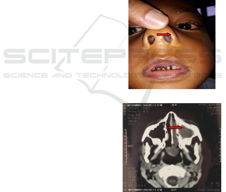
Figure 1: First picture of patient before the surgery
Figure 2: Paranasal sinus CT scan axial view showed
polyp mass on left nasal cavity
Nasal Polyp in Children with Allergic Rhinitis: A Case Report
229
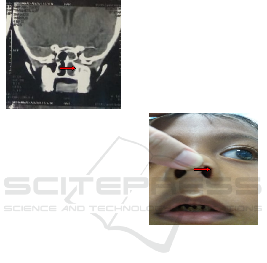
Figure 3: Paranasal sinus CT scan coronal view showed
polyp mass on left nasal cavity
On 22 May 2018, the polypectomy endoscopic
surgery done within general anesthesia. The surgery
start with patient lay on the surgery table under
general anesthesia. Pack was placed in the mouth.
Aseptic and antiseptic procedure was done on the
surgery field. Then xylocain adrenalin package was
placed on both nasal cavity for 15-20 minutes. After
the nasal packages removed, the left nasal cavity
was observed with 0
0
scope, white greyish
gelatinous mass seen fullfil the left nasal cavity dan
seen redness on the posterior side, mobile, and seem
came out from meatus media, inferior and media
nasal concha eutrophy, meatus media blocked by the
polyp mass. Extirpation nasal polyp was done by
cutting forcep until all the nasal polyp in left nasal
cavity was removed. And then nasal cavity was
evaluated until nasopharyng area. No active bleeding
found and nasopharyng area seem clear. Right nasal
cavity also evaluated with 0
0
scope, no mass seen,
inferior and media nasal concha eutrophy,
nasopharyng within normal limit. Bleeding on left
nose was controlled with handscoon nasal package.
Pack in the mouth was removed. Surgery has
finished. Polyp tissue was sent to anatomic
pathology laboratory to find out what type of nasal
polyp. Patient was returned to ward after fully alert.
Post surgery treatment patient was given cefotaxime
injection 500mg twice a day, paracetamol 250mg
twice a day dan tranexamic acid 250mg thrice a day.
On the first post operation day, patient still
complaining pain on the surgery site, but no more
active bleeding found on the left nose. Anterior nasal
package was still applied, no blood seepage seen, no
blood seen on pharyngeal area.
On second post operation day, pain on the
surgery site has reduced. Anterior nasal package was
removed, and then left nasal cavity was evaluated.
No active bleeding, sinechia, or nasal polyp found.
Patient was allowed to discharge with some
medication, cefixime syrup 60mg twice a day and
paracetamol syrup 250mg thrice a day and edvised
to do nasal wash at home.
Patient came for follow up review on ENT clinic
ZAGH on the 6
th
post operation day. Patient
complaining blockage on left nose and stated that
the nose washing has been done as instructed, 6
times a day. On physical examination found bloody
crustae on left nasal cavity, no sinechia found. And
then bloody crustae was extracted followed by nasal
washing until left nasal clear.
Figure 4: First follow up review post surgery, no more
mass seen on left nasal cavity
Next follow up review was done once every 2
days. Nasal blockage complaint has reduced. on
anterior rhinoscopy blood crusta was found less than
before. Patient has come for post operation follow
up review 4 times,and then stopped due to ramadhan
and idul fitri holiday. Patient’s family was advised to
go to the hospital if there’s any nasal bleeding or
nasal blockage happen.
On 4
th
weeks after surgery, patient’s family
informed that patient start to sneezing and have a
runny nose again, also nasal blockage at night. We
advised them to come to ENT clinic of ZAGH and
continue the nose wash, but patient has not come yet
to the ENT clinic.
The anatomy pathology from the polyp tissue
result come as nasal polyp, no malignancies sign
found.
SKIC-MHS 2018 - The 2nd Syiah Kuala International Conference on Medicine and Health Sciences
230

3 DISCUSSION
A nasal polyp case on 5 years old boy reported and
diagnosed by anamnesis, physical examination
through anterior rhinoscopy and radiologic
examination paranasal sinuses CT scan. Nasal polyp
in this case report found in 5 years old boy. This
appropriate with literature reported by Bestari Jaka
Budiman stated that nasal polyp occur more in male
than female, 2-4: 1, and rarely happen in children
with evidence number 0,1%. In Indonesia,
epidemiology study showed comparison between
male and female is 2-3:1 with prevalence 0,2%-
4,3%. Polyp of this patient is antrochoanal polyp,
and according to literature defined antrochoanal
polyp is nasal polyp of maxillary nasal origin, came
out through maxillary sinus ostium to nasal cavity
and extend to choana. Accroding to Khalid,
antrochoanal polyp is polyp that growth from
maxillary sinus mucous and came out through
ostium to nasal cavity. (Budiman, 2010)
The exact cause of antrochoanal polyp is not
known yet, but can caused by allergic facto, cystic
fibrosis infection and mechanical obstruction. In this
patient also found allergic rhinitis that can cause
nasal polyp. To find the type of allergen, patient
should perform the allergic test so patient can avoid
the allergen to decrease the recurrence rate.
(Budiman, 2010)
Surgery was the only feasible treatment for
antrochoanal polyp. Several surgical techniques
were described. In the past, Caldwell-Luc technique
was used. FESS is currently the glod standard
techniques. FESS is less invasive techniques which
permits to restrore drainage of the paranasal sinuses
and ventilation between the nose and sinus cavities
and allows shorter hopsital stay. The antral portion
of an antrochoanal should be removed, together with
the base of its origin, to minimize post-operative
recurrence. The use of micro-debrider may be
indicated, as complementary to endocsopic surgery.
Combining endoscopic surgery and trans-canine
sinuscopy is an alternative technique. The succes
rate was 76,9% in the trans-nasal endoscopic
approach. On the other hand, no recurrence could
happen after long-term follow up if there’s a
correction of a nasal associated nasal anatomic
variation at the time of surgery for antrochoanal
polyp removal. (Chlebna, 2017; Mandour, 2017)
Recurrence rate of nasal polyp after endoscopi
surgery was about 60%.
1
we have informed about
this to patient’s family before the surgery since the
recurrency rate was quite high, followed by some
advised to have a review at ENT clinic if there’s any
nasal blockage found. (Budiman, 2010)
The process of polyp formation due to chronic
inflammation is reversible, so the treatment of
rhinosinusitis should start very early with nasal
washing with saline solution, antibiotic and local
steroid. In the post-operative period the patient hs
been recommended to keep on doing frequent nasal
washing with saline solution. (Chlebna, 2017).
4 CONCLUSION
We described a case report of a 5 years old boy with
rhinitis allergic presenting with nasal polyp.
Diagnosed was made based on anamnesis, physical
examination and radiology (CT scan) finding. The
treatment was done by polypectomy surgery with
FESS technique. Since the recurrence rate quite
high, patient’s family has been advised to control the
rhinitis allergic symptom and seek for medical
treatment if there’s any nasal blockage reported by
patient.
ACKNOWLEDGEMENTS
This paper reports a rare case in children hopefully
can be additional information and knowledge in the
field Ear Nose Throat Head and Neck Surgery.
REFERENCES
Aliyu I, Helen AO. (2013). “Multiple Nasal Polyps in an
11 Year Old Asthmatic Child: A Case Report”,
available from www.sjmms.net. Cited on 23 May
2018
Budiman, BJ, Sari AM. (2010). “Polip Nasi Pada Anak”,
Bagian THT-KL. FK Universitas Andalas. Padang.
Chlebna MO, Trzcinski K, Stelmach I. (2017). “Massive
Nasal Polyposis in a patient with newly Diagnosed
Cystic Fibrosis”, adv respiratory medicine; 85:121-
123. Available from www.journals.viamedica.pl cited
on July 2018-07-23
Chmielik LP, Ryczer T, Chmielik M. (2011). “The clinical
Analysis of Antrochoanal Polyps In Children”,
Available from www.newmedicine.pl/wp-
content/uploads. cited on 23 May 2018.
Mandour ZM. (2017). “Antrochoanal Polyp in Pediatric
Age Group”, Egyptian Journal of Ear, Nose, Throat
and Allied Sciences 18 ;17-21. Available from
www.ejentas.com cited on July 2018
Nasal Polyp in Children with Allergic Rhinitis: A Case Report
231

Mc Clay JE. (2017). “Nasal Polyps”, Available from:
https://emedicine.medscape.com. cited on 23 May
2018
Probst R, Greves G, Iro H. (2006). “Basic
Otorhinolaryngology, A Step by Step Learning guide”,
Thieme. New York. p:57-58
Soepardi EA, Iskandar N, Bashiruddin J, Restuti RD.
(2012).” Buku Ajar Ilmu Kesehatan THT-KL”, edisi
ketujuh, Badan Penerbit FK-UI, Jakarta. Hal: 101-103
SKIC-MHS 2018 - The 2nd Syiah Kuala International Conference on Medicine and Health Sciences
232
