
Oxidative Stress and Inflammation Marker Profiles of White Rat
Pup’s Brain Endosulfan-induced Neurotoxicity in Pregnant Rat
Model
Triawanti
1
, Meitria Syahadatina Noor
2
, Didik Dwi Sanyoto
3
,
Hendra Wana Nur’amin
4
1
Department of Biochemistry, Faculty of Medicine, Lambung Mangkurat University, Banjarmasin, Indonesia
2
Department of Public Health, Faculty of Medicine, Lambung Mangkurat University, Banjarmasin, Indonesia
3
Department of Anatomy, Faculty of Medicine, Lambung Mangkurat University, Banjarmasin, Indonesia
4
Department of Pharmacology, Faculty of Medicine, Lambung Mangkurat University, Banjarmasin, Indonesia
Keywords: Endosulfan, Oxidative Stress, Inflammation, Neurotoxicity
Abstract: Endosulfan is a forbidden insecticide that may cause some nerve related disorders. Some oxidative stress
and inflammation markers may play a role in these neurotoxic events. This study aimed to analyze the effect
of endosulfan exposure in white rat pup’s brain in pregnant rat model. This study used pregnant white rats
induced by endosulfan administered orally and divided to endosulfan treatment and control. After the
female rats gave birth, endosulfan treatment was stopped. The pups were left to suckle on their mothers.
When the pups had reached four weeks, 16 pups were terminated and brains were taken from each group
were taken to examine levels of MDA, SOD, H
2
O
2
, AOPP, TNF-α, and Hsp70. The data collected were
analyzed using Student's T-test or Mann-Whitney U test. The results suggested that MDA, H
2
O
2
, AOPP and
TNF-α levels were higher in endosulfan group compared to control group (p<0.05), SOD levels decrease in
endosulfan group (p<0.05) and no significant difference in Hsp70 levels between the group. This study
concluded that endosulfan interfered the oxidative stress and inflammatory markers of pup’s brain induced
by endosulfan in the pregnant rat model.
1 INTRODUCTION
Endosulfan was used as an organochlorine
insecticide in agriculture globally to improve
farming production, fight against destructing pests
and protection from vector-borne diseases and
related epidemics for humankind. In several
countries, endosulfan is banned permanently due to
high toxicity profiles, but it is still largely used in
some developing countries due to its low cost and
high efficacy against pests (Menezes et al., 2017;
Patočka et al., 2016).
Recent studies have shown that endosulfan
can cause several diseases such as endocrine,
reproduction, genotoxicity, teratogenicity and
neurotoxicity disorders. Endosulfan may cause
neurotoxic effects by overstimulating the central
nervous system, affecting a number of targets on
CNS and also crossing the barrier of the
placenta (Pathak et al., 2008; Silva and Gammon,
2009). Endosulfan is one of the factors that affect
some neuropsychological disorders that occur in
children and adults. Exposure to endosulfan during
pregnancy and lactation in female rats can affect the
neurotransmitter; γ-aminobutyric acid (GABA),
glutamate, serotonin and dopamine (Lafuente and
Pereiro, 2013; Wilson et al., 2014). These findings
suggested that neurotransmitter disorders may cause
neurobehavior disorders caused by endosulfan
exposure (Wilson, 2014). Endosulfan may also
interfere with the ability of the zebrafish nervous
system while swimming because it inhibits the
activity of acetylcholinesterase (AChE) enzyme
(Pereira et al., 2012).
The neurotoxicity manifestation due to
endosulfan exposure may be affected by oxidative
stress disorder and an inflammatory reaction (Jang et
al., 2016; Lakroun et al., 2015). Endosulfan
exposure can cause oxidative stress in humans as
well as rats (Koç et al., 2009; Pathak,
Triawanti, ., Noor, M., Sanyoto, D. and Nur’amin, H.
Oxidative Stress and Inflammation Marker Profiles of White Rat Pup’s Brain Endosulfan-induced Neurotoxicity in Pregnant Rat Model.
DOI: 10.5220/0008790300310038
In Proceedings of the 2nd Syiah Kuala International Conference on Medicine and Health Sciences (SKIC-MHS 2018), pages 31-38
ISBN: 978-989-758-438-1
Copyright
c
2020 by SCITEPRESS – Science and Technology Publications, Lda. All rights reserved
31

2008). Endosulfan had been shown to trigger
systemic toxicity, reactive oxygen species (ROS)
and lipid peroxidation, which can be seen with the
increase of malondialdehyde (MDA) (Jang, 2016;
Ullah et al., 2016). The brain consists of
phospholipids in the cell membranes so that if
damage occurs, it can make some brain
disorders (Kayhan, 2008; Zervos et al., 2011). The
rat liver cells induced by endosulfan exposure in
vitro had reduced antioxidant enzyme activity such
as superoxide dismutase (SOD), glutathione
peroxidase (GPx) and glutathione-S-transferase
(GST) while elevated hydrogen peroxidase (H
2
O
2
)
levels (El-Shenawy, 2010; Jang, 2016). Zebrafish
induced by low concentration endosulfan may
increase the activity of the enzyme SOD and
catalase (CAT), whereas at high concentrations can
lead to the production of reactive oxygen species
(ROS) excess that causes SOD and CAT cannot
handle it anymore (Shao et al., 2012).
Endosulfan exposure may also increase the
activity of inflammatory mediators such as tumor
necrosis factor (TNF-a) in Mouse RAW 264.7 cells
(ATCC) (Terry et al., 2018). Endosulfan exposure
may also increase levels of advanced oxidation
protein products (AOPP) in rabbits. AOPP is one of
the inflammatory markers and oxidative stress in
many diseases (Alagozlu et al., 2013; Ozdem et al.,
2011). The 70 kilodalton heat shock proteins
(Hsp70) are proteins that are formed to protect cells
from oxidative stress and inflammation (Borges et
al., 2012). In endosulfan-induced zebrafish embryos
Hsp70 increase significantly (Moon et al., 2016).
Although endosulfan had been shown to have
many toxic effects; especially neurotoxicity and
prohibited already, many agricultural area use
endosulfan as a weapon against pests legally or
illegally (Patočka, 2016). This study aimed to
determine whether the neurotoxic endosulfan can
affect the levels of some markers of oxidative stress
and inflammatory mediators in the brains of pups.
2 MATERIALS AND METHODS
This research had been received approval
from the ethics committee of Faculty of Medicine,
Lambung Mangkurat University, Banjarmasin,
Indonesia. This study was an experimental study
with posttest-only with control group design.
Materials
The materials used in this study were white
rats (Rattus norvegicus), pup’s brain, distilled water,
deionized water, standard rat feed (comfeed
PARS 53% (12% water, 11% protein, 4% fat, 7%
fiber, 8% ash, 1.1% calcium, 0.9% phosphorus,
antibiotics, 53% coccidiostat), 23.5% wheat flour,
23.5% water ), endosulfan, rat TNF-αelisa kit, rat
HSP70 elisa kit, ether, phosphate buffer saline (PBS)
pH 7, 200 μL 100% TCA, 100 μL 1%, sodium
thiobarbiturate, 250 μL HCl 1 N, EDTA,
dichromate, glacial acetate, H
2
O
2
, olive oil,
adrenaline, sodium bicarbonate (Na
2
CO
3
) and
potassium iodida (KI).
Animal Procedure
Acclimatization
Adult female and male rats were kept for 1
week before being treated to provide the same
physical and psychological conditions. During
maintenance, white rats were given the same
distilled water and food sufficiently.
Endosulfan induction
After a one-week acclimatization period,
female rats were injected by PMSG and HCG in
accordance with estrous cycle. Female rats were
mated with male rats from the same strain. 1 female
was mated to 1 male. After mating, female rats were
individually placed in a polypropylene cage. Female
rats those had been positively pregnant were
weighed and distributed randomly divided into 2
groups. The control group (K) without endosulfan
induction while the treatment group (P) was induced
by endosulfan with a dose of 1 mg/kg BW.
Endosulfan was given by dissolving it in
olive oil and and administered orally during 21 days
of pregnancy. After the female rats gave birth,
endosulfan treatment was terminated. The pups were
left to suckle on their mothers. When the pups had
reached four weeks of age, 16 pups from each group
were terminated and brain tissues were taken to
measure levels of MDA, SOD, H
2
O
2
, AOPP, TNF-α,
and Hsp70 of the brain.
SOD, H
2
O
2,
MDA and AOPP levels assay from
brain homogenate
The brain was pounded with mortar at room
temperature and added with 1 mL of PBS pH 7.4
until it became liquid. Then taken 5 mL and
centrifuged at 8000 rpm for 20 minutes. The
supernatant was then taken for measurement of
H
2
O
2
, MDA, SOD, and AOPP levels.
Measurement of brain SOD levels
Incubation was performed on 3 ml of a
solution containing 0.05 M Na
2
CO
3
, 0.1 M EDTA
pH 10.2. Furthermore, in the solution was added 100
μL brain homogenate and 100 μL adrenaline with
(3.10
-4
) BM 189 M. Initial absorption measurements
SKIC-MHS 2018 - The 2nd Syiah Kuala International Conference on Medicine and Health Sciences
32

(A
0
) was performed with a spectrophotometer at 480
nm wavelength. After that, the sample was incubated
for 5 min at 30
o
C and got the absorbance (A
1
).
Measurement of brain H
2
O
2
levels
Measurement of H
2
O
2
was using a
spectrophotometer. At first, making a standard
curve. A total of 20 μmol H
2
O
2
was added with 2 ml
of dichromate:glacial acetic acid (1:3) mixture. Then
the mixture was heated in boiling water for 10
minutes. Then the cooled mixture was measured for
absorbance at a wavelength of 570 nm. The same
procedure was done for 40,60,80,100,120, 140,160
and 180 μmol H
2
O
2
. A graph was made between the
absorbance on the Y axis with levels of H
2
O
2
on the
X axis to obtain a linear equation.
Preparation of test solution was made with a
total of 1 ml of brain homogenate was added 5 ml of
PBS pH 7.4. A mixture of 1 ml was taken and added
to 2 ml of dichromate:acetate (1:3) mixture and then
wrapped in aluminum foil for 30 minutes. The
mixed solution was heated using a water bath for 10
minutes at 100
°
C. The solution was cooled to room
temperature. The solution was then transferred into
the cuvette and measured its absorbance using UV-
VIS at a wavelength of 570 nm.
Measurement of brain MDA levels
From the last procedure, 200 μL supernatant
was taken for measurement of MDA levels. The first
thing to do was making MDA standard curve. As
many as 0.05 μM MDA standard added 1 mL of
distilled water, then placed in Eppendorf
tube. Thereafter, 100 μL of 100% TCA, 100 μL
sodium thiobarbituric 1%, and 250 μL HCl 1 N were
added respectively. Then heated at 100 °C for 20
minutes, and centrifuged 3500 rpm for 10 minutes.
Subsequently, 450 μL supernatant was taken and the
distilled water added to 3500 μL. Then read with the
spectrophotometer with maximum wavelength 540
nm. The same thing was done to 0.025, 0.0125,
0.00625, 0.003125 and 1.56 x 10
-5
μM MDA. Then
making graphs for the relationship between
absorbance on the Y-axis and MDA levels on the X-
axis to obtain a linear equation.
Measurement of AOPP levels
The test solution was prepared by mixing 200
μL homogenate, 600 L PBS, and 100 L KI 1.16 M.
Then the blank solution was prepared by mixing 800
L of PBS, 100 L KI 1.16 M. Test and blank solution
were placed for 2 min and then add 200 μL of acetic
acid. Absorbance was measured at = 340 nm. The
concentration of AOPPs were expressed through A
= b C with = 26 mM
-1
cm
-1
and b = 1 cm.
TNF-α and Hsp70 assay in brain homogenate
Brain tissues were destroyed and
homogenized with 1 mL of PBS pH 7.4. The
measurement method refers to the rat TNF-α and
HSP70 ELISA Kit (Novateinbio, USA). The
materials and standard reference were placed at
room temperature. As much as 100 μL standard
reference, blank standard, samples were dissolved in
dilution and placed into the well and then incubated
for 3 hours at room temperature. The suspense
washed with PBS and washed up to 4 times. The
conjugates were filled into well as much as 200 μL.
The suspensions were washed with washing buffer
up to 4 times then 200 μL substrate solution was
added to well and incubated 30 minutes at room
temperature. The stop solution was added to every
well and was read for 30 minutes at a 450 nm
wavelength for TNF-α and HSP70.
Data analysis
The data were collected and tabulated. We
used comparative analysis for this study. The data
were tested using Shapiro Wilk for normality test
and Levene for homogeneity test. If the data were
normally distributed and homogenous, Student's T-
test would be performed with 95% confidence level.
If the data were not distributed normally nor
homogenous, the data would be analyzed by
nonparametric statistic Mann-Whitney U test.
3 RESULTS
Effects of Endosulfan Exposure on Oxidative
Stress and Inflammation Markers in The Brain
of Pups
SOD levels
SOD plays an important role as an
antioxidant in almost every cell exposed to oxygen.
It is the first line of defense to fight harms
superoxide radicals. Figure 1 showed the SOD levels
in the experimental pup’s brain.
The control group had higher SOD levels than
endosulfan exposure group. Mann-Whitney U test
showed there was a significant difference between
groups. It meant that endosulfan exposure reduced
the quantity of SOD in the pup’s brain.
Oxidative Stress and Inflammation Marker Profiles of White Rat Pup’s Brain Endosulfan-induced Neurotoxicity in Pregnant Rat Model
33
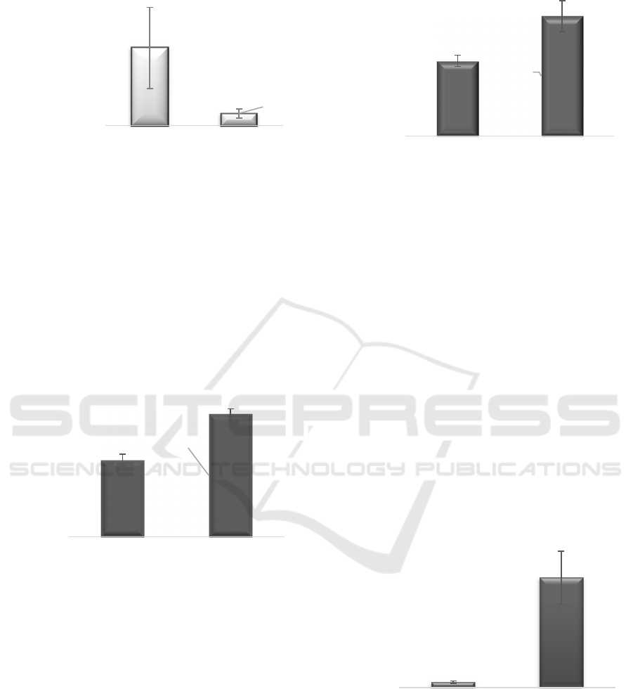
Figure 1. SOD levels of the experimental pup’s
brain
The control group had higher SOD levels than
endosulfan exposure group. Mann-Whitney U test
showed there was a significant difference between
groups. It meant that endosulfan exposure reduced
the quantity of SOD in the pup’s brain.
H
2
O
2
levels
Another marker of oxidative stress was H
2
O
2
levels. H
2
O
2
is a toxic substance to the brain. H
2
O
2
levels were demonstrated in figure 2.
Figure 2. H
2
O
2
levels of the experimental pup’s
brain
H
2
O
2
levels in control group were 5.574 mmol
and endosulfan exposure was 8.906 mmol.
Endosulfan exposure had higher H
2
O
2
levels with a
statistically significant difference between group
(Mann-Whitney U test, p<0.001).
MDA levels
MDA is one of the markers of oxidative
stress, derived from polyunsaturated fatty acids
peroxidation in the cells. MDA levels in the brain of
pups were shown in figure 3.
Figure 3. MDA levels of the experimental pup’s
brain
Figure 3 showed that pups with endosulfan
exposure had higher brain MDA levels compared to
control group. The statistical analysis with Mann-
Whitney U test concluded that there was a
significant difference between endosulfan exposure
and control group (p<0.001). It suggested that
endosulfan exposure may increase lipid peroxidation
in the brain.
AOPP levels
AOPPs are defined as protein aggregates
generated by disulfide bonds created as a result of
oxidative stress. The modified proteins from
oxidative modification are more stable than those of
lipids and making AOPPs better marker for
oxidative stress. AOPP levels of this study were
presented in figure 4.
Figure 4. AOPP levels of the experimental
pup’s brain
Mann-Whitney U test showed a significant
difference between groups (p<0.001) whereas
endosulfan exposure group had worse AOPPs than
the control group. It meant endosulfan may increase
the damage in the brain of pups.
0,012
0,002
0,000
0,005
0,010
0,015
0,020
Control Endosulfan
exposure
SOOLevels(unit/mg
protein)
5,574
8,906
0
2
4
6
8
10
Control Endosulfanexposure
H
2
O
2
levels(mmol)
206,875
330,438
0
50
100
150
200
250
300
350
400
Control Endosulfan
exposure
MDAlevels(μM)
0,563
11,846
0
2
4
6
8
10
12
14
16
Control Endosulfanexposure
AOPPlevels(μM)
SKIC-MHS 2018 - The 2nd Syiah Kuala International Conference on Medicine and Health Sciences
34
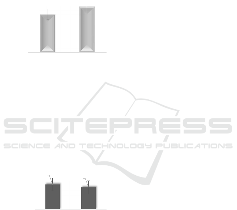
TNF-α levels
TNF-α is a cytokine that plays a role in
systemic inflammation. TNFα levels (ng/L) can be
seen in Figure 5.
Figure 5. TNF-α levels of the experimental
pup’s brain
Figure 5 had shown us that endosulfan
exposure had higher TNF-α levels compared to
control group. Student’s T-test statistical analysis
showed significant difference among groups
(p<0.001).
Hsp70 levels
Hsp70 has a significant role to bind the
receptor in normal condition. Excessive Hsp70 can
make some problems in the stress condition. Hsp70
levels in the experimental pup’s brain were
demonstrated in figure 6.
Figure 6. Hsp70 levels of the experimental
pup’s brain.
The endosulfan exposure gorup had lower
Hsp70 levels than the control group, but statistical
analysis with Student’s T-test showed no significant
difference between group (p=0.218).
4
DISCUSSION
This study used pup’s brain with mothers
induced by endosulfan during gestation. A study in
India showed that endosulfan can be transferred
from mother to fetus through human umbilical cord
as much as 60-70%. This was very dangerous
because it can disrupt the growth and development
of the fetus (Pathak, 2008). Endosulfan was proven
to increase the risk of teratogenicity such as cleft lip,
limb malformation, eye deformity, hands and
feet (Patočka, 2016).
Endosulfan is one of the most harmful
pesticides responsible for environmental damage and
cause disturbances to the nervous system (Kumar et
al., 2014; Wilson, 2014). The nervous system
disorders can occur acutely such as hyperactivity,
tremors, seizures, coordination disorders even
breathing difficulties. A dose of 500 mg/kg can
trigger permanent brain damage until death in
humans. Farmers with chronic endosulfan exposure
show rash and skin irritation (Patočka,
2016). Endosulfan also plays a role in several
diseases such as autism spectrum disorder (ASD)
and schizophrenia (Wilson, 2014).
The incidence of this neurotoxicity may
occur due to impaired GABA function as a major
inhibitory neurotransmitter (Patočka,
2016). Endosulfan inhibits the inhibition of [
35
S]-t-
butylbicyclophosphorothionate (TBPS) in the
picrotoxinin-binding site of the GABA receptor in
the rat brain synaptic membranes, which disrupts
chloride flow through GABA-gated chloride
channels (GABA
A
) resulting in decreased neuronal
excitability (Jang, 2016; Patočka, 2016). Some of
the markers of oxidative stress, antioxidants and
inflammation are thought to play some roles in nerve
damage in endosulfan-induced rats (Jang, 2016;
Lakroun, 2015; Moon, 2016).
SOD is metalloenzymes, which converts
superoxide anion (O
2
-
) become less reactive oxygen
species, ie molecules (O
2
) and hydrogen peroxide
(H
2
O
2
)
.
H
2
O
2
is formed by SOD activity
decomposed into H
2
O and O
2
by CAT and/or GPx in
the presence of reduced CAT and GSH (Bilodeau,
2014; Ighodaro and Akinloye, 2017). In this study,
the pup’s brain with endosulfan exposure had
significantly lower SOD levels than the control
group. In contrast, H
2
O
2
levels increased
significantly in the endosulfan exposure group
compared with control group. This indicated that
existing SOD can not resolve the H
2
O
2
excess in the
brain. H
2
O
2
is a reactive oxygen species (ROS). At
low levels, H
2
O
2
plays a role in normal cellular
348,556
417,306
0
100
200
300
400
500
Control Endosulfan
exposure
TNF‐αlevels(ng/L)
1,174
1,038
0,0
0,5
1,0
1,5
2,0
Control Endosulfan
exposure
Hsp70levels
(ng/L)
Oxidative Stress and Inflammation Marker Profiles of White Rat Pup’s Brain Endosulfan-induced Neurotoxicity in Pregnant Rat Model
35

metabolism, but at high levels, can trigger some
diseases. Increased levels of endogenous H
2
O
2
can
potentiate GABA
A
which may trigger H
2
O
2
-induced
brain dysfunction (Penna et al., 2014).
Lipid peroxidation occurs when oxidants
such as free radicals attack lipids in cells containing
carbon-carbon double bonds, especially
polyunsaturated fatty acids (PUFAs) (Ayala et al.,
2014). Damage to lipids due to oxidative stress can
be seen through MDA; one of the biomarkers of
oxidative stress in various diseases including
neurobehavior (Khoubnasabjafari et al., 2015). The
pups who had received endosulfan exposure in this
study had significantly higher MDA levels
compared to control group. The increased MDA can
lead to neurodegeneration and psychiatry disorders
resulting from lipid peroxidation (Joshi and Pratico,
2014; Sultana et al., 2013). A study on subjects
exposed to endosulfan suggested that women with
preterm delivery had higher levels of α-endosulfan
and oxidative stress markers such as MDA
compared to women with full-term delivery (Pathak
et al., 2010).
AOPP is a marker of oxidative injury and
various inflammatory diseases (Alagozlu,
2013). AOPP is derived from oxidative stress (free
radical) conditions in proteins and may act as a
trigger of inflammatory mediators that will trigger
neutrophils, monocytes and T lymphocytes to
increase dendritic cell stimulation (Škvařilová et al.,
2005). There are many mechanisms for the induction
of protein oxidation resulting in different types of
protein modification. Detection of protein carbonyl
groups is the most commonly used measure, AOPP
is also a marker of protein oxidation. Most AOPPs
are formed due to increased release of
myeloperoxidase (MPO) from activated
phagocytes (Hanasand et al., 2012). This study
showed that endosulfan exposure in pup’s brain can
increase AOPP levels up to 20 times greater than
controls. High levels of AOPP may play a role in the
incidence of neurodegenerative diseases such as
parkinsonism and dementia (Demirbilek et al., 2007;
Miletić et al., 2017).
The pup’s brain exposed by endosulfan in
this study had significantly higher levels of TNF-α
than the control group. TNF-α is a cytokine that
increases in the event of an inflammatory
process. TNF-α can cause neurotoxicity by
triggering the release of glutamate that can damage
the nerves. TNF-α is thought to have a role in
neurodegenerative diseases such as Alzheimer's,
Parkinson's, amyotrophic lateral sclerosis, and
multiple sclerosis (Takeuchi et al., 2006; Ye et al.,
2013). In the children with autism, there are elevated
levels of TNF-α in lymphocytes, cerebrospinal fluid,
and cerebrospinal fluid compared to the control
group (Rose et al., 2014).
In the case of brain injury, biomolecular
process and pathological biochemistry occur that
can cause cell damage, in the form of necrosis and
apoptosis (Kayhan, 2008). This molecular damage
results in the presence of symptoms of prolonged
disability, such as cognitive impairment in the form
of decreased attention, concentration, and
memory (Demirbilek, 2007; Sultana, 2013). More
and more severe the injury a person experiences, the
greater the damage both neurons and glial cells as a
support network. As a result, sequelae generated
even more prolonged. Heat shock protein 70
(Hsp70) includes protein stress, which can be
produced by neuron and glial cells that are under
stress and inflammatory conditions (Borges,
2012). In this study, there was no significant
difference between the groups with endosulfan
exposure and control groups.
5 CONCLUSION
The study concluded that endosulfan
exposure in pup’s brain can trigger some oxidative
stress and inflammation disorders. There were
increases in MDA, SOD, H
2
O
2
, AOPP and TNF-
α levels significantly accompanied by a decrease in
SOD-free radical protection. This can play a role in
some neurodegenerative related
diseases; Alzheimer's, Parkinson's and psychiatry
such as autism and schizophrenia. Endosulfan use
should be restricted and prohibited to prevent the
bad events.
ACKNOWLEDGEMENTS
We would like to thank Faculty of Medicine,
Lambung Mangkurat University for the financial
support and all people for their excellent
contribution.
REFERENCES
Alagozlu, H., Gorgul, A., Bilgihan, A., Tuncer, C.
and Unal, S. (2013): Increased plasma levels
of advanced oxidation protein products (
AOPP ) as a marker for oxidative stress in
SKIC-MHS 2018 - The 2nd Syiah Kuala International Conference on Medicine and Health Sciences
36

patients with active ulcerative colitis, Clin.
Res. Hepatol. Gastroenterol., 37 (1), 80–85.
Ayala, A., Munoz, M. F. and Arguelles, S. (2014):
Lipid peroxidation: Production, metabolism,
and signaling mechanisms of
malondialdehyde and 4-hydroxy-2-nonenal,
Oxid. Med. Cell. Longev.
Bilodeau, J. F. (2014): Review: Maternal and
placental antioxidant response to
preeclampsia - Impact on vasoactive
eicosanoids, Placenta, 35, S32–S38.
Borges, T. J., Wieten, L., Van Herwijnen, M. J. C.,
Broere, F., Van derZee, R., Bonorino, C., et
al. (2012): The anti-inflammatory
mechanisms of Hsp70, Front. Immunol., 3,
1–12.
Demirbilek, M. E., Kilic, N., Komurcu, H. F. and
Akin, K. O. (2007): Advanced Oxidation
Protein Products in Aged with Dementia, Am.
J. Immunol., 3 (2), 52–55.
El-Shenawy, N. S. (2010): Effects of insecticides
fenitrothion, endosulfan and abamectin on
antioxidant parameters of isolated rat
hepatocytes, Toxicol. Vitr., 24 (4), 1148–
1157.
Hanasand, M., Omdal, R., Norheim, K. B.,
Gøransson, L. G., Brede, C. and Jonsson, G.
(2012): Clinica Chimica Acta Improved
detection of advanced oxidation protein
products in plasma, Clin. Chim. Acta, 413 (9–
10), 901–906.
Ighodaro, O. M. and Akinloye, O. A. (2017): First
line defence antioxidants-superoxide
dismutase (SOD), catalase (CAT) and
glutathione peroxidase (GPX): Their
fundamental role in the entire antioxidant
defence grid, Alexandria J. Med., 1–7.
Jang, T. C., Jang, J. H. and Lee, K. W. (2016):
Mechanism of acute endosulfan intoxication-
induced neurotoxicity in Sprague-Dawley
rats, Arh. Hig. Rada Toksikol., 67 (1), 9–17.
Joshi, Y. B. and Pratico, D. (2014): Lipid
peroxidation in psychiatric illness: overview
of clinical evidence, Oxid Med Cell Longev.
Kayhan, F. E. (2008): Biochemical evidence of free
radical-induced lipid peroxidation for chronic
toxicity of endosulfan and malathion in liver,
kidney and gonadal tissues of wistar albino
rats, Fresenius Environ. Bull., 17 (9 A),
1340–1343.
Khoubnasabjafari, M., Ansarin, K. and Jouyban, A.
(2015): Reliability of malondialdehyde as a
biomarker of oxidative stress in
psychological disorders, BioImpacts, 5 (3),
123–127.
Koç, N. D., Kayhan, F. E., Sesal, C. and Muşlu, M.
N. (2009): Dose-dependent effects of
endosulfan and malathion on adult wistar
albino rat ovaries, Pakistan J. Biol. Sci., 12
(6), 498–503.
Kumar, N., Sharma, R., Tripathi, G., Kumar, K.,
Dalvi, R. S. and Krishna, G. (2014): Cellular
Metabolic, Stress, and Histological Response
on Exposure to Acute Toxicity of Endosulfan
in Tilapia (Oreochromis mossambicus),
Environ. Toxicol.
Lafuente, A. and Pereiro, N. (2013): Neurotoxic
effects induced by endosulfan exposure
during pregnancy and lactation in female and
male rat striatum, Toxicology, 311 (1–2), 35–
40.
Lakroun, Z., Kebieche, M., Lahouel, A., Zama, D.,
Desor, F. and Soulimani, R. (2015):
Oxidative stress and brain mitochondria
swelling induced by endosulfan and
protective role of quercetin in rat, Environ.
Sci. Pollut. Res., 22 (10), 7776–7781.
Menezes, R. G., Qadir, T. F., Moin, A., Fatima, H.,
Hussain, S. A., Madadin, M., et al. (2017):
Endosulfan poisoning: An overview, J.
Forensic Leg. Med., 51, 27–33.
Miletić, J., Drakulić, D., Pejić, S., Petković, M., Ilić,
T. V., Miljković, M., et al. (2017):
Prooxidant–antioxidant balance, advanced
oxidation protein products and lipid
peroxidation in Serbian patients with
Parkinson’s disease, Int. J. Neurosci., 7454
(November), 1–8.
Moon, Y. S., Jeon, H. J., Nam, T. H., Choi, S. D.,
Park, B. J., Ok, Y. S., et al. (2016): Acute
toxicity and gene responses induced by
endosulfan in zebrafish (Danio rerio)
embryos, Chem. Speciat. Bioavailab., 28 (1–
4), 103–109.
Ozdem, S., Nacitarhan, C., Gulay, M. S., Hatipoglu,
F. S. and Ozdem, S. S. (2011): The effect of
ascorbic acid supplementation on endosulfan
toxicity in rabbits, Toxicol. Ind. Health, 27
(5), 437–446.
Pathak, R., Suke, S. G., Ahmed, R. S., Tripathi, A.
K., Guleria, K., Sharma, C. S., et al. (2008):
Endosulfan and other organochlorine
pesticide residues in maternal and cord blood
in North Indian population, Bull. Environ.
Contam. Toxicol., 81 (2), 216–219.
Pathak, R., Suke, S. G., Ahmed, T., Ahmed, R. S.,
Tripathi, A., Guleria, K., et al. (2010):
Organochlorine pesticide residue levels and
Oxidative Stress and Inflammation Marker Profiles of White Rat Pup’s Brain Endosulfan-induced Neurotoxicity in Pregnant Rat Model
37

oxidative stress in preterm delivery cases,
Hum. Exp. Toxicol., 29 (5), 351–358.
Patočka, J., Wu, Q., França, T. C. C., Ramalho, T.
C., Pita, R. and Kuča, K. (2016): Clinical
aspects of the poisoning by the pesticide
endosulfan, Quim. Nova, 39 (8), 987–994.
Penna, A., Wang, D.-S., Yu, J., Lecker, I., Brown, P.
M. G. E., Bowie, D., et al. (2014): Hydrogen
Peroxide Increases GABAA Receptor-
Mediated Tonic Current in Hippocampal
Neurons, J. Neurosci., 34 (32), 10624–10634.
Pereira, V. M., Bortolotto, J. W., Kist, L. W.,
Azevedo, M. B. de, Fritsch, R. S., Oliveira,
R. da L., et al. (2012): Endosulfan exposure
inhibits brain AChE activity and impairs
swimming performance in adult zebrafish
(Danio rerio), Neurotoxicology, 33 (3), 469–
475.
Rose, S., Frye, R. E., Slattery, J., Wynne, R.,
Tippett, M., Pavliv, O., et al. (2014):
Oxidative stress induces mitochondrial
dysfunction in a subset of autism
lymphoblastoid cell lines in a well-matched
case control cohort, PLoS One, 9 (1).
Shao, B., Zhu, L., Dong, M., Wang, J., Wang, J.,
Xie, H., et al. (2012): DNA damage and
oxidative stress induced by endosulfan
exposure in zebrafish (Danio rerio),
Ecotoxicology, 21 (5), 1533–1540.
Silva, M. H. and Gammon, D. (2009): An
assessment of the developmental,
reproductive,and neurotoxicity of endosulfan,
Birth Defects Res. Part B - Dev. Reprod.
Toxicol., 86 (1), 1–28.
Škvařilová, M., Bulava, A., Stejskal, D.,
Adamovská, S. and Bartek, J. (2005):
Increased Level of Advanced Oxidation
Products (AOPP) as A Marker of Oxidative
Stress in Patients with Acute Coronary
Syndrome, 149 (1), 83–87.
Sultana, R., Perluigi, M. and Butterfield, D. A.
(2013): Lipid peroxidation triggers
neurodegeneration: A redox proteomics view
into the Alzheimer disease brain, Free Radic.
Biol. Med., 62, 157–169.
Takeuchi, H., Jin, S., Wang, J., Zhang, G.,
Kawanokuchi, J., Kuno, R., et al. (2006):
Tumor necrosis factor-α induces
neurotoxicity via glutamate release from
hemichannels of activated microglia in an
autocrine manner, J. Biol. Chem., 281 (30),
21362–21368.
Terry, A. I., Kruidenier, S. B. and DeKrey, G. K.
(2018): Effects of Endosulfan Isomers on
Cytokine and Nitric Oxide Production by
Differentially Activated RAW 264.7 Cells,
Toxicol. Reports.
Ullah, S., Hasan, Z. and Dhama, K. (2016): Toxic
effects of endosulfanon on behaviour, protein
contents and antioxidant enzyme system in
gills, brain, liver and muscle tissues of Rohu,
Labeo Rohita, Int. J. Pharmacol., 12 (1), 1–
10.
Wilson, W. W., Onyenwe, W., Bradner, J. M.,
Nennig, S. E. and Caudle, W. M. (2014):
Developmental exposure to the
organochlorine insecticide endosulfan alters
expression of proteins associated with
neurotransmission in the frontal cortex,
Synapse, 68 (11), 485–497.
Ye, L., Huang, Y., Zhao, L., Li, Y., Sun, L., Zhou,
Y., et al. (2013): IL-1β and TNF-α induce
neurotoxicity through glutamate production:
A potential role for neuronal glutaminase, J.
Neurochem., 125 (6), 897–908.
Zervos, I. a, Nikolaidis, E., Lavrentiadou, S. N.,
Tsantarliotou, M. P., Eleftheriadou, E. K.,
Papapanagiotou, E. P., et al. (2011):
Endosulfan-induced lipid peroxidation in rat
brain and its effect on t-PA and PAI-1:
ameliorating effect of vitamins C and E., J.
Toxicol. Sci., 36 (4), 423–33.
SKIC-MHS 2018 - The 2nd Syiah Kuala International Conference on Medicine and Health Sciences
38
