
Nevus Hori Treated with Laser Combination: A Case Report
Chesia Christiani Liuwan, M. Yulianto Listiawan
Department of Dermatology and Venerology, School of Medicine, Universitas Airlangga, Dr. Soetomo General Hospital,
Surabaya, Indonesia
Keywords: Nevus Hori, Facial Dermal Melanocytosis, CO
2
Fractional Laser, 1064 nm Nd:YAG laser
Abstract: Introduction: Dermal melanocytosis include the mongolian spot, blue nevus, nevus of Ota, nevus of Ito, and
nevus hori. Nevus hori is also known as acquired bilateral nevus of Ota-like macules (ABNOM). Nevus
Hori is characterized by its bilateral hyperpigmentation on the forehead, eyelids, cheeks, and/or nose and
appears at third decade of life. Objective: To evaluate the clinical manifestation and management of Nevus
Hori. Case: A 16-year-old Javanese female patient, complaint about dark patches in left her cheek since 2
years ago. Histopathology has not been done because patient refused to do the biopsy. Diagnosis of Nevus
Hori is made based on anamnesis and clinical manifestations. The patient is treated with combination CO
2
fractional laser
and 1064 nm Nd:YAG laser for 3 sessions and shows improvement. Conclusion: Among
facial dermal melanocytosis (FDM), nevus of Ota and Nevus Hori are clinically similar and both diseases
cause aesthetic problems as they develop on the face and are not self-limited. This laser combination aimed
to increase the ability to remove pigment. Nevus Hori is diagnosed based on anamnesis and clinical finding.
Laser therapy is the therapy of choice because of excellent results but cost and availability are the limiting
factors.
1 INTRODUCTION
Disorders of melanin pigmentation can be divided
on morphological grounds into two types. The first
is hypermelanosis, where there is an increased
amount of melanin in the skin. The second type is
hypomelanosis, where there is a lack of pigment in
the skin. Furthermore, hypermelanosis can be
divided on histological grounds into epidermal
hypermelanosis, dermal hypermelanosis, and mixed
epidermal and dermal hypermelanosis (Lee et al.,
2004). The dermal hypermelanois due to the
presence of melanin–producing dendritic
melanocytes that lie in the dermis is named dermal
melanocytosis, which includes nevus of Ota, nevus
of Ito, Mongolian spots (Watanabe, 2014).
Dermal melanocytosis is usually localized,
especially common among Asians. Clinically, it
shares bluish–grey coloration. When a sufficient
number of melanin–containing cells are present in
the dermis, various clinical forms are reported,
depending on their onset and distribution (Lee et al.,
2004; Watanabe, 2014). While most dermal
melanocytosis are congenital or have an onset in
early childhood, there is a group that is clearly
acquired, with an onset in adult life. Park et al.,
2014). In this report, dermal melanocytosis
appearing on the face, named facial dermal
melanocytosis (FDM) has been reviewed.
Acquired bilateral nevus of Ota-like macules
(ABNOM), also named Hori nevus, was first
described by Hori et al in 1984. Clinically, ABNOM
is characterized by multiple speckled blue-brown
and/or slate-gray macules occurring bilaterally on
the malar regions or less commonly forehead, upper
eyelids, and cheeks and nose. It most commonly
presents in Asian women after the third decade of
life with 89% described as having Fitzpatrick skin
phototype IV (Lee et al., 2004; Watanabe, 2014).
We report a case of an Indonesian female, aged
16 years old, who suffered Nevus Hori. She
complained about the dark patches which appeared
in her left cheek since 2 years ago. She has been
treated with combination of CO
2
fractional laser and
Nd:YAG laser. This report discusses about the
clinical presentation, diagnosis, and treatment.
The
aim of this study is to evaluate the clinical
manifestation and management of Nevus Hori.
456
Liuwan, C. and Listiawan, M.
Nevus Hori Treated with Laser Combination: A Case Report.
DOI: 10.5220/0008159604560460
In Proceedings of the 23rd Regional Conference of Dermatology (RCD 2018), pages 456-460
ISBN: 978-989-758-494-7
Copyright
c
2021 by SCITEPRESS – Science and Technology Publications, Lda. All rights reserved
2 CASE
16-years-old female, came to Dermato-Venereology
Outpatient Clinic of Dr. Soetomo General Hospital
Surabaya on December 22
nd
2016 with main
complain dark patches at her left cheek since 2 years
ago. The dark patches ware getting wider. She never
complain about itchy, numbness or pain sensation on
her left cheek. She had no complained about visual
disturbances or dizzy.
She already gone to general doctor at Jombang
and got 3 type of cream for morning and night, but
there was no improvement. She usually uses facial
foam that she get from supermarket. She never has
same complaint before. There are no family
members who have the same complaint as her. There
were no history of food and drug allergy in the
patient and her family, no history of atopi in the
patient and her family.
She is a high school student and usually goes to
school by bicycle or on foot. She prefer to stay at
home and helping her mother doing the housework
than playing outside. She only has direct sun
exposure during going to school and going home.
General physical examination was within normal
limit, with no sign of anemic, icterus, cyanotic or
respiratory distress. The blood pressure was 110/70,
pulse rate was 96 times per minute, respiratory rate
20 times per minute and body temperature was 36,3
°C. No abnormalities found on thorax and
abdominal examination. No swelling on his
extremity.
From dermatological examination on left cheek
region, there were hyperpigmented macule, vary in
size, bluish in color, sharply marginated, and from
oculi region there was no episcleral pigmentation.
Laboratory result revealed the complete blood
count all within normal limit. The histology
examination has not been done yet because patient
still refuse to do the biopsy. Patient has been
consulted to ophtalmologist and the result is all
within normal limit. The patient’s visus is normal
(6/6) and there are no pigmentation in her eyes. The
tonometry and funduscopy examination also
revealed normal result and no sign of glaukoma in
this patient.
The patient was treated with CO
2
fractional laser
and Nd:YAG laser 1064 nm for 3 sessions and the
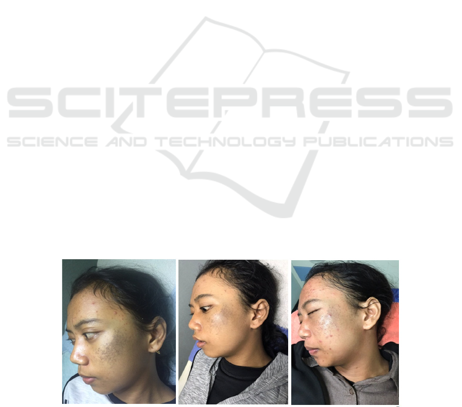
lesion became lighter. The progression and
improvement of the patient can be seen in the figures
3 DISCUSSION
Pigmentation disorders of the skin can either be
hypomelanotic, hypermelanotic, or may present with
a pattern of mixed hypo- and hypermelanosis. The
diagnosis of these disorders can be quite challenging
(Lee et al., 2004). Dermal melanocytosis define a
broad group of congenital and acquired melanocytic
lesions characterized by the presence of intradermal
dendritic, variably pigmented, spindle shaped
melanocytes with or without presence of dermal
melanophages. This group includes Mongolian spot,
nevus of Ota, nevus of Ito and acquired bilateral
nevus of Ota-like macules (ABNOM), and other
unusual cases of dermal melanocytosis that have
been introduced to the literature as part of this
category on the basis of similar histopathological
findings (Watanabe, 2014).
From the standpoint of age of onset, there is
overlap between classical nevus of Ota and Hori’s.
The distribution of pigmentation is identical between
nevus of Ota and Hori’s nevi, although mostly
unilateral in nevus of Ota. The histology of Hori’s is
identical to nevus of Ota.
Figure 1: Comparison of 1
st
Laser session (left), 2
nd
Laser session (middle), and 3
rd
Laser session (right).
Nevus Hori Treated with Laser Combination: A Case Report
457

3.1 Nevus Hori
Acquired bilateral nevus of Ota-like macules
(ABNOM), also named Hori nevus, was first
described by Hori et al in 1984. Clinically, ABNOM
is characterized by multiple speckled blue-brown
and/or slate-gray macules occurring bilaterally on
the malar regions or less commonly forehead, upper
eyelids, and cheeks and nose. It most commonly
presents in Asian women after the third decade of
life (Cho et al., 2009; Park et al., 2014; Watanabe,
2014).
Hori et al hypothesized that the pathogenesis of
ABNOM may be attributed to later reactivation of
preexisting misplaced dermal melanocytes that may
result from faulty migration during embryological
development, dropping off from the basal layer of
epidermis or migration from follicular bulb
melanocytes. Mizoguchi and Mizushima concluded
that there are ‘‘two hits’’ are needed for the
development of ABNOM: the first representing the
ectopic placement of inactive, poorly melanized
dermal melanocytes at birth or soon thereafter and
the second, the activation of these melanocytes in
response to ultraviolet exposure, excessive sex
hormone, chronic inflammation such as atopic
dermatitis, or other unknown triggers (Murakami,
2000; Park et al., 2014).
The diagnosis of ABNOM was made by clinical
appearances, according to the description by Hori et
al.and skin biopsies were not performed. The color
of ABNOM was categorized into one of four groups,
namely brown, slate-gray, brown–blue, and blue
(Cho et al., 2009).
3.2 Nevus Ota
Nevus of Ota or nevus fuscocaeruleus
ophthalmomaxillaris was first described by the
Japanese dermatologist Ota in 1939 as a dermal
melanocytic hamartoma that presents as bluish
hyperpigmentation along the ophthalmic, maxillary
and mandibular branches of the trigeminal nerve
(Metha & Balachandran, 2007; Lapreere et al.,
2012). It is most frequently seen in the Asian
population, has a female predominance, and is
usually congenital, although appearance in early
childhood or at puberty has been described (Kumari
& Thappa, 2006; Lapeere et al., 2012).
The pigmentation of Ota’s nevus is composed of
flat blue black or slate grey macules intermingled
with small brown specks. The intensity of
pigmentation may be influenced by fatigue,
menstruation, insomnia and weather.
Mucosal
pigmentation may occur involving conjunctiva,
sclera, and tympanic membrane (oculodermal
melanocytosis), or other sites.
Ocular melanosis in
22-77% cases is almost always ipsilateral and deep
in the conjunctiva (Metha & Balachandran, 2007;
Lapeere et al., 2012). Pigmentation may also affect
the sclera, cornea, iris, choroid and less commonly
the optic nerve, retrobulbar fat, orbit, periosteum and
extraocular muscles (Metha & Balachandran, 2007).
The pigmentation of mucous membranes of the head
and neck is variable; tympanic membrane being
most frequently affected although nasal, buccal,
pharyngeal and rarely palatine mucosa may be
involved (Sharan et al., 2005). At present, it is
believed that nevus of Ota is caused by heteroplasia
that occurs in melanocyte migration during
embryonic development (Huang et al., 2013).
Nevus of Ota involves innervated areas of the
first branch (V1) and second branch (V2) of the
trigeminal nerve mainly affects the eye region and
pars zygomatica, and the color of the skin lesion is
brown or blue, the diameter of the area is 1–10 cm
or larger.
10
Tanino classified nevus of Ota into 4
types according to the skin lesion involvement area:
Type I was mild, Type II was moderate, Type III
was severe, Type IV was bilateral type (Huang et al.,
2013).
In our report, the patient is female, age 16 years
old with the symptom dark patches at her left cheek
since 2 years ago. There are no patches since she
birth. The dark patches were getting wider, and she
also has ultraviolet exposure since she usually going
to school by bicycle and on foot. According to this
situation is suitable with the clinical manifestation of
Nevus Hori or ABNOM. From the theory, ABNOM
is an acquired dermal melanocytosis which induced
by ultraviolet exposure, sex hormone, and chronic
inflammation.
She never complain about itchy, numbness or
pain sensation on her cheek. She had no complained
about visual disturbances or dizzy. The patient’s
visus is normal (6/6) and there are no pigmentation
in her eyes. The tonometry and funduscopy
examination also revealed normal result and no sign
of glaukoma in this patient. Nevus Hori is said to
have lack mucosal involvement.
From physical examination at left cheek region
there were hyperpigmented macule, vary in size,
bluish in color, sharply marginated. In this case, the
histopathology examination has not been done yet
because the patient still refuse to do the biopsy.
Nevus Hori tends to appear symmetrically at both
cheek (malar area). In the other hands, Nevus Ota
can appear unilaterally in one side of face. In this
RCD 2018 - The 23rd Regional Conference of Dermatology 2018
458

case because the clinical manifestations appear on
the left side only, so based on Tanino classification
included in type I.
Pigmentary disorders appearing on the face, even
if they are benign, frequently cause cosmetic and
psychological problems to many people, especially
women. As with most dermal pigmentary disorders,
single treatment with topical bleaching agents or
superficial-to medium-depth chemical peels is
generally not effective for long-term pigmentary
reduction or elimination.
11
Although methods such
as dermabrasion, cryotherapy, surgical excision, and
cosmetic camouflage had been attempted for FDM
in the past, these have been largely replaced by
pigment-selective lasers given the lower risk of scar
formation and permanent hypopigmentation or
depigmentation with these devices (Kunachak et al.,
1996; Kar et al., 2011).
Three types of Q-switched lasers have been used
widely to treat FDM. These include the Q-switched
694 nm Ruby laser, Q-switched 755 nm Alexandrite
laser and the Q-switched 1064 nm Nd:YAG laser.
Previous studies have shown that all of them were
able to provide excellent results in treating FDM.
Because Q-switched (QS) laser devices have
been widely accepted as the treatment of choice for
nevus of Ota on the principle of selective
photothermolysis and because ABNOM is
histopathologically similar to nevus of Ota, QS
lasers such as the QS ruby laser (QSRL), QS
neodymium:yttrium-aluminum-garnet laser
(QSNYL), and QS alexandrite laser (QSAL) have all
been used for the treatment of ABNOM since the
first report of Hori et al (Lee et al., 2009; Watanabe,
2014).
The laser fluence used was 7 to 10 J/cm2, at a
repetition rate of 1 Hz, and with a spot size of 2 to 4
mm. The number of treatment sessions ranged from
1 to 6 (mean 2.3 sessions) with short treatment
intervals (mean 2.2 weeks) (Watanabe, 2004).
In addition to local thermal destruction and
stimulation, fractionated devices may also play an
important role for drug delivery into the tissue and
for extruding material out of the skin, as in the
studies by Haedersdal et al. This has also been
recently reported by Brian Wei Cheng using a
combination between non ablative fractionated
erbium : YAG laser and Q-switches Nd:YAG laser
ias an effective and safe treatment to treat Nevus
Hori (Sakamoto et al., 2013; Tian, 2015).
After laser treatment, ABNOM showed a higher
degree of erythema as well as a higher incidence and
degree of Post Inflammatory Hyperpigmentation
(PIH) compared to that of nevus of Ota. Several
causes for the increased prevalence of PIH in
ABNOM after laser treatment are considered. First,
the lesion in ABNOM was located in the superficial
dermal layer and there were few epidermal
melanocytes and melanin pigment. In the treatment
of Q-switches laser, melanin acts as a chromophore,
therefore melanin in the epidermis allows laser to be
selectively absorbed so that the epidermal tissue
becomes vacuolized due to the heat. The
melanocytes and melanin pigment of the vacuolated
epidermis are dropped into the dermis, and are
presumed to induce PIH.
Next, melanocytes were especially clustered in
the perivascular area in ABNOM, whereas in nevus
of Ota melanocytes were evenly distributed
throughout the dermal layer in between collagen
fibers. The presence of many melanocytes in the
perivascular area may lead to indirect vascular
damage, increase melanogenesis, induce many types
of inflammatory responses, and produce chemical
substances. These process can induce PIH
(Watanabe, 2014).
Patient already had Q-switched 1064 Nd:YAG
laser combined with fractional CO
2
for three times
and the lesion is having improvement although she
has not satisfied yet. The patient is advised to
continue the laser treatment and observed the
occurrence of PIH. The prognosis of this case is
good.
4 CONCLUSION
Pigmentary disorders appearing on the face
frequently cause cosmetic and psychological
problems to many people, especially women. The
diagnosis of FDM can be quite challenging because
of the similarity manifestation of Nevus of Ota and
ABNOM. Q-switched laser give a promising result
to treat this pigmentary problems. Combination with
CO
2
fractional laser aimed to increase the ability to
remove pigment.
REFERENCES
Cho, S.B., Park, S.J., Kim, M.J., Bu, T.S., 2009.
Treatment of acquired bilateral nevus of Ota-like
macules (Hori’s nevus) using 1064-nm Q-switched
Nd:YAG laser with low fluence. International Journal
of Dermatology 48, 1308–1312. doi:10.1111/j.1365-
4632.2008.04061.x
Huang, W.H., Wang, H.W., Sun, Q.N., Jin, H.Z., Liu,
Y.H., Ma, D.L., Zuo, Y.G., Zheng, H.Y., Wan, K.,
Jing, Q., Zhao, Y.L., 2013. A new classification of
Nevus Hori Treated with Laser Combination: A Case Report
459

nevus of Ota. Chinese Medical Journal 126, 3910–
3914. doi:10.3760/cma.j.issn.0366-6999.20131211
Kar, H.K., Gupta, L., 2014. 1064 nm Q switched Nd:
YAG laser treatment of nevus of Ota: an Indian open
label prospective study of 50 patients. Indian journal
of dermatology, venereology and leprology 77, 565–
70. doi:10.4103/0378-6323.84057
Kumari, R., Thappa, D.M. 2006. Familial nevus of ota.
Indian J Dermatol; 52: 198-9.
Kunachak, S., Kunachakr, S., Sirikulchayanonta, V.,
Leelaudomniti, P., 1996. Dermabrasion is an effective
treatment for acquired bilateral nevus of Ota-like
macules. Dermatologic Surgery 22, 559–562.
doi:10.1111/j.1524-4725.1996.tb00374.x
Lapeere, H. 2012. Hypomelanoses and hypermelanoses In:
Goldsmith, L.A., Katz, S.I., Gilchrest, B.A., Paller.
A.S, Leffell, D.J., Wolf, K., editors. Fitzpatrick’s
dermatology in general medicine. 8
th
ed. New York:
Mc Graw Hill.p. 823.
Lee, B., You, C.K., Won, H.K., Lee, E.S., 2004.
Comparison of characteristics of acquired bilateral
nevus of Ota-like macules and nevus of Ota according
to therapeutic outcome. Journal of Korean Medical
Science 19, 554–559.
doi:10.3346/jkms.2004.19.4.554.
Lee, W.J., Han, S.S., Chang, S.E., Lee, M.W., Choi, J.H.,
Moon, K.C., Koh, J.K., 2009. Q-switched ND:YAG
laser therapy of acquired bilateral nevus of Ota-like
macules. Annals of Dermatology 21, 255–260.
doi:10.5021/ad.2009.21.3.255
Mehta, V., Balachandran, C. 2007. Case report : bilateral
nevus of ota. J Pakistan Association Dermatol; 17: 59-
61.
Murakami, F., Baba, T., Mizoguchi, M. 2000. Ultraviolet-
induced generalized acquired dermal melanocytosis
with numerous melanophages. Br J
Dermatol;142:184-6
Park, J.M., Tsao, H., Tsao, S., 2009. Acquired bilateral
nevus of Ota-like macules (Hori nevus): Etiologic and
therapeutic considerations. Journal of the American
Academy of Dermatology.
doi:10.1016/j.jaad.2008.10.054.
Sakamoto, F.H., Jalian, H.R., Anderson, R.R. 2013.
Understanding lasers, lights, and tissue interactions In:
Hruza, G., Avram, M., editors. Lasers and lights. 3
rd
Ed. New York: Elsevier.p. 6-8.
Sharan S, Grigg JR, Billson FA. Bilateral nevus of Ota
with choroidal melanoma and diffuse retinal
pigmentation in a dark skinned person. Br J
Ophthalmol 2005; 89: 1529. Sharan, S., Grigg, J.R.,
Billson, F.A., 2005. Bilateral naevus of Ota with
choroidal melanoma and diffuse retinal pigmentation
in a dark skinned person [1]. British Journal of
Ophthalmology. doi:10.1136/bjo.2005.070839
Tian, B.C.A., 2015. Novel treatment of Hori′s nevus: A
combination of fractional nonablative 2,940-nm
Er:YAG and low-fluence 1,064-nm Q-switched
Nd:YAG laser. Journal of Cutaneous and Aesthetic
Surgery 8, 227. doi:10.4103/0974-2077.172198
Watanabe S. 2014. Facial Dermal Melanocytosis. Austin J
Dermatolog; 1(2):1-6
RCD 2018 - The 23rd Regional Conference of Dermatology 2018
460
