
Difference of Histamine Expression between Multibacillary Leprosy
with and without Erythema Nodosum Leprosum (ENL) Reaction
Dwi Septiana
1
, Puguh Riyanto
1
, Asih Budiastuti
1
, Dhiana Ernawati
1
, Soejoto
1
, E. S. Indrayanti
1
1
Department of Dermatology and Venereology
Faculty of Medicine Diponegoro University / Dr. Kariadi General Hospital Semarang
Jl. Dr. Sutomo No. 16 Semarang-Indonesia.
Keywords Multibacillary leprosy, erythema nodosum leprosum, histamine expression.
Abstract Leprosy is a chronic infectious disease that poses a problem for public health because it causes disability. One
of the causes for this disability is leprosy reaction. Erythema Nodosum Leprosum (ENL) is a type 2 leprosy
reactions, which were more frequent in borderline lepromatous (BL), and lepromatous (LL) type of leprosy
receiving multi drug therapy (MDT) for multibacillary leprosy (MB). Histamine, primarily H4 receptor-
mediated, plays role in inflammation by increasing vascular permeability and muscle contraction resulting in
leukocytes migration and leakage of plasma containing antibodies and tissue complements. This cascade
finally results in tissue inflammation. To our knowledge, there is no previous study analysing histamine
expression difference between MB leprosy with and without ENL reaction. The purpose of the research was
to understand the difference of histamine expression in MB leprosy patients with and without ENL reactions
who received MDT for at least 6 months. The method of the research was an observational study with cross-
sectional approach. Study samples were 32 people divided into 2 groups, 16 samples in without ENL reactions
group and 16 samples in with ENL reactions group. Histamine expression was measured based on Allred
scoring system. The result from Difference test for histamine expression between two study groups using
Mann Whitney Test showed p < 0.05 (p=0.030 ). The study found that mean histamine expression of ENL
MB patients was higher than non-ENL MB patients.
1 INTRODUCTION
Leprosy is a chronic infectious disease caused by
Mycobacterium leprae (M. leprae) that were initially
attacked peripheral nerves, then attacked the skin and
other tissues, but excepted the central nervous system
(Kosasih, A., Wisnu., I.M., and Daili, E.S., 2011 and
Lee, D.J., Rea, T.H., Modlin, R.L., 2012). Leprosy
reaction is an acute episode in chronic course of
leprosy. It is an immune reaction, an antigen-antibody
reaction. Since they often result in disfigurements,
leprosy reactions have to be treated carefully.
Leprosy reactions consisted of two different types,
specifically type 1 or reversal reaction (RR) and type
2 or erythema nodosum leprosum (ENL) (Odom,
R.B., James, W.D., and Berger, T.G., 2010;
Lockwood, D.N.J., 2010; and Silva, M.R. and Castro,
M.C.R.D, 2012).
Coomb and Gell suggested that type 2 reaction is
a type III hypersensitivity reaction. The remains of
deceased bacilli after multi-drug therapy (MDT)
result in antigen (Odom, R.B., James, W.D., and
Berger, T.G., 2010; Lockwood, D.N.J., 2010; nd
Daili, E.S., 2011 and Lee, D.J., Rea, T.H., Modlin,
R.L., 2012). The antigen involves in reactions with
IgM, IgG, and C3 complement, forming immune
complexes that spread through blood circulation and
attach in the end organs. This cascade results in
complement system activation, producing
anaphylatoxin and promoting degranulation of mast
cell to release histamine. Histamine release will cause
inflammation, primarily after activation of histamine
h4 receptor (Frenzel, I. and Hermine, O., 2013).
After phagocytosis, the macrophage-eaten M
leprae can produce Hsp70 during the stressful
phagolisosom creation. Antileprosy drugs that given
during the course of the therapy, add more pressure to
bacilli inside the macrophages. Hsp70 accumulation
inside the macrophages invites the cytotoxic
lymphocytes to lyse the macrophages and activates
Septiana, D., Riyanto, P., Budiastuti, A., Ernawati, D., , S. and Indrayanti, E.
Difference of Histamine Expression between Multibacillary Leprosy with and without Erythema Nodosum Leprosum (ENL) Reaction.
DOI: 10.5220/0008153001590162
In Proceedings of the 23rd Regional Conference of Dermatology (RCD 2018), pages 159-162
ISBN: 978-989-758-494-7
Copyright
c
2021 by SCITEPRESS – Science and Technology Publications, Lda. All rights reserved
159

Table 1: The difference of mean histamine expression between leprosy patient with and without ENL reaction.
Note: b. Mann-Whitney Test
pro-inflammatory cytokines, such as TNF. After the
lysis, released Hsp70 attach to the TLR4 receptors on
the surface of mast cells. The next process is
activation of cell mast by the TLR4, as indicated by
the histamine formation, TNF, IL-6, and IL-8 then
finally promote the inflammation (Mortaz, E., et al,
2007).
Immunohistochemistry is a method to detect the
existence of specific antigen in a tissue based on
antibody (Ab) and antigen (Ag) binding. ENL is an
inflammatory reaction in tissue. Histamine-bound
histamine h4 receptors will be captured by histamine
h4 receptor antibodies and create histamine
expression on immunohistochemistry technique
(Trufelli, D.C., et al, 2010; Lin, Fan and Shi, Jianhui,
2015; and Park, C.S., Roh, J., and Kim, S.W., 2016).
Previous study have address the role of mast cell
in leprosy reaction have been conducted. They found
higher density of mast cell in leprosy reactions
(Antunes, S.L.G., et al, 2003). Therefore, the
objective of the study was to evaluate the histamine
expression difference between MB leprosy with and
without ENL reaction.
2 METHODS
This cross-sectional observational study was
conducted in outpatient setting of leprosy clinic at
Donorojo Leprosy Hospital, Jepara. The subjects
were patients with multibacillary (MB) infection and
aged 20-65 years old. To be included, the patients
must have been received MDT for more than 6
months. During study period (March 2017- June
2017), 32 patients were included, 16 MB patients
without ENL reaction and 16 MB patients with ENL
reaction. The skin tissue samples were processed and
analysed by immunohistochemistry method in
anatomical pathology laboratory of Medical Faculty,
Gadjah Mada University, Yogyakarta.
Immunohistochemical examination and measurement
of histamine expression can be seen using a light
microscope at 10 fields of view using 400x
magnification. Each field of view will be
photographed and then analyzed using a computer
(image raster software) to calculate the percentage
and intensity of cells that bind histamine h4 receptor
antibodies, which then according to the percentage
and the intensity of the staining will be incorporated
into the semi-quantitative system by using the Allred
scoring system
3 RESULT
In non-ENL group, the number of female and male
patients was equal, the mean age was 42.19 + 2.22
years, mean leukocyte count was 7806.25 + 2094.54
/mm
3
, mean MDT duration was 8.5 + 2.22 months. In
ENL group, all of the patients were males, the mean
age was 32.63 + 11.93 years, mean leukocyte count
was 8587.5 + 1872.92 /mm
3
, mean MDT duration
was 8.96 + 2.024 months. The mean histamine
expression in non-ENL and ENL group were 6.31 +
1.35 and 7.31 + 0,48, respectively.
The present study found that mean histamine
expression of ENL MB patients was higher than non-
ENL MB patients. The Mann-Whitney test for mean
histamine expression found significant difference (p
< 0 .05) with p = 0.030.
4 DISCUSSION
In this study we obtained that male leprosy patients
were more than female where in multibacillary
leprosy group without ENL reactions the number of
men was eight persons (50%) and 16 persons (20%)
in multibacillary leprosy group with ENL reaction.
Gender generally is not a risk factor of ENL.
Sample distribution by age was found to be
statistically significant. Erythema Nodosum
Leprosum based on age group does not represent a
particular group risk for developing ENL.
Non ENL Reaction ENL Reaction P
Mean±SD Median
(Min-maks)
Mean±SD Median
(Min-maks)
Histamine
expression
6,31 ± 1,35 6(4 – 8) 7,31±0,48 7 (7-8) 0,030
b
RCD 2018 - The 23rd Regional Conference of Dermatology 2018
160
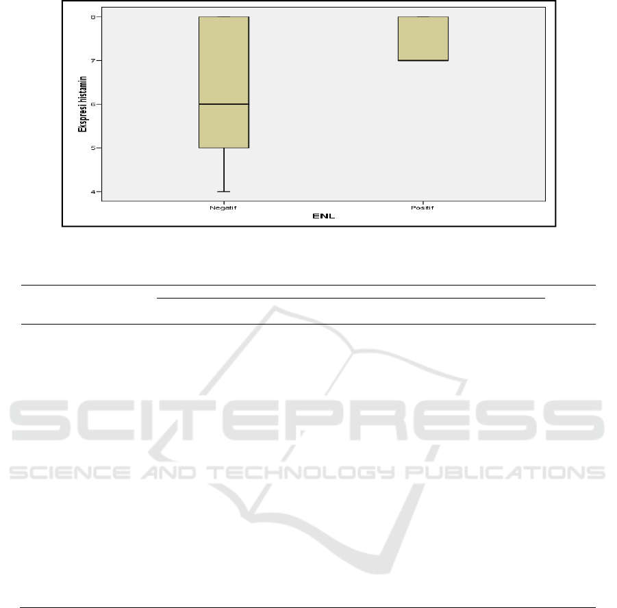
Figure 1: The study found that mean histamine expression of ENL MB patients was higher than non-ENL MB patients.
Table 2: Characteristic subjects of the study.
Non Reaksi ENL Reaksi ENL P
N(%) Mean±SD Median
(Min-maks)
N(%) Mean±SD Median
(Min-maks)
Gender male
8(50) 16(100)
0,002
a
female
8(50) 0(0)
Age
42,19±11,2 40(23-59) 32,63±11,93 31,5(20-59) 0,011
b
leukocytes
7806,25±20
95,54
7750(3700-
10700)
8587,5±187
2,92
9100(4400-
10900)
0,275
c
MDT
Duration
Job
student
0(0)
8,5±2,22
8(6-12)
2(12,5)
8,96±2,024
8(6-12)
0,688
b
non job
farmer
3(18,8)
1(6,3)
7(43,8)
0(0)
Labor
Retirement
0(0)
0(0)
1(6,3)
1(6,3)
0,053
a
driver
fisherman
Housewife
Private
employe
Embankment
farmer
0(0)
1(6,3)
7(43,8)
4(25)
0(0)
1(6,3)
0(0)
0(0)
3(18,8)
1(6,3
Note: a.Uji chi square ; b. Mann Whitney test ; c. Uji t independen
Leprosy is known to occur in all ages ranging
from infancy to old age (3 weeks to over 70 years).
But the most is happened at a young and productive
ages. ENL occurs at any time during leprosy episodes
and most often after 6 months treatment
(Triamchaisri, S., et al, 2015).
Subjects of the study were limited by the number
of leukocytes <11.000 / mmk to exclude
inflammatory and infectious diseases in which
histamine expression increased in the presence of
inflammatory and infectious conditions (Zampeli, E.
and Tiligada, E., 2009). The mean difference of
leukocyte counts of multibacillary leprosy study
subjects with ENL and without ENL was found to be
insignificant ( p = 0.275) showed that the leukocyte
rates of sample was homogeneous.
MDT duration therapy of multibacillary leprosy
group with ENL and without ENL was found to be
insignificant (p = 0,688). Erythema Nodosum
Leprosum can occur at any time during leprosy
course but most often occurs after 6 months of MDT
treatment, since many leprosy bacteria are destroyed,
that means many antigens are released and react with
antibodies, thus activates the complement system and
results in ENL (Odom, R.B., James, W.D., and
Berger, T.G., 2010; Lockwood, D.N.J., 2010; Lee,
Difference of Histamine Expression between Multibacillary Leprosy with and without Erythema Nodosum Leprosum (ENL) Reaction
161

D.J., Rea, T.H., Modlin, R.L., 2012; and Silva, M.R.
and Castro, M.C.R.D, 2012).
Histamine expression value in MB group with
ENL reaction was higher than MB group without
reaction with significant differences (p = 0.030) by
Mann-Whitney test. Histamine plays a role in
immunoinflamatory response particularly in those
with H4 receptor (mast cells, eosinophils, T cells and
dendritic cells) that activates immune cell maturation,
increases cytokine production and increases cellular
chemotactic response (Gutzmer, R., et al, 2011 and
Harvima, I.T. and Nilsson, G., 2011). Histamine
increases in inflammatory states such as ENL.
Histamine expression in MB leprosy group with ENL
reaction was higher than MB leprosy group without
ENL reaction.
5 CONCLUSION
There was a significant difference in histamine
expression between MB leprosy with and without
ENL reaction where the mean of histamine
expression in MB leprosy group with ENL reaction
was higher than leprosy group without ENL reaction.
ACKNOWLEDGEMENT
This study was supported by Faculty of Medicine
Diponegoro University / Dr. Kariadi General Hospital
Semarang, leprosy clinic at Donorojo Leprosy
Hospital, Jepara and anatomical pathology laboratory
of Medical Faculty, Gadjah Mada University,
Yogyakarta.
REFERENCES
Antunes, S.L.G., et al, 2003. Mast Cell Subset and
Neuropeptides in Leprosy Reactions. Arq
Neuropsiquaitr; 61(2-A): 208-219
Frenzel, I. and Hermine, O., 2013 Mast cells and
inflammation. Joint Bone Spine. 80 (2): 141-5
Gutzmer, R., et al, 2011. Pathogenetic and therapeutic
implications of the histamine H4 receptor in
inflammatory skin diseases and pruritus. Frontiers in
Bioscene S3. 985-944.
Harvima, I.T. and Nilsson, G., 2011. Mast cells as
regulators of skin inflammation and immunity. Acta
Derm Venereol. 91 (6): 664-50.
Kosasih, A., Wisnu., I.M., Daili, E.S., 2011. Kusta. In:
Djuanda A ed. Ilmu Penyakit Kulit dan Kelamin. edisi
6. Jakarta: Badan Penerbit Fakultas Kedokteran
Universitas Indonesia 73-83
Lee, D.J., Rea, T.H., Modlin, R.L., 2012. Leprosy. In: Wolf
K, Gold Smith LA, Katz SI eds. Fitzpatrick’s
dermatology in general medicine. Vol 2.ed 8. New
York: Mc Graw Hill. 2253-62
Lin, Fan and Shi, Jianhui, 2015. Standardization of
Dianostic Immunohistochemistry. In: Lin, Fan et al.
Handbook of Practical Immunohistochemistry. Edisi
ke-2. New York: Springer. 17-30
Lockwood, D.N.J., 2010. Leprosy. In: Burns, T.,
Breathnach, S., Neil, C., Griffiths, C., ed. Rook’s
textbook of dermatology. Edisi ke-8. Massachusets :
Blackwell Science Ltd. 32.1
Mortaz, E., et al, 2007. Stimulation of cysteinyl leukotriene
production in mast cells by heat shock and
acetylsalicylic acid. Eur J Pharmacol. 30;561(1-
3):214-19
Odom, R.B., James, W.D., and Berger, T.G., 2010. Hansen
Disease. In : James, W.D., Berger, T.G., Elston, D.M.
Andrew’s disease of the skin Clinical Dermatology.
Edisi ke-11. Philadelphia : WB Saunders Co. Hal. 334-
44
Park, C.S., Roh, J., and Kim, S.W., 2016.
Immunohistochemistry for Pathologists: Protocols,
Pitfalls and Tips. Journal of Pathology and
Translational Medicine; 50(6): 411-418
Silva, M.R. and Castro, M.C.R.D, 2012. Mycobacterial
infections. In : Bolognia, J.L., Jorizzo, J.L., Rapini,
R.P., ed. Dermatology. Edisi ke 3. Edinburg: Mosby.
1221-8.
Triamchaisri, S., et al, 2015. Leprosy Reaction in Thai
Population: A 20 year Retrospective Study.
Dermatology Research and Practice.
Trufelli, D.C., et al, 2010. Immunohistochemistry as an
Important Tool in Biomarkers Detection and Clinical
Practice. US National Library of Medicine National
Institutes of Health; 5:9-20
Zampeli, E. and Tiligada, E., 2009. The role of histamine
H
4
receptor in immune and inflammatory disorders.
British journal of Pharmacology. 157,26-33
RCD 2018 - The 23rd Regional Conference of Dermatology 2018
162
