
Hair Shaft Alteration in Children Undergoing Chemotherapy
Reti Anggraeni, Ardelia Dyah Ayu, M. Eko Irawanto, Suci Widhiati, Muhammad Riza
Faculty of Medicine Sebelas Maret University/ Dr. Moewardi General Hospital, Surakarta
Keywords: alteration, chemotherapy, hair shaft
Abstract: Hairs have no vital function but it may use as an indicator for the human’s health. Hair shaft is a unique
structure which composed of inner cortex and protective outer cuticle. Hair shaft disorders may be inherited
or acquired, as in a local or systemic disease. One of the acquired causals is chemotherapy. Chemotherapy
agent can damage the structure or morphology of the hair including hair shaft pattern. Cross sectional
analytical study was conducted in Dr. Moewardi General Hospital Surakarta. Forty five subjects, aged 1-14
years old were enrolled. The study subjects (n=30) were pediatric patients undergoing chemotherapy, the
control group (n=15) comprised of children who never had chemotherapy. Of the 30 subjects from study
group, 27 (90%) had altered hair shaft. Two (13.3%) of fifteen children in control group had hair shaft
alteration. Statistical analysis using Mann-Whitney U Test obtained p=0.000 which means there was a
significant different between incidents of hair shaft alteration between children undergoing chemotherapy and
children never had chemotherapy. Further study is needed to describe more about the most common group of
chemotherapeutic agents which cause the alteration and the relation between the type of cancer and hair shaft
alteration.
1 INTRODUCTION
Humans have over 2 million hair follicles, which may
have significant positive and negative influences on
skin health.(McElwee and Sinclair, 2008) The hair
shaft is protected by the cuticle layer with its
overlapping cells which resemble shingle on its roof.
If there is damage in cuticle, the exposed hair cortex
is still bonded but it is more susceptible to
environmental damages and fractures.(yu et al., 2006;
Mirmirani et al., 2011)
The most common treatment performed for cancer
is chemotherapy. It has adverse side effects to healthy
tissue and organs including the hair. It affects the hair
in two mainly parts, the hair growth and the hair shaft.
About 65% of patients undergoing chemotherapy will
get chemotherapy-induced hair loss (chemotherapy-
induced alopecia), known as anagen effluvium, which
is usually reversible when the therapy ends.(Trüeb,
2010)
Chemotherapy may also affect the structure and
morphology of the hair leading to the damage of the
hair follicle and later causing hair shaft
alteration.(Lindner et al., 2012) The alteration of hair
shaft reveals in many patterns caused by diminishing
of the follicle function, such as fractured hairs,
narrowings (the most common is Pohl-pinkus
constriction), node-like appearance, curls and twist
hairs, short hairs and disorders on the band hair.
There have been many studies about anagen
effluvium due to chemotherapy agent but there are
only a few studies about hair shaft alteration.
Therefore we performed this study to reveal whether
chemotherapy may cause the hair shaft alteration.
Thus when we have patients undergoing
chemotherapy, especially with altered hair, we can
give more education supports. As it would be a
psychologically problem leading to negative impact
on their perceptions of appearance.
2 METHODS
Cross sectional analytical study was conducted in
pediatric ward of Dr. Moewardi General Hospital
Surakarta between December 2017 and January 2018.
Forty five subjects, aged 1-14 years old were enrolled
in our study by using consecutive sampling method.
The study subjects (n=30) were pediatric patients
undergoing chemotherapy and the control group
(n=15) comprised of children who never had
chemotherapy. Each subject’s scalp was
104
Anggraeni, R., Ayu, A., Irawanto, M., Widhiati, S. and Riza, M.
Hair Shaft Alteration in Children Undergoing Chemotherapy.
DOI: 10.5220/0008151801040107
In Proceedings of the 23rd Regional Conference of Dermatology (RCD 2018), pages 104-107
ISBN: 978-989-758-494-7
Copyright
c
2021 by SCITEPRESS – Science and Technology Publications, Lda. All rights reserved
photographed by using dermoscope, 20 hairs were
removed, put under object glass and examined under
microscope. The photograph and microscopic
features were interpreted by three observers.
3 RESULTS
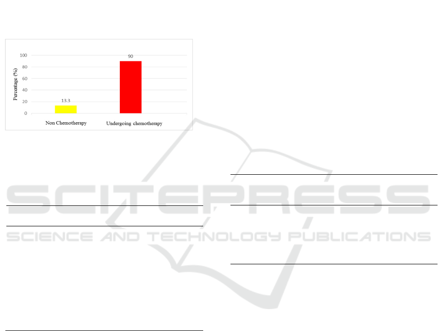
Of the 30 study subjects, there were 27 (90%) had hair
shaft alteration whereas the control group 2 (13.3%)
of 15 subjects experienced altered hair shaft (Figure
1).
Figure 1. The percentage of the hair alteration of the
enrolled subjects
Table 1. The hair shaft alteration of study subject compared
to control group
Total
Percentage
(%)
Study Subject
(n=30)
Altered 27 90
No-
alteration 3 10
Control (n=15)
Altered 2 13.3
No-
alteration 13 86.7
This present study used Mann-Whitney U Test
(p=0.000) which revealed that there was a significant
difference of hair shaft alteration between children
undergoing chemotherapy and children never had
chemotherapy.
The demographic characteristic (Table 2)
obtained that the diagnosis of acute lymphoblastic
leukemia (ALL) was the most common cancer (30%,
n=9) followed by osteosarcoma (16.8%, n=5). The
chemotherapy agents was varied, used as a single or
combination therapy. The most chemotherapy agent
combination used was methotrexate and vincristine
(36.8%, n=11) while the single therapy was mostly
cyclophosphamide (10%, n=3).
Of the 27 altered hair shaft subjects, there were
many patterns with the most common pattern was
Pohl-pinkus (n=9) followed by hair cast node-like
appearance (n=3) (Figure 2).
4 DISCUSSION
The hair consist of two distinct structures, the hair
follicle, the living part located under the skin, and the
hair shaft which is fully keratinized nonliving part
above the skin surface. The follicle is the essential
growth structure of the hair and basically has two
main parts, upper part consists of infundibulum and
isthmus whereas the lower part comprise of hair bulb
and suprabulbar region.(Berker et al., 2012) The hair
shaft has three layers: cuticle, cortex and
medulla.(Berker et al., 2012; McElwee and Sinclair,
2008) Hair has no essential function but if there is a
damage it leads to hair loss or even the alteration of
hair shaft. The individual will have a psychosocial
problem related to his/ her physical performance.
Table 2. Demographic characteristics of study subjects
Total
(n=30)
Percenta
ge
(%)
Age (year)
0 – 5 14 46.7
6 – 11 12 40
12 – 17 4 13.3
Diagnosis
Acute
Lymphoblastic
Leukemia 9 30
Osteosarcoma 5 16.8
Acute Myeloid 3 10
Rhabdomyosarko
ma 2 6.7
Trombositopenia 2 6.7
Systemic Lupus
Erythematosus 2 6.7
Nephtiris Lupus 1 3.3
Retinoblastoma 1 3.3
Nephroblastoma 1 3.3
Pheochromocyto
ma 1 3.3
Genu Cell Tumor 1 3.3
Carcinoma Testis 1 3.3
Nephrotic
Syndrome 1 3.3
Hair Shaft Alteration in Children Undergoing Chemotherapy
105

Chemotherapy Agent
Methotrexate +
vincristine 11 36.8
Etoposide +
carboplatin 8 26.7
Cyclophosphamid
e 3 10
Doxorubicin +
vincristine +
cyclophosphamid
e 2 6.7
Ifosfamide +
etoposide 1 3.3
Cyclophosphamid
e +
metilprednisolone 1 3.3
Methotrexate +
doxorubicin 1 3.3
Etoposide +
bleomycine +
vincristine 1 3.3
Cisplatin +
bleomycine +
vincristine 1 3.3
Vincristine 1 3.3
One of the causal factors for hair disorder is
chemotherapy which is the most common treatment
for cancer. The chemotherapeutic agents either use as
single or combination regimen may damage the
follicle function of the hair which later will destroy
the growth of the hair shaft appearing to the skin
surface. Firstly hair loss which can be seen within
days to weeks of the initiation of chemotherapy and
complete loss occurs in 3 months later. In 60% cases
after the hair loss, patients experiencing differences
in hair color, alteration in thickness, texture or
waviness of their regrowth hair in 1 to 3 months after
chemotherapy ends.(Kanwar and Narang, 2013;
Kanti et al., 2014)
Figure 2. The varieties of hair shaft patterns of the 27
altered hair shaft subjects
The alteration of the hair after hair loss occurs the
shape of the hair shaft. As the hair follicle is
surrounded by dense of capillaries network, growing
hair is sensitive to metabolic imbalances resulted
from internal disease, local inflammation or drug-side
effects.(Rogers, 1995; Tosti and Pazzaglia, 2007)
This remains unclear why chemotherapy agents may
lead to the alteration of hair shaft.(Trüeb, 2010)
The hair shaft alteration reveals in many patterns
consisting of fractured hairs (trichoptilosis,
trichoschisis or trichoclasis, broken (fractured) hairs
and golf tee hairs), narrowings (monilethrix,
monilethrix-like congenital hypotrichosis,
monilethrix-like hairs or Pohl-pinkus constriction,
pseudomonilethrix, exclamation mark hairs and
tapered hairs), node-like appearance (trichonodosis,
trichorrhexis nodosa, trichorrhexis invaginata and
hair casts), curls and twist hairs (pigtail, coiled,
comma, corkscrew, zigzag, pili torti and wooly hairs),
short hairs (upright regrowing, vellus hairs, dark
lines, tulip hairs, block hairs, i-hairs, broom hairs,
broom fibers and flame hairs) and disorders on the
band hair (continuous or interrupted medulla, pilli
annulati, and interrupted hairs).(Itin and Fistarol,
2005; Tosti and Piraccini, 2006; Rudnicka et al.,
2012) The most common alteration is Pohl-pinkus
constriction in which its morphological change
occurs in the hair roots.(Williamson and de Berker,
2005) De Berker et al stated that the monilethrix
pattern caused by cortical defect resulting in a
disorientation of the fibrils which lie along the
cortical cells.(Rogers, 1995) Another study by Ito et
al suggests that the basic cause of the monilethrix
alteration is malfunction of the germinative cells of
the hair matrix leading to abnormalities affecting the
cuticle, internal sheath and mostly affects the
cortex.(Ito et al., 1990) There is an intermediate state
whereas the follicle function decreases but does not
stop the hair fibre production, therefore there is a
short span along the hair of diminished
bore.(Williamson and de Berker, 2005)
In the present study, we found that there was 27
(90%, total n=30) in study subjects got hair shaft
alteration and only 2 (13.3%, total n=15) in the
control group had altered, as it is seen by the result of
Mann-Whitney test with p<0.05 (p=0.000) which
means that there was a significant difference of hair
shaft alteration between children undergoing
chemotherapy and children never had chemotherapy.
Of the 27 study subjects who had altered, there was
many varieties of patterns. The most common pattern
was Pohl-pinkus constriction (n=9), followed by
node-like appearance hair cast (n=3) and Pohl-pinkus
constriction combined with node-like appearance hair
cast (n=3).
A further study is required to determine the most
common group of chemotherapeutic agents which
may cause hair shaft alteration and the relation
RCD 2018 - The 23rd Regional Conference of Dermatology 2018
106

between the type of cancer and the hair shaft
alteration.
5 CONCLUSIONS
Children undergoing chemotherapy experienced a
significant hair shaft alteration. This suggest that
chemotherapy may damage the hair structure leading
to the alteration of hair shaft. The most common
pattern of the hair shaft alteration in our study was
Pohl-pinkus constriction.
REFERENCES
Berker, D.D., Higgins, C.A., Jahoda, C., Christiano, A.M.
2012. Biology of hair and nails. In: Bolognia
Dermatology 3
rd
Edition. Bolognia JL, Jorrizzo JL,
Schaffer JV, editors. United States: Elsevier Saunders
Ltd.; 2012. p. 1075-92.
Itin, P.H., Fistarol, S.K., 2005. Hair shaft abnormalities -
Clues to diagnosis and treatment. Dermatology.
Ito, M., Hashimoto, K., Katsuumi, K., Sato, Y., 1990.
Pathogenesis of monilethrix: Computer stereography
and electron microscopy. Journal of Investigative
Dermatology 95, 186–194.
Kanti, V., Nuwayhid, R., Lindner, J., Hillmann, K., Stroux,
A., Bangemann, N., Kleine-Tebbe, A., Blume-Peytavi,
U., Garcia Bartels, N., 2014. Analysis of quantitative
changes in hair growth during treatment with
chemotherapy or tamoxifen in patients with breast
cancer: A cohort study. British Journal of Dermatology
170, 643–650.
Kanwar, A., Narang, T., 2013. Anagen effluvium. Indian
Journal of Dermatology, Venereology, and Leprology
79, 604.
Lindner, J., Hillmann, K., Blume-Peytavi, U., Lademann,
J., Lux, A., Stroux, A., Schneider, A., Garcia Bartels,
N., 2012. Hair shaft abnormalities after chemotherapy
and tamoxifen therapy in patients with breast cancer
evaluated by optical coherence tomography. British
Journal of Dermatology 167, 1272–1278.
McElwee, K.J., Sinclair, R., 2008. Hair physiology and its
disorders. Drug Discovery Today: Disease
Mechanisms.
Mirmirani, P., Huang, K.P., Price, V.H., 2011. A practical,
algorithmic approach to diagnosing hair shaft disorders.
International Journal of Dermatology.
Rogers, M., 1995. Hair shaft abnormalities: Part I.
Australasian Journal of Dermatology 36, 179–184.
Rudnicka, L., Olszweska, M., Rakowska, A. 2012. Atlas of
Trichoscopy. New York: Springer.
Tosti, A., Piraccini, B.M. 2006. Diagnosis and Treatment
of Hair Disorders. Great Britain: CPI.
Tosti, A., Pazzaglia, M., 2007. Drug Reactions Affecting
Hair: Diagnosis. Dermatologic Clinics.
Trüeb, R.M., 2010. Chemotherapy-induced alopecia.
Current Opinion in Supportive and Palliative Care 4,
281–284.
Williamson, P.J., de Berker, D., 2005. Pohl-Pinkus
constrictions of hair following chemotherapy for
Hodgkin’s disease. British Journal of Haematology
128, 582–582.
yu, M., Finner, A., Shapiro, J., lo, B., Barekatain, A.,
Mcelwee, K.J., 2006. Hair follicles and their role in skin
health. Expert Review of Dermatology 1, 855–871.
Hair Shaft Alteration in Children Undergoing Chemotherapy
107
