
Prevalence of Ancylostomiasis in Pet Cats from Banyuwangi City,
East Java Province
Aditya Yudhana
1
, Ratih Novita Praja
2
, Fifi Anik Suroiyah
3
1
PSDKU Banyuwangi,
Department of Parasitology, Faculty of Veterinary Medicine, Universitas Airlangga, Indonesia
2
PSDKU Banyuwangi, Department of Microbiology, Faculty of Veterinary Medicine, Universitas Airlangga, Indonesia
3
Faculty of Veterinary Medicine, Universitas Airlangga,Wijaya Kusuma Street 113, Banyuwangi, Indonesia
Key
words: Ancylostomiasis, Banyuwangi, Cat, Prevalence.
Abstract: Ancylostomiasis is disease caused by hookworm parasite known as Ancylostoma spp. The number of cat
owners has increased year to year in Banyuwangi City. This condition may also increase the risk factor of
disease transmission related to Ancylostomiasis, which can be transmitted from cat to owner. The purpose
of this research is to determine the prevalence of Ancylostomiasis infection in pet cats. This research used
138 samples divided into 73 male pet cat feces and 65 female pet cat feces using the floatation method.
From 138 samples of examined pet cat feces, in 22 samples (15.9%) the egg of Ancylostoma spp was
positively found. Meanwhile, from 138 samples of examined pet cat feces, 9 samples from male cat (6.5%)
positively contained the egg of Ancylostoma spp and 13 samples from female cat (9.4%) positively
contained the egg of Ancylostoma spp. A control program needs to be applied for diseases caused by an
Ancylostoma spp. infection in cats that are beneficial to the animal and public health aspects. This study’s
results could also be used for further study, such as disease mapping, molecular epidemiology, and
development of new anthelmintic drugs against Ancylostomiasis.
1 INTRODUCTION
Ancylostomiasis is disease caused by a hookworm
parasite known as Ancylostoma spp. Among various
nematode parasites in cats, the hookworm belonging
to family ancylostomatidae is of great importance
due to their blood-sucking activities and chronic
pathogenesis. These parasites are further divided
into two subfamilies: ancylostominae and
necatorinae. The buccal capsule of these worms is
sub-globular, their lips and leaf crown are absent,
their oral opening is unarmed, or with teeth and
cutting plates (Bhatia et al., 2010). Ancylostoma spp.
is a blood-feeding parasitic intestinal nematode
which infects dogs, cats, and other mammals
throughout the temperature and tropical areas in the
world. In addition to the veterinary importance,
Ancylostoma spp can also cause zoonotic disease in
humans (Periago and Bethony, 2012). The larvae of
Ancylostoma caninum hatch from eggs and develop
into infective larvae via two molts. The infective
larvae then infect host animals such as dogs and
cats, migrate into the intestine, and develop into
adult worms following two more molts. If the
infective larvae invade humans, they can cause
cutaneous larvae migrans (CLM) or “creeping
eruptions,” which are hypersensitive reactions in
response to the migration of A. caninum larvae;
however, they cannot develop into adult worms just
by migrating under the skin (Yang et al., 2012).
The infection route of Ancylostoma spp. cannot
be separated from three factors, namely hosts,
agents, and the environment. Infection occurs if
there is an infective larvae of Ancylostoma spp. as a
source of infection and the availability of host that is
sensitive to a condition from environment that
causes contact between both. Cats that live in dirty
and humid areas have more risk of disease
transmission because a dirty environment is a
suitable place to the development of infective larvae
form of Ancylostoma spp. (Borthakur, 2011).
Management system in a cat is one of the factors
that play a role in the transmission of
ancylostomiasis. Pet and stray cats certainly have
different levels of infection risk. Stray cats are more
vulnerable to disease due to dirty environmental
conditions, food that is not always enough, and the
absence of good care from humans or veterinarians.
Yudhana, A., Praja, R. and Suroiyah, F.
Prevalence of Ancylostomiasis in Pet Cats from Banyuwangi City, East Java Province.
DOI: 10.5220/0007547205710574
In Proceedings of the 2nd International Conference Postgraduate School (ICPS 2018), pages 571-574
ISBN: 978-989-758-348-3
Copyright
c
2018 by SCITEPRESS – Science and Technology Publications, Lda. All rights reserved
571
Meanwhile, pet cats have a better environmental
condition, regular feeding, and more care from the
owner, which can minimize risk of disease
transmission (Oktaviana et al., 2014). The number of
cat owners has increased from one year to the next
in Banyuwangi City. This condition may also
increase risk factor of disease transmission regarding
Ancylostomiasis, which can transmit from the cat to
the owner. It is important to conduct research in
prevalence aspect because there is no recording data
or current study about Ancylostomiasis from pet cats
in Banyuwangi City before. Besides,
Ancylostomiasis is transmissible to humans, and cats
are the pets most often found in public or in
communities.
2 MATERIAL AND METHODS
This research was an observational analytic research
with cross sectional design. In this study, 138 feces
samples were obtained from pet cats in their owners’
houses around Banyuwangi City. Cat feces were
obtained by asking directly to the cat owners. Cat
owners were given plastic pots for collecting
samples from their cats. The feces were taken as
fresh as possible and 5% formalin added to soak all
feces as preservatives into plastic pots and then
labels which display the time of taking and the
origin of cats which samples were taken from. The
samples collected were immediately examined in
Laboratory of Instrument Faculty of Veterinary
Medicine Airlangga University PSDKU
Banyuwangi.
The prevalence of Ancylostomiasis infection was
measured by examination of feces using a
concentration method named the flotation method.
The principle of this method is based on the specific
gravity (BJ) of the Ancylostoma spp. egg being
lighter than BJ of solution used, so the eggs can float
to the surface. The procedure of flotation method is
as follows: ± 2 grams of feces were taken, put into a
plastic glass, added with a little aquadest, and stirred
until becoming homogeneous. The solution was then
filtered. After that, it was poured into a centrifuge
tube until ¾ of the tube was filled up. The tube was
then rotated at 1500 rpm for 5 minutes. The
supernatant was discarded, then added with saturated
sucrose for ¾ of the tube volume and re-stirred until
becoming homogeneous. The tubes were rotated
with a speed of 1500 rpm for 5 minutes. Then, the
tube was put on the tube rack perpendicularly, added
with saturated sucrose solution through dropping
using pipette until the surface becomes convex and
left for 3 minutes. Glass cover was put on a convex
surface carefully, then put on a glass object and
examined under microscope with 100x
magnification.
The prevalence of Ancylostomiasis was
calculated using the formula: the number of infected
samples divided with total sample and multiplied
with 100%. The data obtained in this study were
presented descriptively and the proportion of
Ancylostomiasis infection prevalence between male
and female pet cats were divided based on the data.
3 RESULTS AND DISCUSSION
The prevalence of Ancylostomiasis in pet cats in
Banyuwangi City was studied, and and the results
are presented in Table 1. A total of 138 pet cat fecal
samples were examined using floatation methods,
and it was found that 22 (15.9 %) samples positively
contained the egg of Ancylostoma spp. Meanwhile,
from 138 samples of examined pet cat feces, 9
samples from male cats (6.5%) positively contained
the egg of Ancylostoma spp, and 13 samples from
female cats (9.4%) positively contained the egg of
Ancylostoma spp.
Table 1: Prevalence of Ancylostomiasis from Pet Cats in
Banyuwangi City.
Pet Cat
Ancylostomi
asis
Infection
Total
Sam
ple
Pre
vale
nce
(%)
Posi
tive
Nega
tive
Sex
Male 9 64 73 6.5
Female 13 52 65 9.4
Total 22 116 138 15.9
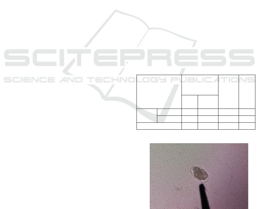
Figure 1: Hookworm Egg Ancylostoma Spp. Found in
Positive Sample (Magnification 100x).
ICPS 2018 - 2nd International Conference Postgraduate School
572

Based on the examination of 138 samples of pet
cats in this study, the prevalence of Ancylostomiasis
infection was 15.9%. Specifically, male pet cats had
a prevalence of 6.5%. This data is lower if compared
to female pet cats because cats which had their
samples taken and had positive Ancylostomiasis
contamination were generally above one year old.
Cats above one year old can be considered as being
in adult phase, which means that they have stronger
body condition and immune system, thus having a
lower risk for infectious diseases. Adult male cats
also play a role as survivors in the environment and
become the top predator. Our results also suggests
that the prevalence of Ancylostomiasis in
Banyuwangi City in female cats is higher than in
male cats. This could happen because the ages of
female cats which had their samples taken from and
had positive Ancylostomiasis contamination were
under six months old. Cats under six months of age
have lower antibody than adult, thus having one of
the risk factors that make pet cats also more
susceptible to Ancylostomiasis infection.
The results from this study were have different
from the study conducted in Brazil by Lorenzini
(2007) which mentions that the prevalence of
Ancylostomiasis in the pet cats under treatment by
veterinarians was 6%. The study was conducted by
taking samples from feces of pet cats which were
regularly checked to the veterinarian. Different data
could also be found from a research conducted in
Australia by Holyoake (2008), which mentions that
the prevalence of Ancylostomiasis in pet cats was
only 0.2%. High results for gastrointestinal parasite
were found in Nigeria by Sowemimo (2012) with
prevalence reaching 57%. The study was conducted
in two urban areas in Nigeria. From each region, 100
samples were taken from pet cats raised in each
urban area, and the total samples obtained amounted
to 200. From the first region, the prevalence of
Ancylostomiasis obtained was 69%, while in the
other region it was 45%. A research conducted in
Bangkok, Thailand, by Jittapalapong et al. (2007)
mentions that the prevalence of Ancylostomiasis
was 9.9%, and that result showed lower prevalence
than previous study. Oktaviana et al. (2014)
conducted a research in Bali using 80 samples
divided into 40 stray cat feces and 40 pet cat feces.
From 40 samples of examined stray cat feces, 19
samples (47.5%) positively contained the egg of
Ancylostoma spp. Meanwhile, from 40 samples of
examined pet cat feces, 10 samples (25.0 %)
positively contained the egg of Ancylostoma spp.
These data become important because Bali is the
nearest place from Banyuwangi, which can increase
risk factor of Ancylostomiasis transmission through
pet cats.
Ancylostomiasis is mostly prevalent throughout
tropical, subtropical and temperate regions
(Mizgajska-Wiktor and Uga, 2006), where visceral
larva migrans is one of the most important parasitic
disease of man transmitted by carnivores (Dalimi
and Mobedi 1992; Fisher 2003). The most reliable
reason for the increased prevalence of
gastrointestinal helminthes in pets is the natural
predator–prey relationship, poor hygiene, and lack
of anthelmintics drug administration (Dryden, 2007).
The major pathogenesis of severe parasitic
infestations is mechanical damage to tissues due to
the migration of the larvae through the organism
(A.tubaeforme), anaemia, decrease of vitamins, and
interference with the immune system. The decline of
physical condition is due to release of such
substances as enzymes and toxins (Behnke 1991;
Loukas and Prociv 2001; Bowman et al. 2003). This
may be the reason for the presented cases of
constipation, anorexia, severe dehydration, and
epilepsy. Increased Hb and PCV and decreased total
protein and albumin may be due to severe parasitic
infestation. Hookworms cause anaemia because
blood loss is the greatest 10–15 days after onset of
the infection and A.tubaeforme may cause fatalities
in heavily infested kittens. Even though the cat is
treated with specific and along with supportive
therapy, it will be dead regardless, which may be
due to the delayed hospital visit by the owner and
thus leads to severity of the concomitant helminthic
infestation.
Abu-Madi et al. (2008) mentions that factors
such as geographical areas may affect the level of
prevalence. Other factors include climate,
consistency of cats staying in place and the roaming
range from the cat itself. In China, A. caninum was
reported twice in Sichuan province, southwest
China, with a prevalence of 25% and 51%,
respectively (Feng et al, 2011), while an overall
higher prevalence (95.1%) of A. caninum infections
in cats was detected in Guangzhou (southern China).
The predominant species of hookworms in cats was
A. caninum in China, while A. tubaeforme was
considered to be the predominant species in
Australia (Silva et al, 2006), which strongly supports
our suggestion that the prevalent species is related to
its geographical distribution. Cats could well be the
main host for Ancylostomiasis in Banyuwangi City.
The prevention of parasitic disease is possible
through health institution care for pets, including
regular anthelmintic treatments, preventing the
contamination of the environment from feces, and
Prevalence of Ancylostomiasis in Pet Cats from Banyuwangi City, East Java Province
573

promoting responsible pet ownership (Overgaauw,
1997).
4 CONCLUSIONS
The prevalence of Ancylostomiasis in pet cats in
Banyuwangi City was 15.9%. From 138 samples of
examined pet cat feces, 22 samples (15.9%)
positively contained the egg of Ancylostoma spp.
Meanwhile, from 138 samples of examined pet cat
feces, 9 samples from male cats (6.5%) positively
contained the egg of Ancylostoma spp, and 13
samples from female cat (9.4%) positively contained
the egg of Ancylostoma spp. A control program
needs to be applied for diseases caused by an
Ancylostoma spp. infection in cats, which are
beneficial to the animal and public health aspect. Pet
cats need to be given more attention for its health. In
addition, further research about epidemiology of
Ancylostomiasis in cats needs to be done.
ACKNOWLEDGEMENTS
The authors thank those who have assisted in this
research process at the Parasitology Laboratory
Faculty of Veterinary Medicine Airlangga
University PSDKU Banyuwangi, family and
colleagues who have been willing to help in the
process of research and writing of this article.
REFERENCES
Abu-Madi MA, DA Al-Ahbabi, MM Al-Mashadani, R Al-
Ibrahim, P Pal, JW Lewis. 2008. Patterns of parasitic
infections in faecal samples from stray cat
populations in Qatar. J. Helminth 81: 281-286.
Behnke JM. 1991. Immunology In: Gilles HM, Ball PAJ
(eds) Human parasitic diseases: hookworm
infections, Amsterdam Elsevier 4: 93–155.
Bhatia BB, Pathak KML, Juyal PD. 2010. A text book of
veterinary parasitology. Kalyani Publishers, New
Delhi.
Borthakur SK. 2011. Gastrointestinal Helminthes in Stray
Cats (Felis catus) from Aizawl, Mizoram, India.
Department of Parasitology, College of Veterinary
Sciences and Animal Husbandry, Central
Agrilcultural University, Selesih, Aizawl, Mizoram,
India.
Bowman DD, Barr SC, Hendrix CM, Lindsay DS. 2003.
Gastrointestinal Parasites of Cats In: Bowman DD
(ed) Companion and exotic animal parasitology.
International Veterinary Information Service, Ithaca.
Dalimi AH and Mobedi I. 1992. Helminthes parasites of
carnivores in northern Iran. Ann Trop Med Parasitol
68: 395–397.
Dryden MW. 2007. Total parasite control—piecing the
puzzle together. The North American Veterinary
Conference, 992–994.
Feng Y, He Q, and Li JP. 2011. Investigation of canine
and felid parasite in Tongjiang County, Chinese.
Modern Agricultural Sciences and Technology 11:
354–355.
Fisher M. 2003. Toxocara cati: an underestimated
zoonotic agent. Trends Parasitol 19:167–170.
Holyoake CS. 2008. A National Study of Gastrointestinal
Parasites Infecting Dogs and Cats in Australia.
Division of health science. Murdoch University,
Western Australia.
Jittapalapong S, I Tawin , P Nongnuch , K Chanya ,
Arkom Sa and Sirichai. 2007. Gastrointestinal
Parasites of Stray Cats in Bangkok Metropolitan
Areas, Thailand. Wongnakphet Kasetsart J. Nat. Sci.
41: 69 – 73.
Lorenzini G. 2007. Prevalence of Intestinal Parasites in
Dogs and Cats Under Veterinary Care in Porto
Alegre, Rio Grande Do Sul, Brazil. Pontificia
Universidade Catolica do Rio Grande do Sul, Porto
Alegre-RS.
Loukas A and Prociv P. 2001. Immune responses in
hookworm Infections. Clin Microbiol Rev 14(4):
689–703.
Mizgajska-Wiktor H and Uga S. 2006. Toxocara cati eggs
in the environmental contamination. Exposure and
environmental contamination. In: Holland CV, Smith
HV (eds) Toxocara: the enigmatic parasite.
Oxfordshire, CABI Publishing, 211–227.
Oktaviana A, Dwinata M, Oka MBI. 2014. Prevalensi
Infeksi Cacing Ancylostoma Spp Pada Kucing Lokal
(Felis catus) Di Kota Denpasar. Buletin Veteriner
Udayana 6(2): 161-167.
Overgaauw PAM. 1997. Aspects of Toxocara
epidemiology: toxocarosis in the human. Crit Rev
Microbiol 23: 215–231.
Periago MV and Bethony JM. 2012. Hookworm virulence
factors: making the most of the host. Microbes Infect.
Elsevier 14(15): 1451-1464.
Silva LMC, Miranda RRC, Santos HA, and Rabelo EML.
2006. Differential diagnosis of dog hookworms based
on PCR-RFLP from the ITS region of their rDNA.
Veterinary Parasitology 140(3): 373–377.
Sowemimo A. 2012. Prevalence and intensity of
gastrointestinal parasites of domestic cats in Ode –
Irele and Oyo communities, Southwest Nigeria.
Department of Zoology, Faculty of Science, Obafemi
Awolowo University, Ile-Ife, Osun, Nigeria.
Yang Y, Zheng J, and Chen J. 2012. Cloning, sequencing
and phylogenetic analysis of the small GTPase gene
cdc-42 from Ancylostoma caninum. Experimental
Parasitology 132: 550.
ICPS 2018 - 2nd International Conference Postgraduate School
574
