The Effect of Pakoasi (Chromolaena odorata L.) Leaf Extract in
Curing Open Wound of Rabbit Skin (Oryctolagus cuniculus)
F. Ramdani
1
, M. Sriasih
2
, A. S. Dradjat
2
Postgraduate student at Magister Program (Manajemen Sumber Daya Peternakan) of Animal Science Faculty, University
of Mataram. Jalan Majapahit No. 62 Mataram, NTB
2
Faculty of Animal Science, University of Mataram, Jl. Majapahit No. 62 Mataram, NTB
Keywords:
Open Wound, Pakoasi, Rabbit
Abstract: Pakoasi (Chromolaena odorata. L) is a weed that is often used in treating wounds, but its effectiveness has
not been widely studied. The purpose of this research was to evaluate the effect of Pakoasi leaf extract on
open wound healing process in rabbit. Twenty healthy male rabbits were wounded with diameter of 2 cm
under local anaesthesia, and were divided into four groups: G0 (placebo), G1 (treated with 10% extract), G2
(treated with 20% extract) and G3 (positive control). Macroscopic and microscopic observations were
carried out, and the obtained data were analysed using analysis of variance. The macroscopic results showed
that on day 4, there was a significant difference (p <0.05) of wound diameter among groups, with G1
showing the shortest wound diameter (1.78 ± 0.08cm). Seventeen days after treatment, the wound diameter
(cm) in G1, G2, G3 and G0 were 0 ± 0; 0.08 ± 0.04; 0.17 ± 0.06 and 0.35 ± 0.08, respectively. The
complete healing process of open wound occurred as fast as 16 days after treatment in
G1 based on macroscopic and microscopic observation. This study concluded that ointment
containing 10% o f p a k o a s i extract is effective for curing open wound.
1 INTRODUCTION
Pakoasi (Chromolaena odorata L.) is a weed
belongs to Compositae family. This weed is easy to
grow, widespread and fast in tropical areas
(Madhavan, 2015) so that grassland cannot be
overgrown with grass. Some societies in West Nusa
Tenggara commonly use pakoasi as a traditional
medicine to treat open wounds, burns and other
traumas. However, the effectiveness of pakoasi as a
traditional medicine has not been widely studied yet.
Previous studies showed that the application of
pakoasi leaf extract of 2.5%, 5% and 10% in ethanol
has no effect in curing open wound in rabbits (Arif,
2016). The findings might be influenced by the
small number of samples, the number of wound
made in each rabbit and the absence of microscopic
assessment. Hence in this study, we evaluated
pakoasi (C. odorata L.) leaf extract effectiveness by
examining pakoasi ointment (10% and 20%) on the
wound healing process in male rabbits (Oryctolagus
cuniculus) skin. We made macroscopic and
microscopic evaluations.
2 MATERIALS AND METHODS
2.1 Leaf Extraction
The pakoasi leaves were removed by breaking the
stalk. They were washed thoroughly, dried and
powdered. The powder was then extracted with 96%
ethanol solution. The process was followed by
phytochemical test by gas chromatography-mass
spectrometry (GCMS) to evaluate the extract
chemical content.
2.2 Ointment Preparation
Ointment containing different concentration of
pakoasi extract was prepared as described by
Goeswin (2006). The formulation is presented in
Table 1.
Table. 1 : Component and content of pakoasi ointment.
No Component
Pakoasi extract
10 % 20%
1 Pakoasi Extract 1 2
Ramdani, F., Sriasih, M. and Drajat, A.
The Effect of Pakoasi (Chromolaena odorata L.) Leaf Extract in Curing Open Wound of Rabbit Skin (Oryctolagus cuniculus).
DOI: 10.5220/0007544904570461
In Proceedings of the 2nd International Conference Postgraduate School (ICPS 2018), pages 457-461
ISBN: 978-989-758-348-3
Copyright
c
2018 by SCITEPRESS – Science and Technology Publications, Lda. All rights reserved
457
2 Vaselin Albu
m
7.65 6.8
3 Adeps Lanae 1.35 1.2
4 Ointment 10 10
2.3 Rabbit Skin Incision
Twenty male healthy rabbits were wounded 2 cm in
diameter. Before making skin incision, the fur was
shaved around 5 cm. The skin was cleaned using
70% alcohol and was then anesthetized using
lidocaine. After 5 minutes, a circum incision of 2 cm
in diameter was performed on the top of vertebra.
The wounded rabbits were randomly divided into
four groups, with each group consisting of 5 rabbits
as follows; G1: the wounded rabbit was treated with
ointment of 10% pakoasi leaf extract, G2: treated
with 20% of pakoasi leaf extract, G3: treated with
povidone iodine as a positive control, while G0: no
treatment was given as a negative control. The
treatment was given twice a day in each group until
the wound was completely closed. The observations
were done macroscopically on day 4, 11 and 17, and
microscopically on day 7
and 21. The results were
then analysed by analysis of variance.
3 RESULTS AND DISCUSSION
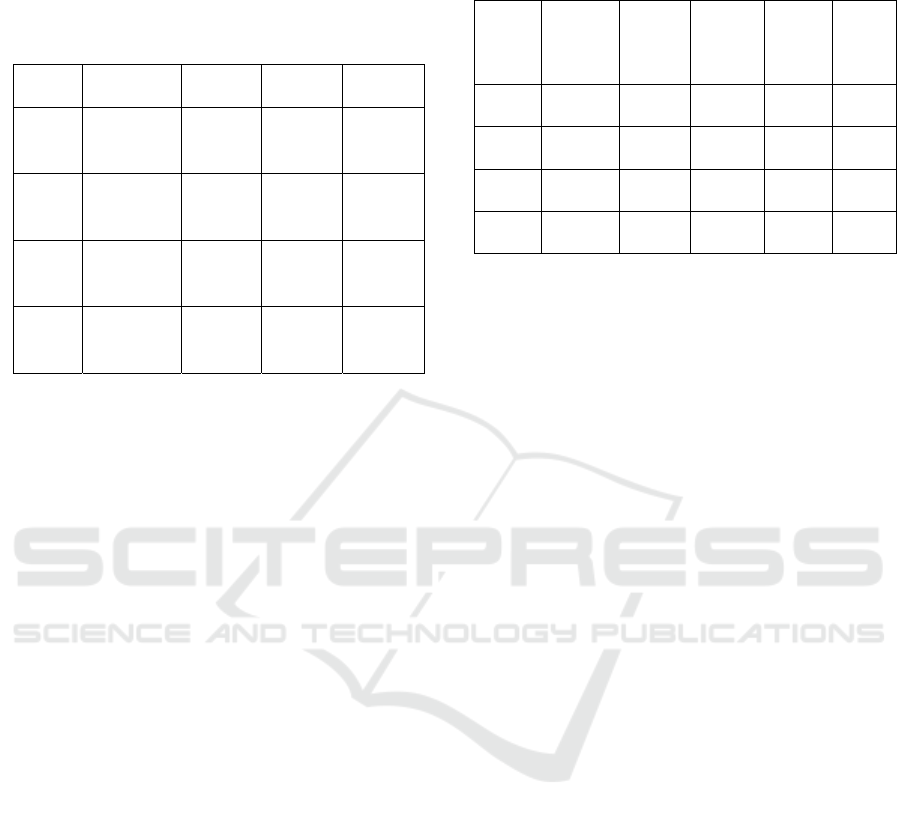
3.1 Phytochemical Evaluation of C.
odorata L.
Figure 1: The results of phytochemical test of C.
odorata L. leaf extract.
Figure 1 shows that there were five main
components, showing more than 5%, derived from
the extract. Those were Germacrene D (23.86%) and
seemed to be the highest content of the extract.
Germacrene D is derived from terpenoid as a
precursor of hydrocarbons (Nils et al., 2000).
Second, Trans (beta) –caryophyllene, which can be
classified as terpenoids sesquiterpen (21.07%). The
compounds have antimicrobial, antioxidant,
anesthetic and anti-inflammatory activities
(Ghelardini et al., 2001). Third, Cadinene which is a
class of terpenoids sesquiterpen (14.30%).
Sesquiterpene compounds have considerable effects
as antimicrobial, antifungal and antibiotic (Ali et al.,
2008; Guo et al., 2008). Other component was
Hexadecanoic acid (CAS) Palmitic acid (12.07%), a
derivative of saponin which has anti-inflamation and
antifungal, by damaging the structure of walls and
cell membranes of fungi. This synergistic
mechanisms is led by various compounds such as
terpenoids, which may increase the effect of
antifungal activity (Padmin et al., 2010). Last
component was Octadecatrienoic acid methyl ester
(6.30%), a class of fatty acids and has an anti-
inflammatory function (Mei Dong et al., 2000).
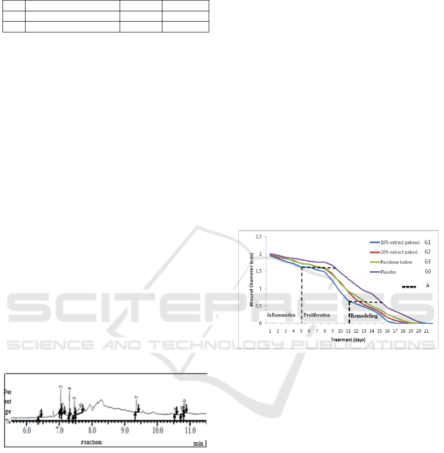
3.2 Macroscopic Evaluation
Figure 2 shows the measurement results of open
wound diameter in rabbits after treatment with
pakoasi leaf extract ointment
Figure 2: Healing process of each group folowing
treatment.
Data presented in Figure 2 show that healing
process began with inflammatory phase during day 1
to 4, then proliferative phase from day 4 to 11. The
process was followed by remodelling phase that
began from day 11 to 21. With 58% terpenoid and
12.07% saponin found in pakoasi extract, the wound
healing process observed from the inflammatory
phase could be accelerated by a difference of 0.35
cm within 4.5 days between G1 and G0. The
inflammatory phase causes neutrophil migration,
before progressing to the wound area that is replaced
by monocytes (Yuliani, 2012). Thus, the
inflammation process to the proliferation stage
becomes faster, and accelerates the healing process.
Terpenoids sesquiterpen on pakoasi leaf extract
has a function as an antioxidant. Antioxidant will
bind unstable free radicals that can damage cell
membranes. With this bond, free radicals are
expected to be stable, so that cell membrane damage
ICPS 2018 - 2nd International Conference Postgraduate School
458
can be reduced, and proliferation phase can be more
rapid (Ardiana et al., 2015).
Table 2. Wound diameter (mean cm ± SD) on day 4, 11
and 17 after treatments.
Note: Value with different superscript in the same
column is significantly different (p <0.05).
On day 4, there was a significant difference (p
<0.05) of wound diameter between G1 (1.75 ± 0.04)
and G2, G3 and G0. The mean and standard
deviation of wound diameter obtained by G1 on day
11 after treatment was significantly different from
mean diameter of G2 and G3 (p <0.05). The
treatment given in G2 and G3 did not cause any
significant difference (p >0.05), whereas G2 and G3
were significantly different with G1 and G0. On day
17, the average diameter and standard deviation in
G1, G2, G3 and G0 was 0 ± 0, 0.08 ± 0.04, 0.17 ±
0.06 and 0.35 ± 0.08, respectively, and all treatments
showed significant differences (p >0.05).
3.3 Microscopic Evaluation
The results of histopathologic examination on day 3
and 7 (Table 3) showed significant increases in
blood vessels and fibroblasts in G1 compared with
other groups (p <0.05).
Table 3. Scoring of epiteilization, collagenation,
fibrolast development, PMN response, and blood
vessel number of all groups on day 3, 7 and 21.
G
roups Epithel
isation
Colla
gen
Fibro
blast
PMN Bloo
d
vesse
l
G1 1.33±
0.57
a
0.67±
1.15
a
2.33 ±
1.15
a
2.00±
1.00
a
3.67±
0.57
a
G2 1.67±
0.57
b
0.00±
0.00
b
1.33±
0.57
b
1.67±
0.57
b
1.00±
0.00
b
G3 1.67 ±
0.57
b
0.67±
0.57
a
0.67±
0.57
c
1.67±
0.57
b
1.33±
0.57
c
G0 1.33 ±
0.57
a
0.67±
0.57
a
1.33±
0.57
b
1.33±
0.57
c
0.66±
0.57
d
Note: Value with different superscript in the same
column is significantly different (p <0.05).
Macrophages and neutrophils play roles during
inflammatory phase. Neutrophil immediately exits
blood vessels, and its number increases from 24 to
48 hours (Regan and Barbul, 1994). The number
decreases on day 3 as it is replaced by macrophages.
Macrophages and neutrophils have functions in
preventing infection by phagocytosis of
microorganisms that enter the wounded area. In the
absence of infection in the wounded area,
polymorphonuclear leukocytes are relatively short in
time, and the number decreases rapidly after day 3
(Regan and Barbul, 1994).
The results of histopathology examination on day
3 and 7 in Table 3 show a significant increase in the
number of blood vessels and fibroblasts in G1
compared to other groups. On day 3, the average
number of blood vessels that are at that time blood
vessel buds or endothelial progenitor cells go to the
blood circulation to the granulation tissue become
mature endothelium that will initiate angiogenesis
(Icha et al., 2016).
The next phase is proliferation or migration. The
cells that play roles in this phase are macrophages,
lymphocytes, fibroblasts and endothelial cells.
Macrophages have a longer life span than
polymorphonuclear leukocytes, and they remain
until healing process has complete (Yuliani and
Viktor 2015).
Observation on day 7 showed the
highest number of blood vessels due to endothelial
cells experiencing the peak of the proliferation
phase. Saponin content in pakoasi leaf extract
increases monocyte proliferation. This leads to
increasing number of macrophages. Macrophages
produce and secrete growth factors to attract more
fibroblasts to the wounded area, to synthesize
collagen and to increase proliferation of capillary
blood vessels (Ardiana et al., 2015). Therefore, G1
had a high proportion of fibroblasts and blood
Groups Treatments
The day
4
The day
11
The day
17
G1
10%
Pakoasi
extract
1.72±
0.04
a
0.65±
0.05
a
0±0
a
G2
20%
Pakoasi
extract
1.81±
0.02
b
0.91±
0.07
b
0.08±
0.04
b
G3
Povidone
Iodine
1.79±
0.05
b
0.93±
0.08
b
0.17±
0.06
c
G0 Placebo
1.87±
0.04
b
1.28±
0.08
c
0.35±
0.08
d
The Effect of Pakoasi (Chromolaena odorata L.) Leaf Extract in Curing Open Wound of Rabbit Skin (Oryctolagus cuniculus)
459

vessels in microscopic examination compared to
other groups (Table 3).
The presence of growth factors may increase cell
propagation or proliferation, and this will become a
frame of fibroblast receptorization and proliferation.
When fibrin clumped, fibroblasts will proliferate,
and the number of fibroblasts will increase
(Diegelmann, 2004). According to Thang et al.
(1998), the active components present in C.odorata
can stimulate fibroblast proliferation .
In the remodeling phase, the wound surface
restores epithelial integrity and epithelialization
from the basement membrane. Wound epithelial
cells begin to show increasing mitotic activity and
migrate the living connective tissue. Marginal basal
cells on the edges of the wound become loosely
bonded from the dermis nearby, enlarge and migrate
to the surface of the wound that has filled the
previous matrix (Singer and Clark, 1999).
The
microscopic results showed that the treatment given
in G1 influenced re-epithelization, collagen
formation, fibroblast formation, PMN cell
inflammation, and blood vessel formation faster than
G2, G3 and G0. This is consistent with a study
carried out by Henshaw et al. (2017) that showed
that the ethanol extract of C. odorata L. significantly
increased the number of red blood cells,
haemoglobin concentration and cell volume in white
mouse animal experiments.
4 CONCLUSIONS
Pakoasi leaf extract ointment with concentration of
10% cure open wound faster than 20% concentration
ointment, positive control (povidon iodine) and
negative control (without treatment).
ACKNOWLEDGEMENTS
Acknowledgments are given to Mr. Suparman for
technical assistance, Kholik DVM, M.Vet and
Chandra Dwiatma DVM, M.Si for assisting in
macroscopic and microscopic evaluations.
REFERENCES
Ardiana T, Andina R. P. dan Muhammad D F. 2015.
Efektivitas Pemberian Gel Binahong (Anredera
Cordifolia) 5% Terhadap Jumlah Sel Fibroblast Pada
Soket Pasca Pencabutan Gigi Marmut (Cavia
Cobaya). ODONTO Dental Journal. Volume 2. Nomer
1. Juli 2015.
Arif M. Z. 2016. Formulasi Sediaan Salep Ekstrak Etanol
Daun Kirinyuh (Euphatorium Odoratum L.) Sebagai
Penyembuh Luka Terbuka Pada Kelinci. Fakultas
Farmasi Universitas Muhammadiyah Surakarta
Ali N.A, Martina W, Arnold N, Lindequist U, Wessjohan
L. 2008. Essential Oil Composition from Oleogum
Resin of Soqotraen Commiphora kua, Rec. Nat. Prod.
2 (3): 70- 75
Diegelmann 2004. Wound Healing: An Overview of Acute,
Fibrotic, and Delayed Healing. Frontiers in
Bioscience, hal 283-289.
Ghelardini C, Galeotti N, Di Cesare Mannelli L, Mazzanti
G, and Bartolini A. 2001. Local anaesthetic activity of
β-caryophyllene/ Farmaco, vol. 56, no. 5-7, pp. 387–
389.
Goeswin A. 2006. Pengembangan Sediaan Farmasi.
Institut Teknolgi Bandung. Bandung.
Guo L, Jin-zong W, Tin H, Tong C and Khalid R. 2008.
Chemical Composition, Antifungal and Antitumor
Properties of Ether Extracts of Scapania verrucosa
Heeg. and its Endophytic Fungus Chaetomium
fusiforme, Molecules, 13: 2114-2125, DOI:
10.3390/molecules 13092114
Henshaw U O, Ifeyinwa M.O, Emmanuel K.U, Item J.A.
2017. Some hematological parameters of Wistar rats
treated with Chromolaena odorata leave extracts.
JoJournal of Biological Research; volume 90:6210
Icha Nofikasari, Afifah Rufaida, Chynintia Dewi
Aqmarina, Failaso, Annisa Rahmi Fauzia, Juni
Handajani. 2016. Efek aplikasi topikal gel ekstrak
pandan wangi terhadap penyembuhan luka gingiva.
Majalah Kedokteran Gigi Indonesia Vol 2 No 2 –
Agustus 2016 ISSN 2460-0164 (print), ISSN 2442-
2576
Madhavan M. 2015. Quantitative Estimation of total
phenols and antibacterial studies of leaves extracts of
Chromolaena odorata (L.) King & H.E. Robins
International Journal of Herbal Medicine 2015; 3(2):
20-23
Mei Dong, Yukiko Oda, Mitsuru Hirota. 2000. 10E,12Z-
90Hdroxy-10,12,15-octadecatrinic acid Methyl Ester
as an Anti-Inflammatory CompoundFrom Ehretia
dicksonii. The United Graduate Sholl of Agricultural
Science, Gifu University, Japan. Biosci. Biotecnol,
Biovhem., 64 (4), 882-886.
Nils Bulow, Wilfried A Konig. 2000. The role of
germacrene D as a precursor in sesquiterpene
biosynthesis: investigations of acid catalyzed,
photochemically and thermally induced
rearrangements. Phytochemistry Volume 55, Issue 2,
September 2000, Pages 141-168
Regan, M.C., and Barbul, A., 1994. The Cellular Biology
of Wound Healing in Fibrin Sealing in Surgical and
Nonsurgical Fields.
Singer, J.A. and Clark, R.A.F., 1999. Cutaneous Wound
Healing. Departements of Emergency and
Dermatology. State University of New York. Stony
ICPS 2018 - 2nd International Conference Postgraduate School
460

Brook. New York. The New England Journal of
Medicine; Vol 341.
Thang, T.P., Hughes, M.A., and Cherry, G.W.,1998.
Enhanced Proliferation of Fibroblast and Endothelial
Cells Treated with an Extract of the Leaves of
Chromolaena odorata (Eupolin), an Herbal Remedy
for Treating Wound. Plasttic Recontr. Surgery. 101.
Padmin E.A, Valarmathi A, and Rani M.U. 2010.
Comparative Analysis of Chemical Composition and
Antibacterial Activities of Mentha spicata and
Camellia sinennsis. Asian J. Exp. Biol. Sci, 1 (4) : 772
– 781
Yuliani S H. 2012. Formulasi Sediaan Hidrogel
Penyembuhan Luka Ekstrak Etanol Daun Binahong
(Anredera Cordifolia (Ten) Steenis) Program Pasca
Sarjana. Program Study Ilmu Farmasi Fakultas
Farmasi UGM. Yoyakarta.
Yuliani N.S dan Viktor L. (2015). Pengaruh Ekstrak Daun
C.Odorata Terhadap Proses Kesembuhan Luka Insisi
Pada Tikus Sprague-Dawley. Jurnal Kajian Veteriner
Desember 2015 Vol. 3 No. 2 : 93-99.
The Effect of Pakoasi (Chromolaena odorata L.) Leaf Extract in Curing Open Wound of Rabbit Skin (Oryctolagus cuniculus)
461
