
The Effects of Environmental Temperature Exposure of Blood
Spatter Towards Protein and Agglutinations Level in ABO Blood
Typing
Amanovitasari, D.
1
, Kahar, H.
2
, Prasetya, R.A.
3
, Lestari, T.P.
3
and Yudianto, A.
4
1
Master of Forensic Science, School of Postgraduate, Airlangga University, Surabaya, Indonesia
2
Clinical Pathology Department, Faculty of Medicine, Airlangga University, Indonesia
3
Surabaya Pharmacy Academy
4
Forensic Medicine and Medicolegal Department, Faculty of Medicine, Airlangga University, Indonesia
Keywords: Blood spatter, Environmental temperature, Protein level, Agglutination level, ABO Blood Typing
Abstract: The time needed for gathering evidence of a crime is an obstacle in the process of identifying the blood type
in the blood spots on the cloth. The blood will dry out after contact with the outside air within 3-5 minutes
and change the color from red to dark brown. Hence, the aim of this study was to investigate the effect of
exposure to environmental temperature on the levels of protein and agglutination in ABO blood typing of
blood spots after 0 (1 hour after blood drops on cloth), 5, 10, 15, and 20 days. Time series design was used
in this study using 30 blood spots on cotton from 1 individual subject (blood type A). Blood samples were
incubated at room temperature (23-24
o
C) (15 samples) and other ones were incubated at ambient
temperature (exposed to sunlight and rain). Determination of protein level was performed with a UV
spectrophotometer using trizole reagent. The agglutination level was examined by elution absorption
method using antisera A and read macroscopically. One Way ANOVA and Kruskal Wallis test were
performed in this trial and conclude that there was no significant effect to protein level (p>0.01). Based on
the trial ABO blood typing can still be performed for all blood ages (0, 5, 10, 15, 20 days).
1 INTRODUCTION
Murder cases in Indonesia tended to increase from
1,277 cases in 2014 to 1,491 cases in the next year
(BPS, 2016). Examining the crime scene, the
evidence commonly found in cases of murder is
blood spots. Blood spots on the objects around the
victim are often disguised or even removed by the
perpetrator by throwing the victim’s clothes away.
Hence, the blood spots could become vague or
unseen (Yudianto, 2013). The blood will dry out
within 3-5 minutes after contact with the outside air.
Once blood dries up, the color changes from red to
dark brown (Princess and Ketut, 2015). In the
forensic investigation, one of the most common
blood spot examinations is determining the blood
type (Knight, 2001). Blood typing can be done
quickly and cheaply; however, it can provide
accurate data to assist an investigation process
(Yudianto, 2013).
The human blood type is grouped according to
several blood typing systems. In 1900, the man who
first discovered the ABO blood typing was
Landsteiner. With the development of medical
science, many blood typing systems are found,
namely Rhesus (Rh), M and N, Kell, Duffy, and
Lewis (Knight, 2001). ABO blood type examination
of a sample of blood spots is still an important
technique for the identification of corpses in
criminal cases. Blood type examination in blood
spots can be done by direct agglutination or elution
absorption method (Nishi et al., 2005). The former
method will become more difficult when the red
blood cells lysis because of the sunlight exposure.
However, as long as the antigen in the blood spots is
still attached to the clothes, the antigen can still be
detected.
Different levels of protein in dried blood over
periods of time can be affected by several factors
such as the occurrence of microbes that lead to cell
degradation, UV exposure of the sun, and ambient
temperature. Putri and Ketut (2015) stated that the
D., A., K., K., R.A., P., T.P., L. and A., Y.
The Effects of Environmental Temperature Exposure of Blood Spatter Towards Protein and Agglutinations Level in ABO Blood Typing.
DOI: 10.5220/0007542303290332
In Proceedings of the 2nd International Conference Postgraduate School (ICPS 2018), pages 329-332
ISBN: 978-989-758-348-3
Copyright
c
2018 by SCITEPRESS – Science and Technology Publications, Lda. All rights reserved
329
decreasing protein level in dried blood can occur due
to the activity of microorganisms as time goes by.
Protein in the dried blood can stand up to four
months, but there is no further data regarding the
effect of environmental temperature exposure to the
dried blood. Hence, the aim of this study was to
determine the effect of the exposure on the level of
protein and agglutination in ABO blood typing of
blood spots after 0 (2 hours after blood drops on a
cloth and exposed to the room temperature), 5, 10,
15, and 20 days. The maximum exposure time of 20
days was chosen based on Article 24 paragraph (1)
of Indonesian Criminal Procedural Code, which
regulates the detention duration of a suspect in the
interest of the examination shall only be valid for a
maximum period of twenty days.
2 MATERIAL AND METHOD
This study was laboratory experiments using control
time series design. In this study, the independent
variable is defined as the exposure time of 0 days (1
hour after blood drops on the cloth), 5 days (5x24
hours), 10 days (10x24 hours), 15 days (15x24
hours) and 20 days (20x24 hours), while the
dependent variable is the protein and agglutination
level in ABO blood typing. The confounding
variable is room temperature (23–24
o
C) and the
ambient temperature (exposed to sunlight and rain).
Samples of 30 blood spots on cotton were obtained
from one individual subject (blood type A). Blood
samples were incubated at room temperature (15
samples) and the others were incubated at ambient
temperature. This study was conducted at Human
Genetic Laboratory Institute of Tropical Disease
Center Airlangga University Surabaya. Samples
which had been exposed to room or ambient
temperature at a given period of time were then
tested for the protein level with a UV
spectrophotometer using trizole reagent. While the
agglutination level was examined by elution
absorption method using antisera A and read
macroscopically. The data from samples incubated
at room temperature was analyzed using One Way
ANOVA, while the others were analyzed using
Kruskall Wallis.
3 RESULT AND DISCUSSION
Measurement of protein concentration using UV
spectrophotometer from sample of blood spatter of
both room (23-24
o
C) and climate exposure (exposed
to sun and rain) for 0 days (1 hour after blood drip
on cloth), 5th day, 10th day, 15th day and 20th day.
Shapiro Wilk normality test is performed to
determine whether the data is normally distributed or
not.
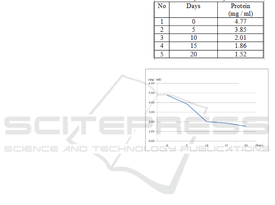
Table 1: Means of protein concentration at room
temperature. (23 – 24
o
C)
Figure 1: Protein concentration graphic at room
temperature. (23 – 24
o
C)
Based on Shapiro Wilk normality test, results
obtained all times of exposure significance (0 =
0.484, 5 = 0.888, 10 = 0.618, 15 = 0.502, 20 =
0.619) variables show the significance value (Sig)>
0.01, all the is normally distributed
Based on the homogeneity test, the value of
significance (sig) was 0.109> 0.01, it can be
concluded that the protein content from the sample
at room temperature (23-240C) from all times of
exposure (0, 5, 10, 15, 20 days) fulfills the
assumption of homogenity in One Way Anova test.
The results of One Way Anova test, prove that
the significance value (sig) is 0.119> 0.01.
Alternative hypothesis is rejected, it can be
concluded that there is no effect of long exposure at
room temperature (23-24
o
C) significantly to protein
concentration for all time variables (0, 5, 10, 15, 20
days).
ICPS 2018 - 2nd International Conference Postgraduate School
330
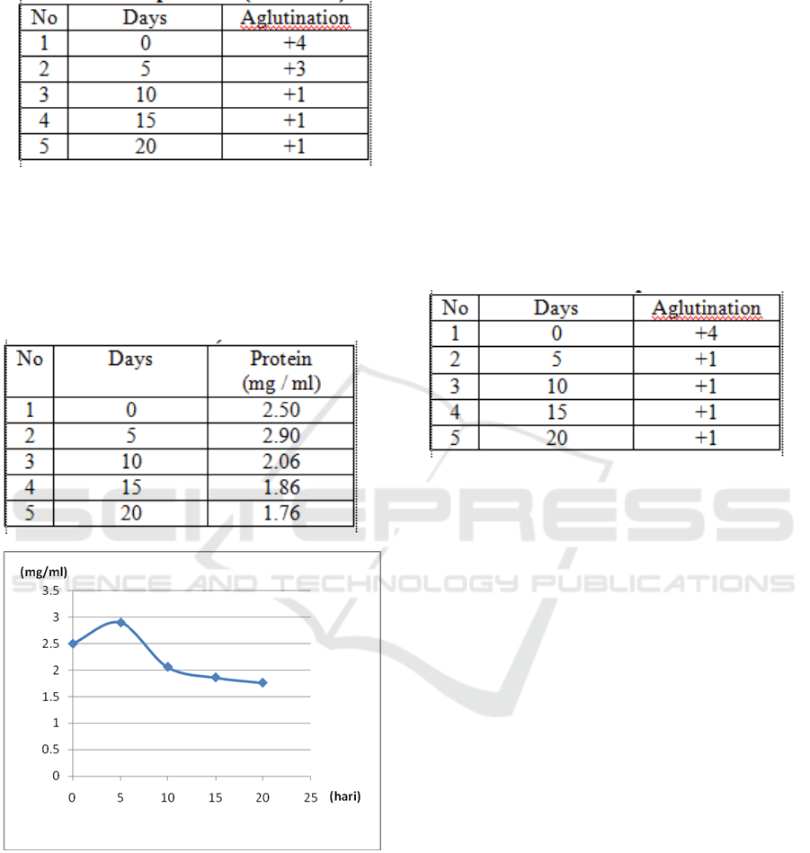
Table 2: Means of Agglutination level at room
temperature (23-24
o
C)
Shapiro Wilk normality test is performed to
determine whether there is correlation of exposure
time’s effect on the sample of blood spatter on the
cotton fabric left at ambient temperature (exposed
to sun and rain) to the protein content.
Table 3: Means of protein concentration at ambient
temperature (exposed to sun and rain)
Figure 2: concentration graphic at ambient temperature
(exposed to sun and rain)
Shapiro Wilk normality test shows all the
variable exposure times value significance (sig)>
0.01, (0 = 0.054, 5, 10 = 0.172, 15 = 0.605, 20 =
0.301). The data distribution of this research is
normally distributed.
Based on homogeneity test, the value of
significance (sig) is 0.003. Protein level from the
sample of blood spatter left at ambient temperature
(exposed to sunlight and rain) at 0, 5, 10, 15, 20 days
is heterogeneous. The assumption of homogenicity
in One Way Anova test is not fulfilled. The results
of protein concentration were tested by non-
parametric test using Kruskall Wallis test.
Kruskall Wallis test obtained significance value
(sig) of 0.833> 0.01, then null hypothesis is accepted
and alternative hypothesis is rejected, it can be
concluded that there is no effect of exposure time at
ambient temperature (exposed to sunlight and rain)
protein levels in the blood spots on the cloth left
within 0, 5, 10, 15, 20 days.
Table 4: Means of agglutination level at ambient
temperature (exposed to sun and rain)
The result show that the protein level of blood
spots exposed to the room temperature slightly
decreased over the given period of time. However, a
statistical test of One Way Anova did not show
significant difference (p > 0,01). It means that
protein denaturation process can occur at room
temperature very slowly; therefore, the decrease will
not be significant indeed.
On the other hand, the other samples, which are
exposed to the environmental temperature depicted a
fluctuation level of protein. Thus, Kruskall Wallis
statistical test yielded no significant difference
(p > 0,01), which means there is no correlation
between exposure time and the protein level of
blood spots. The uncertain environmental
temperature in the presence of exposure to sunlight
and rain can accelerate the process of decay. The
most optimal degradation process occurs at a
temperature of 70-100
o
F or equivalent to 21-38
o
C.
At those temperatures, the chemical breakdown
process of tissue and the development of
microorganisms will help the occurrence of decay.
At temperatures below 50°F (0°C) or above 100°F
(45°C) the decomposition process becomes slower
due to inhibition of microorganism growth (Aziz,
2014).
The Effects of Environmental Temperature Exposure of Blood Spatter Towards Protein and Agglutinations Level in ABO Blood Typing
331

4 CONCLUSIONS
From the discussion above, we can conclude that the
room temperature exposure did not affect the protein
level of the blood spots significantly. Furthermore,
there is no correlation between ambient temperature
exposures to the blood spots. The blood type of the
samples can still be detected after 20 days of
exposure both at room and ambient temperature.
REFERENCES
Afifah, Ratna, Nurul, 1986, Praperadilan Dan Ruang
Lingkupnya, Akademika Pressindo, Jakarta.
Albertolle, Matthew E; Hassis, Maria E; Ng, Connie Jen;
Et Al. Mass Spectrometry-Based Analyses Showing
The Effects Of Secretor And Blood Group Status On
Salivary N-Glycosylation. Clinical Proteomics
Journal.2015. Diakses Januari 2017.
Alfanie Iwan, Nirmalasari Nila, Arizal Hendi Muhammad.
2017. Ilmu Kedokteran Forensik dan Medikolegal.
Jakarta : Rajawali Pers.
Asthia T 2007 Studi Perbandingan Hasil Pemeriksaan
Golongan Darah terhadap Sample Saliva Segera dan
Saliva disimpan selama 1 jam padaTemperature 15º C.
Skripsi. Universitas Indonesia. Jakarta.
De Almeida, Et Al.Saliva Composition And Functions: A
Comprehensive Review. The Journal Of
Contemporary Dental Practice.2008.Diakses Januari
2017.
Fawles, J et al. 2000. The Chemical Constituent In
Cigarettes And Cigarette Smoke. New Zealand: New
Zealand Ministry Of Health. Diakses Februari 2017.
Gani, 2002. Ilmu Kedokteran Forensik. Fakultas
Kedokteran Universitas Andalas. Padang
Gerard J. Tortora, Bryan Derrickson.2009.Principles Of
Anatomy And Physiology 912th Edition. USA; John
Wiley And Sons,Inc. Diakses Januari 2017.
Guyton dan Hall.2008.Buku Ajar Fisiologi Kedokteran
Edisi 11.Jakarta: EGC
Hardjanto Pudji, 2015. TKP Bicara. Surabaya: PT. Refka
Petra Media.
Haque, amdadul M; Adhikari, Benu. Drying and
Denaturations of proteins in spray drying process.
Handbook of industrial drying.2015. taylor and
francis Group, LLC.Diakses Januari 2017.
Hold. K.M, Boer. D, Juidema. J. 199. Saliva as an
analytical tool in Toxicology. International Journal
of drug Testing. P : 1-35 diakses Februari 2017
Idries, 2008. Penerapan Ilmu Kedokteran Forensik dalam
Proses Penyidikan. Jakarta : Sagung Seto.
Jellinghaus K, Blasser L, Scheller C Bohnert M. Drying
time for Human Saliva. Arch Criminal Mart-Apr;
235(3-4) : 99-109. 2015.
https://www.ncbi.nlm.nih.gof/pubmed/26419084.
diakses februari 2017.
Joachim Klimek. Saliva and Oral Health. Lecture Handout
for Undergraduate Student of Dentistry. 2004
Germany : 4-37
Leffondre et al 2002. Modelling Smoking History-
American Journal of Epidemilogy.
http://aj.oxford.journal.org/contant/156/9/813.full.pdf.
diakses Februari 2017
Martiman Prodjohamidjojo.2009.Komentar atas KUHAP:
Kitab Undang-Undang Hukum Acara Pidana, Jakarta:
Pradnya Paramitha.
Michael Bowers dan Gary Bell.1995 .Journal Of Indian
Academy Of Oral Medicine And Radiology, July-
September 2011.Diakses Februari 2017.
Tery Martin. 2012. Harmful Chemicals in Cigarettes.
Journal of smoking Effect.
http//quitsmoking.about.com .Diakses februari 2017.
Rogers, Debora : Newton, Mery. Sexual Assault
Examination. Clinical Forensic Medicine a Pisician’s
Guide2nd Edition. 2005. Human press Inc. New
Jersey.
O’Mullane et al.. Saliva and Oral Health. 3rd edition.
British Dental Association. 2004.London.
Takahashi M, Kamiyama S. 2011. Imunological Studies
on ABH-Activites in Secretory Cel of Human Major
Salivary glands-Corelation between ABH activity in
Secretory Cel and Secretor-non Secretor. Diakses
February 2017
Tarigan R, 2010. Studi Penggolongan Darah A, B, O, AB,
melalui Analisa Biokimiawi Klinis. Skripsi.
Universitas Sumatera Utara : Medan
Tjiptomartono Legowo Agung. 2008. Ilmu Kedokteran
Forensik dalam Proses Penyidikan. Jakarta : Agung
Seto .
Sitopoe, M. 2000. Kekhususan Rokok Indonesia. Jakarta:
Gramedia Widiasarana Indonesia.
Sastroasmoro S, Ismail S. 2002. Dasar-Dasar Metode
Penelitian Klinis. Edisi kedua. Jakarta : Sagung Seto.
Yudianto, Ahmad. Panduan Praktis Serologi Forensik.
Global persada press. Surabaya. 2013.
ICPS 2018 - 2nd International Conference Postgraduate School
332
