
Soft-tissue Artefact Assessment and Compensation in Motion
Analysis by Combining Motion Capture Data and
Ultrasound Depth Measurements
Azadeh Rouhandeh and Chris Joslin
Systems and Computer Engineering, Carleton University, 1125 Colonel By, Ottawa, Canada
Keywords: Soft-tissue Artefacts, Soft-tissue Motion Capture, Hip Joint Centre Correction.
Abstract: Accurately determining the hip joint centre is a necessary component in biomechanical human motion
analysis to measure skeletal parameters and describe human motion. The hip joint centre can be estimated
using functional methods based on the relative motion of the femur to pelvis using reflective markers attached
to the skin surface through an optical motion capture system; but this suffers inaccuracy due to the soft tissue
artefact. A key objective in movement analysis is the assessment and correction of this artefact; in this case
we present a non-invasive method to assess and reduce the soft tissue artefact effects using optical motion
capture data and tissue thickness from ultrasound measurements during flexion, extension, and abduction of
the hip joint. Results show that the displacement of markers is non-linear and larger in areas closer to the hip
joint. The marker displacements are dependent on the movement type, being relatively larger in abduction
movement. The quantification of soft tissue artefacts is used as a basis for a correction procedure for hip joint
centre and minimizing effects. Results show that our method for soft tissue artefact assessment and
minimization reduces the error in the functional hip joint centre approximately from 13-23mm to 7-14mm.
1 INTRODUCTION
Human hip joint is generally considered as a ball-and-
socket joint that connects the hip bone and femur. The
accurate location of the Hip Joint Centre (HJC) is a
necessary component in functional analysis of the hip
to measure skeletal parameters and describe human
motions. The location of the hip joint can be
estimated through various methods which can be
divided into three categories: image-based
techniques, predictive methods, and functional
methods (Kirkwood et al., 1999; Leardini, 1999).
Imaged-based determination of the hip centre
requires a medical imaging modality such as x-ray
radiographs, CT scans and magnetic resonance
imaging (MRI). In these techniques, standardized
images of the pelvis are obtained and the HJC
location is considered as the geometrical centre of the
head of the femur modeled as a circle in 2D images
and a sphere in 3D images. One error in determination
of HJC location using image-based techniques is
caused by the assumption of the femoral head as a
sphere although it is not perfectly spherical. The use
of image-based determination of the HJC is limited as
MRI-based techniques require expensive medical
imaging and the other modalities in this category
expose the subject to ionizing radiation (Speirs et al.,
2012; Bell et al., 1989). Predictive methods estimate
the HJC based on regression equations between
palpable bony landmarks and the joint centre (Bell et
al, 1989). These methods need the exact locations of
bony landmarks in the calculations of HJC. The
accuracy of them depends on identification of the
anatomical landmarks and the error range of them in
able-bodied adults was reported to be between 25-
30mm (Camomilla et al., 2006). This error is higher
in people with pelvic deformities due to the
assumption of hip symmetry for both legs in these
methods (Bouffard, 2012). The error associated with
the predictive methods has led to an increased interest
in identifying hip joint centres using the functional
methods. Functional methods are based on the
relative motion of the femur to the pelvis. In order to
have the functional centre of the hip joint, the relative
motion of the femur to the pelvis must be accurately
measured. Optical motion capture systems are the
Rouhandeh, A. and Joslin, C.
Soft-tissue Artefact Assessment and Compensation in Motion Analysis by Combining Motion Capture Data and Ultrasound Depth Measurements.
DOI: 10.5220/0006624205110521
In Proceedings of the 13th International Joint Conference on Computer Vision, Imaging and Computer Graphics Theory and Applications (VISIGRAPP 2018) - Volume 4: VISAPP, pages
511-521
ISBN: 978-989-758-290-5
Copyright © 2018 by SCITEPRESS – Science and Technology Publications, Lda. All rights reserved
511

most used systems in the study of human movement
which are non-invasive. In optical motion capture,
reflective markers are attached to the skin of the body
and cameras track 3D trajectories of the markers. In
this technique of movement recording, the internal
bone is inaccessible and markers are not rigidly
placed on the bone; thus, there is the relative motion
between the markers and bone due to muscles
activities and skin deformation which is known as
Soft Tissue Artefact (STA). One of the main
objectives in human movement analysis is the
assessment and correction of the soft tissue artefact,
as it is the main source of error.
Several techniques have been presented to assess
STA which are separated into five categories: intra-
cortical pins, external fixators, percutaneous trackers,
radiographic examinations, and magnetic resonance
imaging (Leardini et al., 2005). Techniques based on
intra-cortical pins, external fixators, and
percutaneous trackers can represent relatively
accurate measurements of the bone motion; but the
use of these techniques is limited as the procedures of
applying them are invasive and subjects may
experience pain. The main drawbacks of techniques
based on radiographic examinations are these
methods are invasive due to radiation exposure, the
3D measurements of the STA are estimated from two
planes which provide 2D information, and these
techniques require extensive processing of image data
(Sangeaux et al., 2006). MRI-based techniques
require expensive medical imaging and they are not
suitable for everyday clinical measurements and
analyses (Yahia-Cherif et al., 2004).
Several methods have been proposed to reduce the
STA effects: the solidification model, multiple
anatomical landmark calibration, pliant surface
modelling, dynamic anatomical landmark calibration,
point cluster technique, global minimization, and
techniques based on MRI (Leardini et al., 2005;
Yahia-Cherif et al., 2004). The solidification model
does not compensate the STA effects well as it can
only identify erroneous frames (Leardini et al., 2005;
Cheze et al, 1995). Dynamic calibration and multiple
anatomical landmark calibration are based on invalid
assumptions (linearity assumptions) and they are time
consuming because they require additional data
acquisitions (Cappello, 2005). The limitations of the
point cluster technique are an overabundance of
markers and instability (Alexander and Adriacchi,
2001; Ceratti et al., 2006). The drawback of the global
optimization technique is that it simplifies joints
structures that are not subject-specific which cannot
be applied to people with hip joint disorders (Lu and
O’Connor, 1999; Stagni et al., 2009). MRI-based
techniques are expensive and consequently they are
inappropriate for everyday clinical uses.
Despite the numerous methods proposed, the
objective of a reliable non-invasive and clinical
assessment and correction of STA in human hip joint
kinematics is still being investigated, and this is the
domain where our work lies in. We proposed a
method for assessing STA using optical motion
capture analysis and ultrasound depth measurements
(UDM) (Rouhandeh et al., 2014a). To quantify STA,
we processed the motion capture data using principal
component analysis (PCA) to align the central axis of
the bone in each movement type (Rouhandeh et al,
2014a). In this study, we present our mathematical
method for assessing and correcting STA using
optical motion capture analysis and ultrasound depth
measurements based on finding three key markers,
which is the basis for our previous study (Rouhandeh
et al., 2014b).
2 MATERIALS
2.1 Overview
We propose a method consisting of ultrasound
measurements of tissue thickness and motion capture
analysis to quantify and minimize STA non-
invasively to determine the HJC using a functional
method. Our solution is to first record each marker’s
position placed on the thigh and pelvis for a range of
motions of the hip joint (standing, flexion, extension,
and abduction). When the thigh moves, the muscles
of the upper thigh area contract and relax which cause
change in the muscle thickness. These changes affect
the positions of the markers attached to the skin
relative to the underlying bone and introduce an STA
error in the calculation of the HJC. We propose using
ultrasound imaging to measure the changes in tissue
thickness, UDM, at the marker positions for the same
standing and extended positions. This information is
used to select three markers having less change in
their tissue thickness. These markers are considered
as three key markers and will be further used in
mathematical analysis on the data to assess and
eliminate STA effects. Next step is fitting curves to
the markers’ positions and applying UDM
information in order to determine bone positions at
the positions of three key markers. In fact, by
determining these positions, we eliminate the error in
markers positions caused by changes in tissue
thickness. We use these positions on the bone to
assess STA during several movements of the hip joint
as the. Therefore, once the bone positions at three key
VISAPP 2018 - International Conference on Computer Vision Theory and Applications
512
markers of all motions of the hip joint (standing,
flexion, extension, and abduction) have been
determined, we attempt to find a rotation matrix and
translation vector which transform the bone positions
at three key markers of standing position to each of
the other movement types as the bone is rigid body.
By applying the matrix and the vector to the markers
trajectories of standing position and comparing with
the trajectories of markers of the other movement
types, the STA can be quantified. The next step is the
HJC calculation; we calculate the HJC using a
coordinate transformation technique, SCoRE algo-
rithm (Ehrig et al., 2006). In order to have an accurate
HJC location, we use the displacement of the markers
from the previous step and recalculate the markers’
positions to eliminate STA effects used in the SCoRE
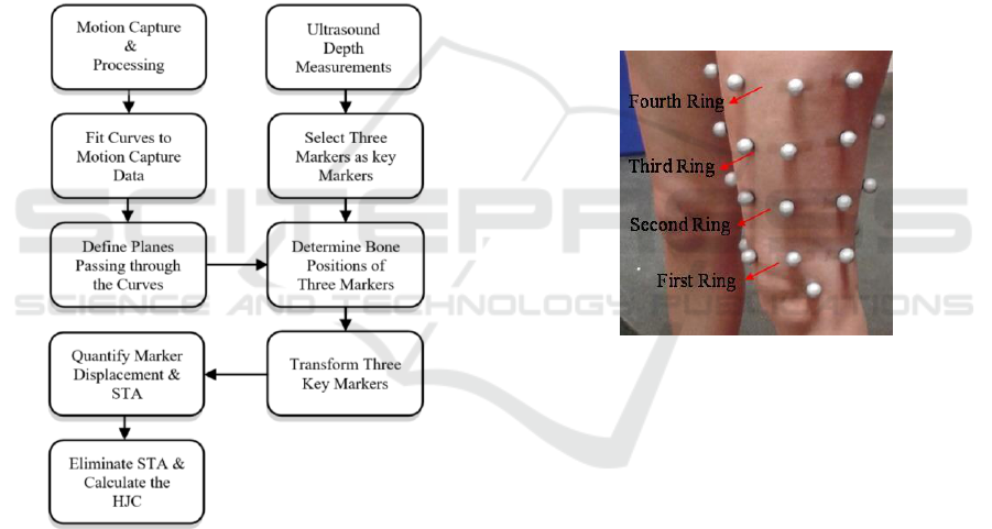
algorithm. Our method is outlined in Figure 1 and
each step is described in the following subsections.
Figure 1: Overall Process for STA Assessment and
Compensation.
2.2 Motion Capture
Ten healthy adult volunteers participated in this study
after signing an informed consent. Optical motion
capture systems are the most used systems in human
movement studies. Our optical motion capture system
is a Vicon MX system consisting of 10 wall-mounted
near-infrared cameras. The subject is surrounded by
the cameras while small reflective markers placed on
the skin surface. To capture the movement of the hip
joint, we use two groups of markers attached to the
skin of the subjects. The first group of markers
consists of 8 spherical reflective ones at palpable
bony landmarks where the bone is very close to the
skin surface and thus the soft tissue artefact is
minimal. These locations include three on the hip
area, left and right anterior superior iliac spine and the
lower spine, two on either side of the knee, medial
and lateral femoral epicondyles, and two on either
side of the ankle, medial and lateral malleolus, and
one on grater trochanter. As our goal is soft tissue
artefact assessment in hip joint kinematics, the other
group consists of the markers which are distributed
over the skin of the thigh. These markers are affixed
to the skin surface of the subjects in four ring
formations. The rings are placed approximately 5cm
apart, with eight markers per ring. These positions are
marked on the thigh and used for the ultrasound depth
measurements in the second stage of our experiments
in this thesis. The markers configuration of the thigh
is illustrated in Figure 2.
Figure 2: Subjects’ Thigh Markers Configuration.
Once the markers have been attached to the subject’s
skin surface, we can capture and track the movements
of the hip joint. The first step in our motion tracking
is capturing the markers trajectories in standing
position as a reference for subsequent processing.
Participants are requested to move their leg which is
equipped with the reflective markers in three key
motions, flexion, extension, and abduction, starting
from standing position. Markers trajectories are
captured for these positions as shown in Figure 3.
To have the same range of motion of the hip joint
for ultrasound depth measurements, the positions are
determined using non-reflective blocks that are setup
ahead of capture with a specific configured distance.
2.3 Ultrasound Measurements
There Ultrasound is one of the preferred imaging
modalities because this modality is non-invasive and
poses no harm to human bodies and, in addition, it is
Soft-tissue Artefact Assessment and Compensation in Motion Analysis by Combining Motion Capture Data and Ultrasound Depth
Measurements
513
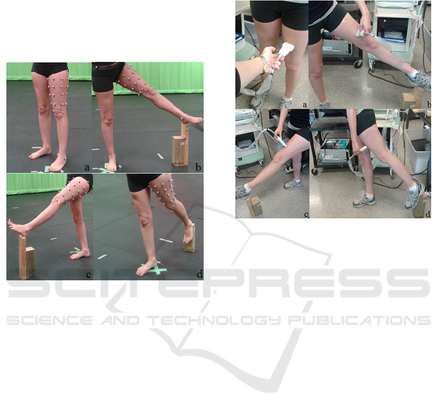
a low cost and portable imaging modality. In our
proposed method, to improve the determination of the
HJC location, ultrasound imaging is used to measure
the tissue thickness. Depth measurements were
obtained using an ultrasound imaging machine
(Picus, Esaote Europe) and a standard linear probe
(L10-5, 5MHz operating frequency, 4cm wide).
Figure 3: Subject Positions during Optical Motion Capture,
a) Standing, b) Abduction, c) Flexion, and d) Extension
Using the ultrasound imaging to measure
thickness of the tissue from the bone position,
ultrasound echoes pass through tissues. As soft
tissues and the underlying bone having different
acoustic impedances, their reflected echoes are
different. In fact, ultrasound echoes reflected from the
bone surface are very strong and cause high intensity
pixels in the image representing the bone surface.
Detecting the desired edges in ultrasound images is
not easy as they are extremely noisy and consist of
various artefacts and unrelated high contrast noise.
In our application, the echoes reflected from the
layered structures of different muscles cause
relatively high intensity pixels and consequently error
in detecting the desired bone surface. When we
observe unrelated edges that make the bone surface
detection difficult, we give a little push to the
ultrasound probe to distinguish the unrelated edges.
As mentioned, these unrelated edges are caused by
the layered structures of different muscles; therefore,
pushing the ultrasound probe changes the thickness of
the muscles and structures of corresponding high
intensity pixels in the image however the pixels
correspond to the bone surface are not changed. After
detecting the desired edge, the probe is released to
measure the real thickness of soft tissues.
Figure 4: Subject Positions during Ultrasound Depth
Measurements, a) Standing, b) Abduction, c) Flexion, and
d) Extension.
3 DATA ANALYSIS & METHOD
3.1 Overview
In this section, we explain our proposed approach
consisting of five steps to analyze data for STA
assessment. The first step is finding three of the
markers which have less depth changes during all
positions (standing, flexion, extension, and
abduction). The second step is passing a curve
through the ring formation of each of the key markers.
The third step is defining planes passing through the
curves from the previous step. Then we propose a
mathematical method to determine the projection of
the key markers on the underlying bone. These
positions on the bone are considered as references for
the later processing in the STA assessment. All the
steps are explained in detail in the following sections.
3.2 Determining Key Markers
Once the tissue thickness of the indicated markers on
the thigh has been measured, we need to find three of
the markers which have less depth changes during all
positions (standing, flexion, extension, and
abduction). To this aim, the coefficient of variation
VISAPP 2018 - International Conference on Computer Vision Theory and Applications
514
(CV) of each marker’s depth measurements during all
positions is obtained and three markers with less
value of CV will be selected as three key markers for
next steps of our method. As the coefficient of
variation measures relative variability and describe
the variation relative to mean of a set of data, it is
useful to compare data variation among two or more
sets of data. The low value of the CV shows that the
dispersion in the variable of a set of data is not great.
The coefficient of variation of each marker is
calculated using Equation (1).
(1)
Where
,
and
is the depth measurements for all four positions.
3.3 Curve Fitting
The next step is generating smooth curves which pass
through the key data points of the ring formation of
the motion capture data. To this end, we use a
piecewise polynomial cubic spline interpolation. In
piecewise polynomial cubic spline interpolation, a
cubic polynomial is fitted between each pair of
markers data of the ring formation to create a smooth
curve. If we consider the markers data of motion
capture, eight markers per each ring formation, are
the sampled points from our desired curve, our goal
is to find an approximated function between each
consecutive pair of these eight points. For one
dimension of the points, we have distinct nodes
such that:
. Equation (2) gives the
cubic polynomial in each subinterval to have a closed
interpolated curve.
(2)
Where
, as given by Equation (3), is a cubic
polynomial that will be used on the subintervals.
,
(3)
To define the spline,
, four unknown parameters
of each
should be found based on the
interpolation conditions and continuity conditions in
both the first and second derivatives which are
expressed in Equation (4) and (5) for .
(4)
To have a closed curve, the cubic polynomial in
subintervals
and
,
and
should satisfy the following conditions in
Equation (5).
(5)
By applying the conditions, the cubic polynomials in
the subintervals and consequently
are
determined. As cubic spline interpolation is
continuous in both the first and second derivatives
everywhere in subintervals and at the merging points,
it is a useful interpolating method in our application
to produce smooth interpolated functions.
3.4 Defining a Plane
To determine the bone position at the three key
markers, we need to define a plane containing the
bone which passes through each curve from the
previous step. To define the plane, we need to provide
three non-collinear points on the plane: one of the
three key markers, P, one data point on the curve
which is very close to the marker, Q, and one other
marker data on the opposite side of the first marker
data, R. Figure 5. shows these three points.
Figure 5: Passing a Plane through Each Curve.
The general equation of a plane is defined by
Equation (6).
(6)
Given the coordinates of these three points in space,
,
, and
, we can find the parameters of the
equation of the plane using Equation (7).
(7)
Soft-tissue Artefact Assessment and Compensation in Motion Analysis by Combining Motion Capture Data and Ultrasound Depth
Measurements
515
3.5 Bone Position at Key Markers
Once the plane has been defined, we apply the
ultrasound depth measurements at the positions of
three key markers to determine three points on the
bone. To determine these points on the bone, they
should satisfy three conditions:
• This point should lie on the plane from the
previous step
• The distance between the bone position and
the key marker data on the position that the
ultrasound depth is measured should be equal
to the ultrasound depth measurement
• If we define two vectors, one between the key
marker data and the data point on the curve
which is very close to the marker and the other
vector between the key marker data and the
bone point, these two vectors should be
perpendicular; as the UDM is the minimal
distance between the skin surface and the
bone.
Figure 6 illustrates the curve fitted to the markers’
data and a point on the underlying bone at the position
of one of the key markers.
Figure 6: Determining Points on the Bone.
The conditions for the points on the femur bone can
be written as Equations 8, 9 and 10, respectively. In
the following equations,
is the
desired point on the bone and is the ultrasound
depth measurement. The coordinate of the bone point
satisfies Equation (8) so that it is on the plane passing
through the key marker point.
(8)
The distance between the bone position and the key
marker which is equal to the ultrasound depth
measurement is given by Equation (9).
(9)
Vector and are perpendicular if the dot product
is equal to zero as given by Equation (10).
(10)
3.6 Transformation of Key Markers
In the previous step, the bone positions at the three
key markers of all movement types of the hip joint
were determined. Determining these positions on the
bone, the errors associated with the changes in tissue
thickness at markers trajectories are eliminated;
therefore, these bone positions are considered as data
without the STA. By having these points, we can find
a rotation matrix and a translation vector which
transform the bone positions at the three key markers
of the standing position to each of the other
movements. We derive the matrix and vector by
solving a linear least square problem recursively. Our
objective function for each movement (compared
with standing position) is given by Equation (11).
(11)
Where is the rotation matrix , is the
translation vector ,
is the vector of key
marker in standing position , and
is the
corresponding key marker of the other movements
.
3.7 Quantification of Soft-tissue
Artefact
The most important aspect of STA is to determine
how the markers are displaced relative to the
underlying bone due to the movement. Due to muscle
contractions and skin deformation, markers move
during different range of motions of the hip. We
propose a method to determine three points on the
bone which are the projection of positions of three
markers having less change in their tissue thickness
during all range of movements of the hip. We propose
an approach to determine the transformation matrix
and translation vector of the bone positions from the
standing position to the other types of movements.
VISAPP 2018 - International Conference on Computer Vision Theory and Applications
516
Determining the transformation matrix and
translation vector, we have the rigid movement of the
bone. If no STA error exists in the markers’
trajectories, then they would move as rigidly as the
bone. Therefore, to quantify STA, we apply the
matrix and vector to the trajectories of the markers of
standing position, compare with the trajectories of
markers of the other movements, and compute the
displacement of the markers.
3.8 Determining Hip Joint Centre
To determine the HJC, we use the SCoRE algorithm
(Ehrig et al, 2006) which considers both joint
segments, femur and pelvis, in the CoR estimation. In
this algorithm, a local coordinate system for each
moving segment of the joint (pelvis and femur head)
is defined, and then these local systems for all time
frames are transferred into a global reference system
to estimate the HJC.
4 EXPERIMENTS AND RESULTS
4.1 Setup
The acquisition was performed at Carleton
University’s Motion Capture Studio and Ultrasound
Imaging Laboratory. The study was carried out on 10
volunteers (5 females and 5 males) aged between 21
and 30 years (Mean: 27.2 years; Std. dev.: 2.7 years)
with a mean body weight 64.1 (Std. Dev.: 13.9) of kg
and a mean height 172.1cm (Std. Dev.: 8.7).
By processing the motion capture data using
MATLAB and curve-fitting toolbox, we could fit the
curves passing through the markers data and
determine the bone positions at three key markers
from the previous step.
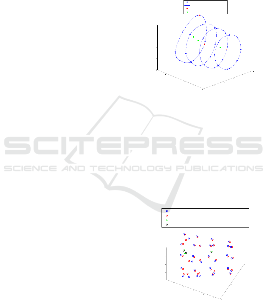
Figure 7 illustrates the trajectories of all markers
placed on the skin surface of one of the subjects
during standing position in optical motion capture,
the curves fitted to motion capture data, secondary
points on the curves used in determination of the
points on the bone, and the trajectories of key markers
projections on the underlying bone.
After the determination of the bone positions at
the three key markers, we used these locations as
references and we found rotation matrices and
translation vectors that transformed the bone
positions at the three key markers of the standing
position to each of the other movements, flexion,
extension and abduction. We derived them by solving
linear least square problems recursively in
MATLAB. If the markers locations didn’t suffer from
soft tissue artefacts, they would have the same
movement as the bone from standing to the other
movement types. Based on this fact, we applied the
rotation matrix and translation vector (corresponding
to each movement) to the markers trajectories of
standing position and compared with the trajectories
Figure 7: Curve Fitting to Motion Capture Data and
Determination of Bone Positions at 3 Key Points Positions
of Standing Position.
of markers of that corresponding movement, and then
computed the displacement of the markers.
4.2 Trajectory Results
Figure 8 illustrates the trajectories of markers from
optical motion capture which suffer from STA, and
the corresponding trajectories after applying rotation
matrix and translation vector to the markers
trajectories of standing position to have data without
STA effects. This figure shows the 3D displacements
of the markers during abduction movement.
Figure 8: Transformation of Standing Markers to
Abduction Movement.
4.3 Displacements Due to STA
-300
-250
-200
-150
-100
-50
500
550
600
650
700
750
-200
-150
-100
-50
0
A
H
B
G
A
C
H
B
F
D
A
H
G
E
C
B
H
A
D
F
G
C
B
E
G
D
C
F
E
F
D
E
Markers Data
Curves Fitted to the Markers
Secondary Points on the Curves
Bone Positions at 3 Key Markers
-200
-150
-100
-50
0
600
650
700
750
800
-50
0
50
100
Data without STA - Abduction
Data with STA - Abduction
3 Key Markers before Transformation - Abduction
3 Key Markers after Transformation - Abduction
Soft-tissue Artefact Assessment and Compensation in Motion Analysis by Combining Motion Capture Data and Ultrasound Depth
Measurements
517
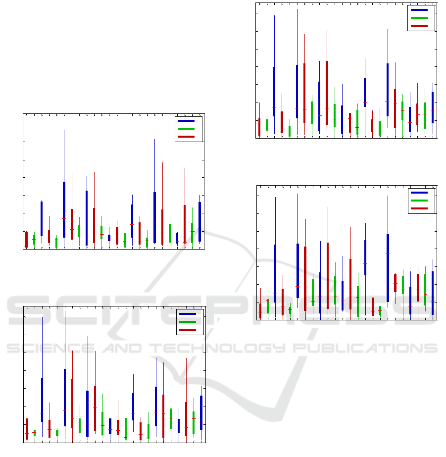
As the study was carried out on 10 subjects, to show
the results of the markers’ displacements of all the
subjects, we used box-plots. In the box-plot
representation of markers displacements, the lowest
value, highest value, median value, and the size of the
first and third quartile of each marker displacement
for all participants were illustrated. Figures 9, 10, 11,
and 12 show the displacements of the markers of each
ring during abduction movement.
Figure 9: Box-plots of STA Components of the First Ring
of Markers Configuration during Abduction.
Figure 10: Box-plots of STA Components of the Second
Ring of Markers Configuration during Abduction.
Figure 11: Box-plots of STA Components of the Third Ring
of Markers Configuration during Abduction.
Figure 12: Box-plots of STA Components of the Fourth
Ring of Markers Configuration during Abduction.
The assessment of STA was used to correct STA
errors to more accurately determine the HJC location
using the SCoRE algorithm. Two groups of markers
consisting of three non-collinear markers were
required to determine HJC using SCoRE algorithm,
one group placed on the thigh and the other placed on
the pelvis. As previously discussed, three key
markers have less change in their corresponding
tissue thickness during all movements; therefore, they
were considered as the first group of markers attached
to the thigh. The second group of markers included
the trajectories of markers on the left and right
anterior superior iliac spine and the lower spine. The
second group of markers were placed on the bony
landmarks and they were not affected by the STA. In
this part, at first, we transferred all the markers in a
way that the markers on the left and right anterior
superior iliac spine and the lower spine match the
same markers locations in the other movements. Then
we applied the SCoRE algorithm (Ehrig et al., 2006)
using Equation (12) on the 3 key markers, once on the
markers positions before reducing STA and once
1A 1B 1C 1D 1E 1F 1G 1H
0
5
10
15
20
25
30
35
Markers
STA [mm]
z
y
x
2A 2B 2C 2D 2E 2F 2G 2H
0
5
10
15
20
25
30
35
Markers
STA [mm]
z
y
x
3A 3B 3C 3D 3E 3F 3G 3H
0
5
10
15
20
25
30
35
Markers
STA [mm]
z
y
x
4A 4B 4C 4D 4E 4F 4G 4H
0
5
10
15
20
25
30
35
Markers
STA [mm]
z
y
x
VISAPP 2018 - International Conference on Computer Vision Theory and Applications
518
when we recalculated the markers positions (the
positions on the bone) based on the STA
quantification.
(12)
Where
,
are the joint centres of the femoral and
pelvic segments in the local coordinate systems,
,
are rotation matrices and
,
are translation
vectors to transform the local coordinate systems of
the pelvis and femur to an appropriate global system.
We calculated
,…,
and
,…,
based on the
markers attached to the thigh and
,…,
and
,…,
based on the markers attached to the pelvis as
discussed before. The indices of these parameters
indicate four frames that correspond to the frames of
standing position, flexion, extension and abduction.
For each participant during each movement, the
SCoRE algorithm returned two centres and the
distance between them showed the effectiveness of
our method in minimizing STA effects (Ehrig et al.,
2011).
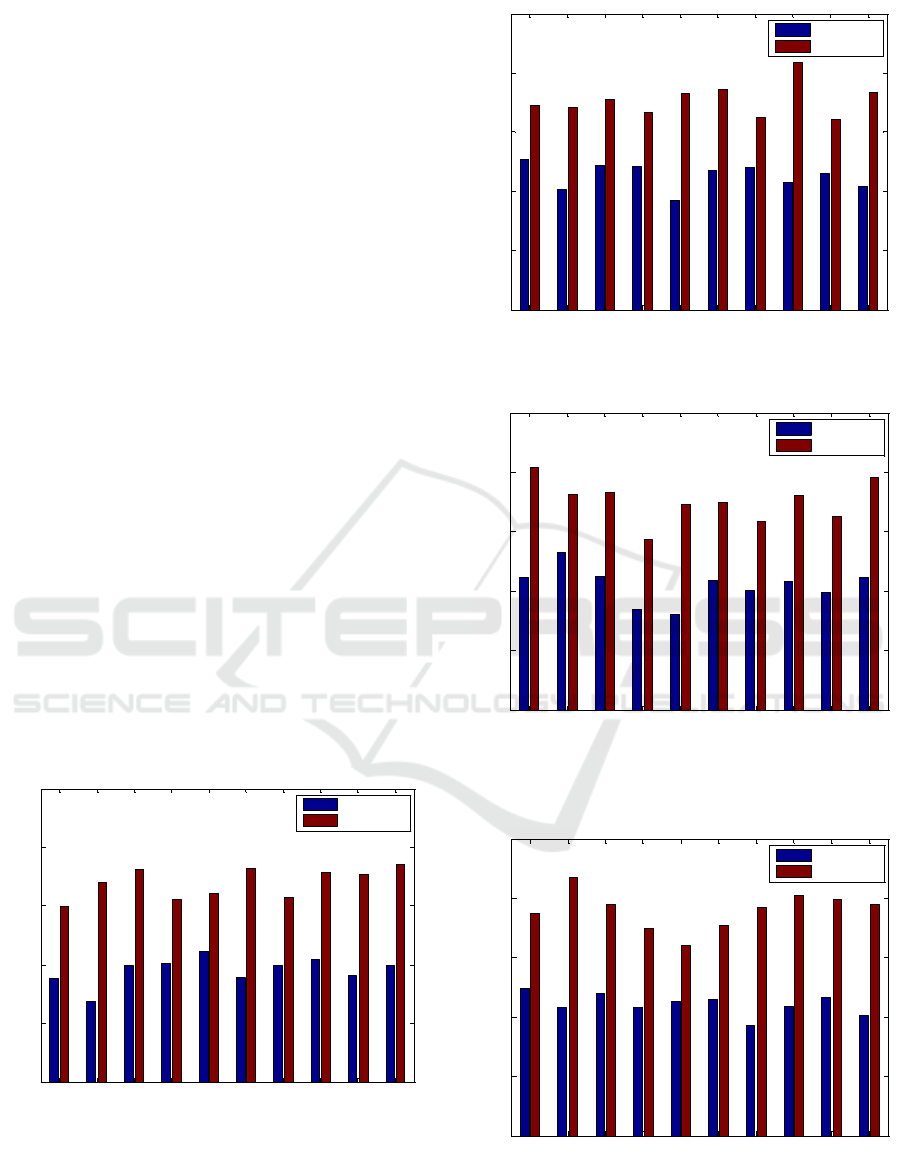
4.4 Hip Joint Centre Error
Figures 13, 14, 15, and 16 show the error in
determination of the hip joint centre for all subjects
during standing position, flexion, extension and
abduction. Each subject has two levels of error; one
based on the markers positions before reducing STA
and one based on the recalculated positions of the
markers after eliminating STA.
Figure 13: Hip Joint Centre Location Error Using SCoRE
Algorithm, Standing Position.
Figure 14: Hip Joint Centre Location Error Using SCoRE
Algorithm, Flexion.
Figure 15: Hip Joint Centre Location Error Using SCoRE
Algorithm, Extension.
Figure 16: Hip Joint Centre Location Error Using SCoRE
Algorithm, Abduction.
1 2 3 4 5 6 7 8 9 10
0
5
10
15
20
25
Subject
Error (mm)
Without STA
With STA
1 2 3 4 5 6 7 8 9 10
0
5
10
15
20
25
Subject
Error (mm)
Without STA
With STA
1 2 3 4 5 6 7 8 9 10
0
5
10
15
20
25
Subject
Error (mm)
Without STA
With STA
1 2 3 4 5 6 7 8 9 10
0
5
10
15
20
25
Subject
Error (mm)
Without STA
With STA
Soft-tissue Artefact Assessment and Compensation in Motion Analysis by Combining Motion Capture Data and Ultrasound Depth
Measurements
519

5 CONCLUSION
Soft tissue artefact is the most significant source of
error in human movement analysis. In this study, we
have proposed a combined experimental setup of
optical motion capture system and ultrasound
imaging system. The optical motion capture system is
the most common used system in human movement
studies as it tracks trajectories of the markers to have
realistic motions of the body non-invasively.
Ultrasound is one of the preferred imaging modalities
because this modality is non-invasive and poses no
harm to human bodies and, in addition, it is a low cost
and portable imaging modality. As the optical motion
capture system and ultrasound imaging system are
non-invasive, our proposed experimental setup is
non-invasive and appropriate for clinical daily uses in
contrast to the previous studies on STA assessment
and compensation which were invasive.
Using optical motion capture system along with
ultrasound depth measurements data, we quantified
STA on ten subjects during three ranges of motions
of the hip joint, flexion, extension, and abduction
comparing with natural position which was
considered standing position. At first, we recorded
each marker’s position placed on the thigh and pelvis
for a range of motions of the hip joint. We used
ultrasound imaging to measure the changes in tissue
thickness at the marker positions for the same
standing and extended positions. Three markers were
selected as three key markers based on the ultrasound
depth measurements. Then we proposed using a
piecewise polynomial cubic spline interpolation to fit
curves to the markers’ positions and applying UDM
information to determine bone positions at the
positions of three key markers. We used these
positions on the bone to assess STA during several
movements of the hip joint as the.
The results showed the markers’ displacements
were non-linear, subject and task dependent, and
generally larger in areas closer to the hip joint. The
hip is surrounded by several muscles linked to bones
via tendons. These muscles provide the joint stability
and control body movements. As different muscles of
the hip and thigh produce different movements of the
hip, the markers displacements are dependent on the
movement. Most of the subjects had relatively larger
STA in abduction movement; because different
subjects had muscles with different levels of strength.
This STA assessment was used to correct STA
errors to more accurately determination of the HJC
location using the SCoRE algorithm. For each subject
during each movement, two centres of rotation were
obtained; one based on markers trajectories before
minimizing the STA and one centre after minimizing
the STA and recalculating markers trajectories. The
error associated with the data before minimizing the
STA and after minimizing the STA effects was
approximately in the range of 13-23mm and 7-14mm,
respectively. The results obtained from our proposed
method shows improvements over previous studies
reported at 15-26mm (Ehrig, 2011; Piazza, 2004).
ACKNOWLEDGEMENTS
The work in this paper was funded and supported by an
NSERC Collaborative Health Research Project.
REFERENCES
Kirkwood, R. N., Culham, E. G., Costigan, P., 1999.
Radiographic and Non-invasive Determination of the
Hip Joint Center Location: Effect on Hip Joint
Moments. In Clinical Biomechanics, Vol. 14, No. 4, pp.
227-235
Leardini, A., 1999. Validation of a Functional Method for
the Estimation of Hip Joint Centre Location. In Journal
of Biomechanics, Vol. 32, No. 1, pp. 99-103
Speirs, A. D., Benoit, D. L., Beaulieu, M. L., Lamontagne,
M., Beaulé, P. E., 2012. The Accuracy of the Use of
Functional Hip Motions on Localization of the Center
of the Hip. In Journal of Hospital for Special Surgery,
Vol. 8, No. 3, pp. 192-197
Bell, A. L., Brand, R. A., Pedersen, D. R., 1989. Prediction
of Hip Joint Centre Location from External Landmarks.
In Human Movement Science, Vol. 8, No. 1, pp. 3-16
Camomilla, V., Cereatti, A., Vannozzi, G., Cappozzo, A.,
2006 An Optimized Protocol for Hip Joint Centre
Determination Using the Functional Method. In
Journal of Biomechanics, Vol. 39, No. 6, pp. 1096-1106
Bouffard, V., 2012. Hip Joint Center Localisation: A
Biomechanical Application to Hip Arthroplasty
Population. In World Journal of Orthopedics, Vol. 3,
No. 8, pp. 131
Leardini, A., Chiari, L., Croce, U. D., Cappozzo, A., 2005,
Human Movement Analysis Using
Stereophotogrammetry: Part 3. Soft Tissue Artifact
Assessment and Compensation. In Gait & Posture, Vol.
21, No. 2, pp. 212-225
Sangeux, M., Marin, F., Charleux, F., Dürselen, L., Ho Ba
Tho, M. C., 2006. Quantification of the 3D Relative
Movement of External Marker Sets vs. Bones Based on
Magnetic Resonance Imaging. In Clinical
Biomechanics, Vol. 21, No. 9, pp. 984-991
Yahia‐Cherif, L., Gilles, B. Molet, T., Magnenat‐
Thalmann, N., 2004. Motion Capture and Visualization
of the Hip Joint with Dynamic MRI and Optical
Systems. In Computer Animation and Virtual Worlds,
Vol. 15, No. 3-4, pp. 377-385
VISAPP 2018 - International Conference on Computer Vision Theory and Applications
520

Cheze, L., Fregly, B. J., Dimnet, J., 1995. A Solidification
Procedure to Facilitate Kinematic Analyses based on
Video System Data. In Journal of Biomechanics, Vol.
28, No. 7, pp. 879-884
Cappello, A., Stagni, R., Fantozzi, S., Leardini, A., 2005.
Soft Tissue Artifact Compensation in Knee Kinematics
by Double Anatomical Landmark Calibration:
Performance of a Novel Method during Selected Motor
Tasks. In IEEE Transactions on Biomedical
Engineering, Vol. 52, No. 6, pp. 992-998
Alexander E. J., Andriacchi, T. P., 2001. Correcting for
Deformation in Skin-based Marker Systems. In Journal
of Biomechanics, vol. 34, no. 3, pp. 355-361
Cereatti, A., Della Croce, U., Cappozzo, A., 2006.
Reconstruction of Skeletal Movement Using Skin
Markers: Comparative Assessment of Bone Pose
Estimators. In Journal of NeuroEngineering and
Rehabilitation [Online], Vol. 3
Lu, T. W., O’Connor, J. J., 1999. Bone Position Estimation
from Skin Marker Co-ordinates Using Global
Optimisation with Joint Constraints. In Journal of
Biomechanics, Vol. 32, No. 2, pp. 129-134
Stagni, R., Fantozzi, S., Cappello, A., 2009, Double
Calibration vs. Global Optimisation: Performance and
Effectiveness for Clinical Application. In Gait &
Posture, Vol. 29, No. 1, pp. 119-122
Rouhandeh, A., Joslin, C., Qu, Z. and Ono, Y., 2014a,
August. Non-invasive Assessment of Soft-tissue
Artefacts in Hip Joint Kinematics Using Motion
Capture Data and Ultrasound Depth Measurements. In
Engineering in Medicine and Biology Society (EMBC),
36th Annual International Conference of the IEEE, pp.
4342-4345
Rouhandeh, A., Joslin, C., Qu, Z., Ono, Y., 2014b. Soft-
tissue Artefact Assessment and Compensation in Hip
Joint Kinematics Using Motion Capture Data and
Ultrasound Depth Measurements. In International
Conference on Biomedical Engineering and Systems,
Prague
Ehrig, R. M., Taylor, W. R., Duda, G. N., Heller, M. O.,
2006. A Survey of Formal Methods for Determining the
Centre of Rotation of Ball Joints. In Journal of
Biomechanics, Vol. 39, No. 15, pp. 2798-2809
Ehrig, R. M., 2011. The SCoRE Residual: A Quality Index
to Assess the Accuracy of Joint Estimations. In Journal
of Biomechanics, Vol. 44, No. 7, pp. 1400-1404
Piazza, S., Erdemir, A., Okita, N., Cavanagh, P., 2004,
Assessment of the Functional Method of Hip joint
Center Location Subject to Reduced Range of Hip
Motion. In Journal of Biomechanics, Vol. 37, No. 3, pp.
349-356
Soft-tissue Artefact Assessment and Compensation in Motion Analysis by Combining Motion Capture Data and Ultrasound Depth
Measurements
521
