
Osteoclast and Osteoblast Quantity Change in Guinea Pig’s Tooth
Movement
Erliera
1
, Aditya Rachmawati
1
, Veranyca Chiuman
1
1
Orthodonti Department, Faculty of Dentistry, Universitas Sumatera Utara,
Alumni Street No. 2 Universitas Sumatera Utara, Medan, Indonesia
Keywords: Remodelling, Osteoclast, Osteoblast.
Abstract: An orthodontic treatment is defined by the quantity and the quality of the remodelling process including bone
resorption by osteoclast, and deposition by osteoblast. The activities of osteoclast and osteoblast in
orthodontic tooth movement can be seen by quantitative analysis done to the alveolar bone histologic tissue
on guinea pig. The aim of this research is to observe when the cells’ activities in orthodontic tooth movement
reach its optimum to move the tooth. This is an experimental research which includes intervension and control
groups. Guinea pigs were divided into five groups by the time they be observed and were given new separator
on each session to produce continuous force. This research showed that osteoclast activities reach its
maximum at day 7, whereas the osteoblast activities is at day 21. The amount of osteoclast cells in five
timesperiod is not statistically different (p=0,061), while the amount of osteoblast cell is statistically different
(p=0,006). The difference of osteoclast cells’ amount in control groups is statistically different with the
intervention group at day 28 (p=0,028), whereas the osteoblast is statistically different at day 7 (p=0,015). In
conclusion, osteoclast cells reach its maximum amount at day 7, whereas the osteoblast at day 21. The amount
of osteoclast cells was highly increased when the intervention begins and decreased significantly through the
end of the research (day 28), whereas the amount of osteoblast cell in control group is decreased drastically
when the force was being applied (day 7), and had started to increase after day 14.
1 INTRODUCTION
Orthodontic treatment is one of the treatments which
is done in dentistry to achieve an aesthetic dentofacial
appearance by correcting the inter-incisal alignment,
producing a good occlusion, removing the
arrangement of crowded and rotated teeth (Ardhana,
2013). The movement of teeth during orthodontic
treatment causes an intercourse reaction between
periodontal tissue, alveolar bone, and causes the
release of certain substances from within the teeth and
the systems around it (Ariffin, 2011). Histologically,
the periodontal tissue receives forces which are
generally known in orthodontic as pressure area,
which is the area where the resorption of alveolar
bone happens due to the pressure on periodontal
ligament, and the tension area, which is the area
where the formation of the alveolar bone takes place.
Both of these forces are the ones that make the
movement of teeth possible (Harry and Sandy, 2004).
The basic orthodontic treatment can be done by
repositioning the teeth by either using fixed or
removable appliances (Hikmah, 2015). There are few
concepts describing the mechanism of orthodontic
tooth movement: pressure tension theory, blood flow
theory, bone bending and piezoelectric theory
(Bhalajhi, 2004). When an orthodontic force is
applied, the space of periodontal ligament in the
pressure area becomes smaller which causes vascular
constriction, reduction of cells replication and
collagen production, followed by resorption of the
bone (Meikle, 2006). Whereas in the tension area, the
space between periodontal ligament becomes wider,
vascularization increases, replication of cell and
production of collagen increases, thus causes
deposition of bone (Graber and Vanarsdall, 2012).
Orthodontic tooth movement are divided into
three phases: initial phase, lag phase, and post lag
phase (Krishnan and Davidovitch, 2006). Initial
phase is the phase which involves rapid movement of
the teeth. This phase causes teeth to move inside the
space of periodontal ligament and bending of alveolar
bone (Ariffin, 2011). In lag phase, there are no
movement of the teeth. But if it happens, it only
Erliera, ., Rachmawati, A. and Chiuman, V.
Osteoclast and Osteoblast Quantity Change in Guinea Pig’s Tooth Movement.
DOI: 10.5220/0010076405050508
In Proceedings of the International Conference of Science, Technology, Engineering, Environmental and Ramification Researches (ICOSTEERR 2018) - Research in Industry 4.0, pages
505-508
ISBN: 978-989-758-449-7
Copyright
c
2020 by SCITEPRESS – Science and Technology Publications, Lda. All rights reserved
505
occurs in a short distance (Krishnan and Davidovitch,
2006). This movement is caused by the hyalinization
tissue process of the periodontal ligament that receive
force. The movement of teeth will not happen until
the cell completely resorb the whole necrotic tissue
area (Krishnan and Davidovitch, 2015). In post lag
phase, the speed of tooth movement increases
gradually. Tooth movement will reoccur after all the
hyaline area is eliminated and then the resorption will
then begin (Bhalajhi, 2004).
A successful orthodontic treatment is based on the
remodelling process which involves the process of
bone resorption by osteoclast, and bone deposition by
osteoblast (Proffit, 2007). These processes are based
on the quantity and quality of osteoclast and
osteoblast (Hikmah, 2016). Boulpaep and boron (cit
Kini and Nandeesh) stated that after putting the force
on ligament periodontal for a few hours, osteoclast
will be multiplied and resorption process will begin
on the bone surface, whereas for the involving tension
area, osteoblast will replicate and forms a new layer
of bone. Based on this situation, a successful
orthodontic treatment is based on the effectiveness of
remodelling process, which is based on the quality
and quantity of the osteoclast and osteoblast (Kini and
Nandeesh, 2012).
Activation of osteoblast and osteoclast cells can
be observed in a few ways: histology examination
through phatology anatomy with biomarker protein
coloring, or with the coloring of Hematoxylin Eosin
(HE). Samples which are going to be examined is
firstly fixated using formalin, then going through
decalcification, dehydration, clearing, and
embedding processes. Samples are then cut
longitudinally by using microtom to identify the
histologic image of osteoblast and osteoclast cell.
Generally, cells that are analyzed histologically is
done on guinea pig (Cavia cobaya) since it has high
similarity to human biologically (Legendre, 2016).
2 MATERIAL AND METHOD
This research was approved by the ethics committee
of Biology Department, University of Sumatera
Utara. Thirty guinea pigs weighing 250-400 grams
aged 2-4 month were kept under standard condition 3
days before the research begins and were all in good
condition during the experimental period. Guinea pig
which lost the separator or die before the observation
time is excluded in this research. Guinea pigs were
randomly divided into five groups of 6 animals each:
control group (no treatment), and four experimental
groups (observed on day 7, 14, 21, and 28) and were
then fitted with 0,5 mm Ormco elastomeric separator
in the left maxilla incisors on day 0. The fitted
separator is then removed and replaced with the new
separator in both maxilla incisors on day 7 to produce
continuous force. Six guinea pigs in each group were
euthanized using 75 mg/kg ketamine on days 0, 7, 14,
21, and 28 after the orthodontic force is applied. After
euthanasia, the anterior maxilla with two incisors
were cutted longitudinally and fixed in a 10%
buffered formalin solution for 24 hours. After
washing with water, the specimens were dehydrated
and embedded in paraffin. Slices measuring 4-6 μm
in thickness were obtained and stained with
haematoxylin- eosin. The quantitative analysis of
osteoclast and osteoblast cells is then done by
observing the histologic slide under microscope with
400x magnification. Cells count is done in five field
of view to get the average value in each pressure and
tension side and is done by two observers. Data were
expressed as mean and standard deviation values. The
data’s normality was tested with Saphiro-Wilk and
independent t test was done as an intern rater test. The
data were then analyzed using Repeated
Measurement Test of ANOVA.
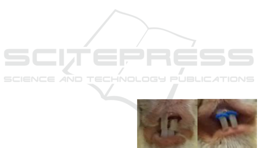
Figure 1: Placement of 0,5 mm Ormco elastomeric
separator in the left maxilla incisor on day 0, and the
replacement of new separator in both maxilla incisors on
day 7, 14, and 21 to produce continuous force.
ICOSTEERR 2018 - International Conference of Science, Technology, Engineering, Environmental and Ramification Researches
506

Figure 2: Microscopic view of osteoclast and osteoblast cells with 400x magnification.
3 RESULTS
Five guinea pigs were excluded from this study.
Three guinea pigs died and the other two has been lost
for the separator before observation time.
Saphiro Wilk is used as a normality test and
resulting p>0,05 indicates that the data were normally
distributed. Independent t test showed no significant
differences between the observers’ data (p>0,05).
Table 1: Osteoclast Cells Distribution in Five Timesperiod
Observation Time
Mean±SD
Day 0
0,64±0,26
Day 7
1,32±0,77
Day 14
1,20±1,03
Day 21
0,56±0,38
Day 28
0,32±0,36
Table 2: Osteoblast Cells Distribution in Five Timesperiod
Observation Time
Mean±SD
Day 0
7,04±0,36
Day 7
5,80±0,24
Day 14
6,04±0,90
Day 21
6,44±0,74
Day 28
5,04±0,93
Table 3: Osteoclast Quantity Differences of Five
Timesperiod
Timesperiod
(Days)
Mean
Differences
Significance
p-
value
0-7
+0,680
1,000
0,061
7-14
-0,120
1,000
14-21
-0,640
1,000
21-28
-0,240
1,000
Table 4: Osteoblast Quantity Differences of Five
Timesperiod
Timesperiod
(Days)
Mean
Differences
Significance
p-
value
0-7
-1,240
0,015
0,006
7-14
+0,240
1,000
14-21
+0,400
1,000
21-28
-1,400
0,220
Table 5: Quantity Differences of Osteoclast between
Control and Experimental Groups
Days
Mean
Differences
Significance
0 7
-0,680
1,000
14
-0,560
1,000
21
0,080
1,000
28
0,320
0,028
Table 6: Quantity Differences of Osteoblast between
Control and Experimental Groups
Days
Mean
Differences
Significance
0 7
1,240
0,015
14
1,000
0,601
21
0,600
1,000
28
2,000
0,232
Figure 3: Activities of Osteoclast and Osteoblast
Osteoclast and Osteoblast Quantity Change in Guinea Pig’s Tooth Movement
507

4 DISCUSSION
Figure 3 illustrate the activities of osteoclast and
osteoblast on the pressure and tension side. The
quantity of osteoblast cell found much more than the
osteoclast cells. This result is corresponding with
Sutantyo’s research that state out osteoclast reactivity
is not only based on the quantity, but also the quality
of the cell itself (Patil and Jayade, 2006).
Table 1 and table 2 illustrate the osteoclast and
osteoblast cells distribution in five times period. A
number of osteoclast and osteoblast cell was observed
on day 0, which prove that resorption and deposition
of bone as a remodelling process always happen
although no orthodontic force is given. A research by
Miyoshi et al., state that control samples with no
treatment undergone a physiologic bone remodelling
process showed by the movement of the tooth (Patil
and Jayade, 2006). Table 3 and table 4 illustrate the
osteoclast and osteoblast quantity differences in five
times period. Data analysis shows no significant
differences of osteoclast quantity in five times period
(p=0,061), but there were significant differences of
osteoblast quantity (p=0,006). Table 5 and table 6
illustrate the quantity differences of osteoclast and
osteoblast between control and experimental groups.
Post Hoc test with Bonferroni correction showing
significant differences of osteoclast cell quantity
between control group and the group observed on
days 28 (p=0,028), and significant differences of
osteoblast cell quantity between control group and
group observed on days 7 (p=0,015).
5 CONCLUSIONS
In conclusion, the finding of the present study
show that the activities of osteoclast and osteoblast
cells are not only depends on its quantity, but also its
quality. The present study also concludes that cells’
activities in orthodontic tooth movement reach its
optimum to move the tooth in a cycle of less than 28
days as the osteoclast and osteoblast cells quantity
will drop after days 28.
ACKNOWLEDGEMENTS
We are very extremely privileged this
completion of our projects addressed to our Research
Institution from Universitas Sumatera Utara at the
expense of this study from TALENTA funding in
year 2018 and our very great appreciation for
Hospital of University Sumatera Utara, Animal
House of Biology’s Department and Patology
Anatomy Department from our beloved Universitas
Sumatera Utara.
REFERENCES
Ardhana, W., 2013, ‘Identifikasi perawatan ortodontik
spesialistik dan umum’, Maj Ked Gi, vol. 20, no. 1, p.
232-7.
Ariffin, SHZ., Yamamoto, Z., Abidin, IZZ., Wahab, RMA.,
Ariffin ZZ., 2011, ‘Cellular and molecular changes in
orthodontic tooth movement’, Sci World J, vol. 11, p.
1788-803.
Bhalajhi, SI., 2004, The art and Science, 3
rd
ed, New Delhi,
Arya (MEDI) Publishing House.
Graber, TM., Vanarsdall, RL., 2012, Orthodontics current
principles and techniques, 5
th
ed, Missouri, Mosby.
Harry, R., Sandy, J., 2004, ‘Orthodontics. Part 11:
Orthodontic tooth movement’, British Dent J, vol. 196,
no. 7, p. 391-4.
Hikmah. N., 2015, ‘Profil osteoblas dan osteoklas tulang
alveolar pada model tikus diabetes tahap awal dengan
aplikasi gaya ortodonti yang berbeda’, El-Hayah, vol.
5, no. 2, p. 97-102.
Hikmah, N., Dewi, A., Maulana, H., 2016, ‘Rasio osteoklas
dan osteoblas pada tulang alveolar model tikus diabetes
dengan aplikasi gaya ortodonti’, Jurnal Ked Brawijaya,
vol. 29, no. 1, p. 54-8.
Kini, U., Nandeesh, BN., 2012, Physiology of bone
formation, remodeling, and metabolism. In: Fogelman
et al (eds.). Radionuclide and hybrid bone imaging,
Berlin, Springer.
Krishnan, V., Davidovitch, Z., 2006, ‘Cellular, molecular,
and tissue-level reactions to orthodontic force’, Am J
Orthod Dentofacial Orthop, vol. 129, p. 1-32.
Krishnan, V., Davidovitch, Z., 2015, Biological
mechanisms of tooth movement, 2
nd
ed, Chichester,
Wiley Blackwell.
Legendre, L., 2016, Anatomy and disorders of the oral
cavity of guinea pigs, Vancouver, Elsevier.
Meikle, MC., 2006, ‘The tissue, cellular, and molecular
regulation of orthodontic tooth movement: 100 years
after carl sandstedt’, European J Orthod, vol. 28, p.
221-38.
Patil, A., Jayade, VP., 2006, ‘Advances in biology of
orthodontic tooth movement – a review’, J Ind Orthod
Soc, vol. 39, p. 155-64.
Proffit, WR., Fields, HW., Sarver, DM., 2007,
Contemporary orthodontics, 4
th
ed, Missouri, Elsevier.
ICOSTEERR 2018 - International Conference of Science, Technology, Engineering, Environmental and Ramification Researches
508
