A Low-power Color Mosaic Image Compressor Based on Optimal
Combination of 1-D Discrete Wavelet Packet Transform and DPCM for
Wireless Capsule Endoscopy
Kinde A. Fante, Basabi Bhaumik and Shouri Chatterjee
Department of Electrical Engineering, Indian Institute of Technology Delhi, Hauz Khas, New Delhi-110016, India
Keywords:
Wireless Capsule Endoscopy, Discrete Wavelet Packet Transform, Mosaic Image Compressor, Low-power.
Abstract:
A novel low-power endoscopic image compressor is designed that occupies small silicon chip area, gives a
high compression rate and maintains acceptable image quality. By utilizing unique properties of human gas-
trointestinal tract images, computationally simple and elegant methods are employed. The employed methods
are lifting scheme based two level 1-D discrete wavelet packet transform, uniform quantization, chrominance
component sub-sampling, differential pulse code modulation and Golomb-Rice entropy encoder. All the mod-
ules are highly optimized from computational complexity, efficiency and memory requirement perspectives.
The proposed algorithm requires neither demosaicking nor de-interleaving operations that require large mem-
ory and consume a significant amount of power. The proposed image compression scheme achieves a com-
pression rate of 81.31 % with peak signal to noise ratio of 39.45 dB. The implementation of the algorithm
in 130 nm standard CMOS process technology occupies a core area of 0.342 mm×0.342 mm. It consumes
48.4 µW of power for encoding two color mosaic frames, with a resolution of 512×512, per second. The
proposed endoscopic image compression scheme gives a power consumption reduction of about two orders
less than the realizations proposed in literature.
1 INTRODUCTION
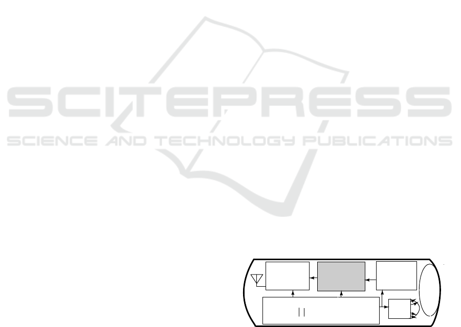
Wireless Capsule Endoscope (WCE) (Idden et al.,
2000) was introduced by Given Imaging Ltd in 2000.
Since then it has been widely used to diagnose gas-
trointestinal abnormalities with minimal invasiveness.
Its main electrical components are image sensor, RF
transmitter, button battery and structured light source
(LEDs) as shown in Figure.1. During the diagnos-
tic procedure, the capsule is swallowed by the patient
through the mouth. As it travels through the gastroin-
testinal(GI) tract with the aid of peristalsis, it cap-
tures high resolution color images of the GI tract wall
which cannot be reached by the wired conventional
endoscopy. The images are then transmitted by the
wireless RF transmitter to a receiver worn round the
patients waist for an average of eight hours before the
battery runs out (Moglia et al., 2008). The capsule
comes out of the digestive system along with the stool
and it is discarded. The images are downloaded onto
a workstation (PC) with appropriate image process-
ing software to make a video. The diagnosis is done
offline by Gastroentrologist.
+
_
RF
transmitter
CMOS
image
LEDs
Battery
L
e
n
s
Image
compressor
sensor
Figure 1: Block diagram of the wireless capsule endoscopy.
In order to reduce the power consumption of the
RF transmitter without significant overhead, the im-
age compressor inside WCE should have small chip
area, low power consumption and maintain high im-
age quality. Previous works on this topic were pre-
dominantly based on discrete cosine transform (DCT)
and differential pulse code modulation (DPCM). The
works in (Lin et al., 2006; Wahid et al., 2008; Dung
et al., 2008; Lin and Dung, 2011; Chen et al., 2009;
Turcza and Duplaga, 2011; Xie et al., 2007; Tur-
cza and Duplaga, 2013) have used block-based DCT
in order to encode endoscopic color mosaic images.
These block-based transforms require huge memory
to buffer data when they work with image sensor
190
Fante K., Bhaumik B. and Chatterjee S..
A Low-Power Color Mosaic Image Compressor Based on Optimal Combination of 1-D Discrete Wavelet Packet Transform and DPCM for Wireless
Capsule Endoscopy.
DOI: 10.5220/0005284701900197
In Proceedings of the International Conference on Biomedical Electronics and Devices (BIODEVICES-2015), pages 190-197
ISBN: 978-989-758-071-0
Copyright
c
2015 SCITEPRESS (Science and Technology Publications, Lda.)
that sends the pixel values in raster scan fashion.
The DPCM based algorithms were proposed for both
mosaic and full color image formats. The works
proposed for mosaic image compression (Xie et al.,
2006; Cheng et al., 2010) have used JPEG-LS algo-
rithm which requires to store at least one row of the
previously encoded pixels for prediction. The context
variables also require 1.9KB of memory (Cheng et al.,
2010). The DPCM based algorithms which were pro-
posed for full color image compression (Khan and
Wahid, 2011b; Khan and Wahid, 2011a) are sim-
ple. However, these algorithms require the imple-
mentation of the demosaicing algorithms inside the
image sensor which consumes considerable amount
of power and need memory. Memory consumes high
power and occupies large silicon area which increases
the overhead of the image compressor. Demosaicing
the mosaic image increases its size by threefold which
reduces the effective compression rate by the same
amount. Despite its success in many applications,
wavelet based image compression algorithm has not
been explored for WCE application to the best of the
authors’ knowledge. In this work we present the study
and design of wavelet based mosaic image compres-
sion algorithm which does not require huge memory
buffer and is computationally simple.
The proposed image compressor combines mul-
tiple methods which are computationally simple to
achieve high compression efficiency. These meth-
ods are: lifting scheme based discrete wavelet packet
transform (DWPT), DPCM, uniform quantization,
sub-sampling and Golomb-Rice encoding. The nov-
elty of this work lies in modifying the wavelet fil-
ter coefficients to utilize the unique properties of hu-
man GI tract images. It also combines DWPT with
DPCM, uniform quantization and sub-sampling op-
timally to achieve its target. We have experimen-
tally determined the optimal combinations of the sub-
sampling and uniform quantization parameters that
can achieve the desired performance for WCE appli-
cation. The final contributionof this work is optimiza-
tion of Golomb-Rice encoder from memory require-
ment and efficiency perspective. The Golomb-Rice
encoder parameter,k, is determined dynamically from
local image properties using only four contexts in or-
der to reduce the memory requirement and still main-
tain its adaptability property. The context variables
require only 34 bytes of memory. We get a mem-
ory reduction of about 1.866 KB as compared to the
method proposed in (Xie et al., 2006; Cheng et al.,
2010). The image compressor can elongate the life-
time of the WCE significantly so that it can cover the
whole GI tract.
The rest of the paper is organized as follows. The
detailed discussion of the proposed algorithm is given
in section 2. The performance evaluation of the algo-
rithm is presented in section 3. The hardware imple-
mentation is discussed in the section 4. The conclu-
sion is given in the section 5.
2 ANALYSIS OF SUITABLE
METHODS FOR EFFICIENT
COMPRESSION OF WCE
IMAGE
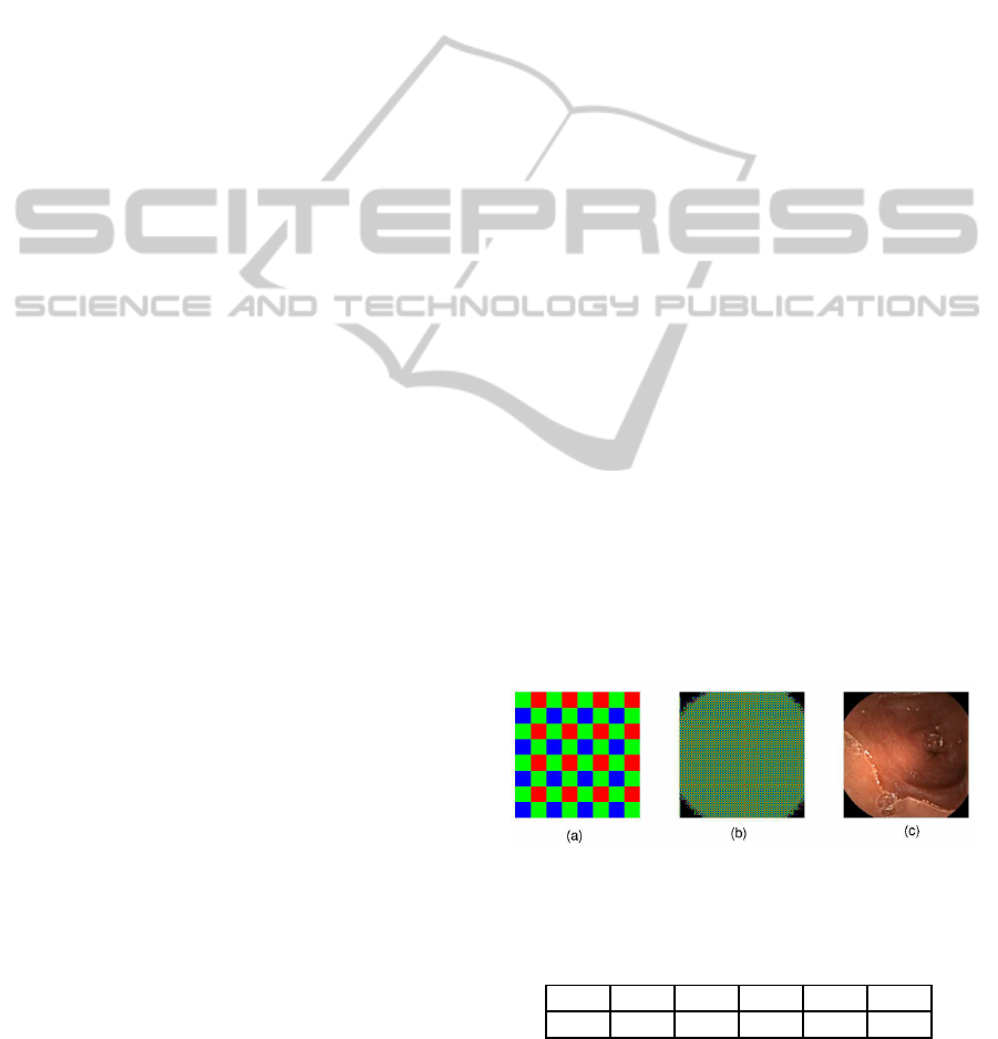
Mosaic image is captured using color filter arrays
(CFA) (Bayer, 1976) shown in Figure 2 (a). The
spectral bands are interleaved in mosaic image. The
pixel values have high frequency content in its neigh-
borhood. Hence, conventional image compression
methods fails to give high compression efficiency for
mosaic images. In the following sections we will
showhowcomputationallysimple methods can be op-
timally combined to achieve high compression effi-
ciency for WCE mosaic image.
2.1 Analysis of Color Mosaic Image
Using Wavelet
Consider a pair of rows of color mosaic image which
has six elements per row as shown in table 1, where
the spectral band samples are given as R (red), G
(green) and B (blue). Let’s apply 1-D DWPT on this
image using 5-3 integer wavelet (Angelopoulou et al.,
2007) with lifting scheme (Sweldens, 1995). We use
the horizontal filtering operation as shown in Figure
3.
Figure 2: The Bayer arrangement of color filters on the pixel
array of an image sensor (a), mosaic WCE image (b) and
typical WCE full color image (c).
Table 1: Two rows of color mosaic image.
G
0,0
R
0,1
G
0,2
R
0,3
G
0,4
R
0,5
B
1,0
G
1,1
B
1,2
G
1,3
B
1,4
G
1,5
If we directly apply the lifting scheme 1-D DWT
on the color mosaic image which is given in table 1,
we will get the followingoutputs. For the first row, the
ALow-PowerColorMosaicImageCompressorBasedonOptimalCombinationof1-DDiscreteWaveletPacketTransform
andDPCMforWirelessCapsuleEndoscopy
191
Original
image
Level 0 Level 1
H LH HH
Level 2
L HLLL
Figure 3: Diagrammatic representation of dyadic decompo-
sition for two decomposition levels using horizontal filter-
ing.
high-pass filter (H
R
) and low-pass filter (L
G
) outputs
after horizontal filtering are given by:
H
R
[2i, 2j+ 1] = R
2i,2j+1
− ⌊
G
2i,2j
+ G
2i,2j
+ 2
2
⌋ (1)
L
G
[2i, 2j] = G
2i,2j
+
⌊
H
R
[2i, 2j− 1] + H
R
[2i, 2j+ 1] + 2
4
⌋ (2)
where i and j are row and column indexes of the image
array. Similarly, for the second row of image given in
table 1, the high-pass filter (H
G
) and low-pass filter
(L
B
) outputs after horizontal filtering are given by:
H
G
[2i+ 1, 2j + 1] = G
2i+1,2j+1
− ⌊
B
2i+1,2j
+ B
2i+1,2j+2
2
⌋
(3)
L
B
[2i+ 1, 2j] = B
2i+1,2j
+
⌊
H
G
[2i+ 1, 2j − 1] + H
G
[2i+ 1, 2j + 1] + 2
4
⌋ (4)
Our simulation result on 120 endoscopic images taken
from Gastrolab (Gastrolab, 2014) shows that the
smoothness of the low-pass filter outputs (L
G
and L
B
)
are worse than the G and B color channels when they
are de-interleaved. For this reason, the low-pass fil-
tering operation of (2) is modified as:
L
G
[2i, 2j] = G
2i,2j
(5)
Similarly, the low-pass filtering operation given in (4)
is modified as:
L
B
[2i+ 1, 2j] = B
2i+1,2j
(6)
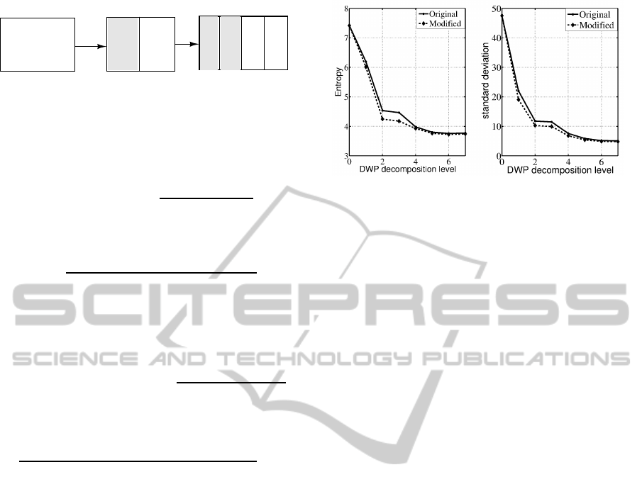
Figure 4 shows the statistical measurements (entropy
and standard deviation) of the decomposed mosaic
image given in Figure 2(b) using 1-D DWPT when
the low-pass filtering operation is done using the orig-
inal equations (2) and (4), and the modified equations
(5) and (6). The modification of the low-pass filtering
operation gives two advantages: It reduces computa-
tional resources and improves the compression effi-
ciency.
We observed from Figure 4 that the average sta-
tistical measurements of the subbands of the decom-
posed color mosaic image do not show significant
change after second level decomposition. Therefore,
Figure 4: Average entropy (left) and standard deviation
(right) of 120 endoscopic images at different wavelet packet
decomposition level.
the level of decomposition is limited to two in order
to save computational resources.
After the second level DWP decomposition, three
quarter of the subbands of the mosaic image are low-
pass signals. Figure 5 shows that the histogram of
detail subbands (HH and HL subbands) of the second
level DWPT is narrow and the values are small, near
zero with high probability. The LH subband shows
narrower histogram than the LL subband. The sub-
bands which have narrow histogram can be efficiently
encoded using computationally simple image com-
pression techniques such as DPCM.
The 2-D decomposition of color mosaic images
using Mallat wavelet packet was discussed in (Zhang
and Wu, 2004). The authors have shown the simul-
taneous de-correlation of spectral and spatial redun-
dancies of mosaic image using the convolution based
5-3 integer wavelet. The convolution based wavelet
decomposition has higher computational complexity
as compared to the lifting scheme (Sweldens, 1995).
The 2-D decomposition of the mosaic image enables
to de-correlate the image in both the vertical and hor-
izontal direction. Hence, it gives higher compression
efficiency than its 1-D counterpart. However, the 2-D
based operation requires huge memory to buffer data
during computation especially when the image com-
pressor works with image sensor which sends pixel
values in raster-scan fashion. Since memory occupies
large area and consumes high power we use the 1-
D based decomposition for this area and power con-
strained application. In the subsequent sections we
show that 1-D DWP decomposition of mosaic endo-
scopic image can be optimally combined with DPCM,
uniform quantization and sub-sampling to achieve a
high compression rate.
BIODEVICES2015-InternationalConferenceonBiomedicalElectronicsandDevices
192
−80 −40 0 40 80
0
2000
4000
(a) Histogram of HH band.
0 50 100 1250 200
0
250
500
750
1,000
(c) Histogram of LH band.
−80 −40 0 40 80
0
2000
4000
(b) Histogram of HL band.
0 50 100 150 200
0
200
400
600
800
(d) Histogram of LL band.
Figure 5: Histogram of the subbands of mosaic image
shown in Figure 2 (b) after second level wavelet packet de-
composition.
2.2 Uniform Quantization and
Sub-sampling
The detail subbands of the first level DWP decompo-
sition of color mosaic image, which are given by (1)
and (3), represent the chrominance component of a
color image. These subbands are the color difference
images (R-G and G-B) which are low-pass signals
because of the high correlation between RGB color
channels. Human GI tract has a reddish color. There
is a little sharp color transition in human GI tract. The
absence of sharp color transitions in human GI tract
enables to sub-sample chrominance components of
the GI tract images without much loss of information.
After first level discrete wavelet decomposition of
color mosaic image, the high frequency wavelet sub-
bands (H
R
and H
G
) are down-sampled to reduce the
size of image. For example, image is down-sampled
by two by removing every other column from the im-
age. Sub-sampling is a computationally simple and
efficient image compression technique.
In addition to DWPT, the high frequency content
of the mosaic image can be reduced using a compu-
tationally simple way of low-pass filtering operation
(Pattanaik et al., 2006) which uses quantization. The
quantization operation can be done using only addi-
tion and shift operation if the quantizer is in the form
of power-of-two. It has very simple hardware realiza-
tion. For a given pixel value P and a quantizer value q,
where q is positive integer, the quantized pixel value
P
q
is given by:
P
q
= ⌊
P
q
+ 0.5⌋ (7)
From the statistics point of view it is obvious that the
standard deviation of the a set of pixel values is re-
duced by
1
q
when the set is uniformly quantized using
quantizer value q. Whereas the maximum error intro-
duced when the values are dequantized is limited to
q
2
.
If a set of pixels have low standard deviation, then it
will have higher spatial correlation which is important
for efficientcompression. Note that the uniform quan-
tization supports the DWP decomposition in reducing
the high frequency components of mosaic image. As
shown in table 2, the proper combination of quantizer
and sub-sampler gives optimal performance in terms
of compression rate and image quality. We have cho-
sen quantizer value of four and the down-sampling
factor of four as optimal parameters.
2.3 Differential Pulse Code Modulation
(DPCM)
After second level DWP decomposition of color mo-
saic image, two subbands (LL and LH) have large
values and the other two subbands (HH and HL)
have small values near to zero as shown in Figure
5. The small values can be efficiently encoded us-
ing Golomb-Rice entropy encoder. We use DPCM
scheme subband-wise to remove spatial redundancy
in the LH and LL subbands. In DPCM scheme, the
current pixel, X, is estimated from previously en-
coded neighborhood pixels and then the estimated
pixel value, X p, is subtracted from current pixel value
to get prediction error, dX = X − X
p
. If the predic-
tion accuracy is high, then the prediction error will
be small and can be efficiently encoded. In baseline
lossless JPEG standard (JPEG, 1998), up to six dif-
ferent prediction modes are recommended. We have
chosen the simplest and static prediction mode which
estimates the current pixel from a previously encoded
pixel in the same row and same subband in order to
reduce the computational complexity and to avoid the
memory required to buffer one row of previously en-
coded pixels. As shown in Figure 6 the prediction er-
ror of LL and LH subbands have small values which
can can be efficiently encoded using computationally
simple entropy encoding techniques such as Golomb-
Rice encoder as shown in the next section.
−80 −40 0 40 80
0
1000
2000
3000
4000
(a)
−80 −40 0 40 80
0
500
1000
1500
2000
2500
(b)
Figure 6: Histogram of the DPCM prediction error of LH
(a) and LL (b) subbands for the mosaic image shown in Fig-
ure 2(b).
ALow-PowerColorMosaicImageCompressorBasedonOptimalCombinationof1-DDiscreteWaveletPacketTransform
andDPCMforWirelessCapsuleEndoscopy
193
2.4 Locally Adaptive Optimized
Golomb-Rice Encoder
After second level DWPT decompostion of mosaic
image, the detail subbands (HH and HL subbands)
have two-sided laplacian distribution around zero as
shown in Figure 5 (a) and (b). Similarly, the predic-
tion error of the DPCM scheme applied on approx-
imate subbands (LL and LH subbands) have a two-
sided laplacian distribution around zero as shown in
Figure 6. Golomb-Rice (GR) coding (Golomb, 1966;
Rice, 1991) is simple and efficient for encoding pos-
itive integers that form two-sided geometric distri-
bution around zero (Gallager and Voorhi, 1975) and
has been implemented in baseline JPEG-LS standard
(JPEG, 1998). In JPEG-LS two modes of encoding
are employed: regular mode and run-length encoding
mode. Experiments show that the runs are too short
in endoscopic image except at the corners. Hence, we
do not use the run-length coding mode to save silicon
area and power consumption.
In the regular mode of JPEG-LS encoding, the
Golomb-Rice encoder parameter k, is determined
from the global image statistics using 365 contexts.
The context variable requires a memory of size
1.9 KB (Chen et al., 2009) which is potentially expen-
sive for power and area constrained device like WCE.
Non-adaptiveparameter k based Golomb-Rice encod-
ing was used in (Khan and Wahid, 2011b). The fixed
parameter based Golomb-Rice encoding gives lower
compression rate than the adaptive one.
We have modified the regular mode of JPEG-LS
encoder in order to reduce the memory requirement.
In our case, we have used only a single context to
determine the parameter k for each subbands. By us-
ing single context for each subband, the Golomb-Rice
parameter, k, can adapt to local statistics of the pixel
values. We use two registers in order to store the ac-
cumulated values and to count the number of contexts
encountered so far for each of the subbands. By us-
ing single context in Golomb-Rice encoder we have
simplified the encoding process and still maintain its
adaptability property. Totally, we have used four con-
texts, one for each subband in order to make the pa-
rameter adaptation intra-subband. Each subband is
encoded separately because of the difference in their
statistical properties. The maximum number of pre-
viously encoded values to be stored for the param-
eter k estimation is determined using a constant N
0
.
This value is determined experimentally for the opti-
mal hardware cost and compression efficiency and it
is found to be 8 (64 in JPEG-LS). The context vari-
ables require only 34 bytes of memory which is very
small.
2.5 The Proposed Image Compression
Algorithm
The block diagram of the proposed image compres-
sor is depicted in Figure 7. Due to the square shape
of image sensor and circular shape of the lens inside
endoscopic capsule, the corner region pixels have no
important information as shown in Figure 2(b). This
regions can be cropped without any loss of informa-
tion. The corner pixels are cropped using the algo-
rithm proposed in (Khan and Wahid, 2011a). The
mosaic image pixel values are quantized to increase
the spatial correlation in the neighborhood pixels as
described in section 2.2. The the first level DWPT
is applied on the quantized pixel values as described
in section 2.1. The chrominance (H subband) com-
ponents of the first level wavelet decomposition of
the mosaic image is down-sampled to reduce the im-
age size. Then L subband of the first level wavelet
decomposition and the down-sampled H subband are
further decomposed into four subbands (LL, LH, HH
and HL) by applying second level wavelet decompo-
sition. DPCM is applied on the LL and LH subbands
of the second level DWPT as given in section 2.3. Fi-
nally, locally adaptive Golomb-Rice encoder is used
as an entropy encoder as described in section 2.4. The
decoding operation is the inverse of the encoding pro-
cess.
3 PERFORMANCE EVALUATION
The performance of image compression algorithm is
evaluated using compression rate (CR) and given by:
CR = (1−
Image size after compression
Image size before compression
) × 100 % (8)
The proposed image compression algorithm is lossy
due to the quantization and sub-sampling operations.
The quality of the decompressed image is evaluated
using peak signal to noise ratio (PSNR)(Korhonen
and Junyong, 2012) which is given by:
PSNR = 10log
10
(
255
2
1
MN
∑
M
x=1
∑
N
y=1
( f(x, y) −
¯
f(x, y))
2
) (9)
where M and N are width and height of the original
image f(x, y) and the noisy image
¯
f(x, y), x and y are
coordinates of the pixel. All the experiments in this
work are done using 120 endoscopic images obtained
from Gastrolab (Gastrolab, 2014). The test images in-
clude the images of the whole GI tract, from esopha-
gus to colon. Therefore, we believe that these images
are a good representative of the whole human diges-
tive system images. The images are originally avail-
able in RGB full color format. For our experiment, we
BIODEVICES2015-InternationalConferenceonBiomedicalElectronicsandDevices
194
Corner
clipper
Uniform
quantizer 1D
DWPT
DPCM
AGR
Output
Input
Sub-
1D
DWPT
Level 1 Level 2
encoder
sampling
Figure 7: Block diagram of the proposed endoscopic image compressor.
Table 2: The performance of the proposed image compres-
sion algorithm for different parameters.
Parameters
Quantizer
H subband
sub-sampling
factor
CR (%)
PSNR
(dB)
1 - 55.02 ∞
2 - 65.49 51.3719
4 - 73.57 46.6142
8 78.35 41.0004
- 2 63.22 46.2585
- 4 68.05 41.2555
- 8 71.01 36.75
4 2 78.59 40.6736
4 4 81.31 39.4471
4 8 82.95 36.6333
have convertedthe full color images into color mosaic
images using matlab. For visual comparison (subjec-
tive image quality assessment) both original mosaic
images and the decompressed images are converted
to full color images using a demosaicing algorithm
(Getreuer, 2011). The performance of the proposed
image compression algorithm is shown in table 2.
As shown in Table 2, the lossless mode of the
proposed image compressor gives a compression rate
of 55.02 %. The nearly-lossless and lossy versions
of the algorithm give up to 82.95 % compression
rate. This implies that the proposed image compres-
sor (quantizer=4 and H subband down-sampling by 4)
reduces the size of image by an average factor of 5.5.
It has an average peak signal to noise ratio (PSNR)
of 39.45 dB which is greater than minimum PSNR
(35dB)(Istepanian et al., 2008; Cosman et al., 1994)
required for accurate diagnosis of medical images.
4 HARDWARE REALIZATION
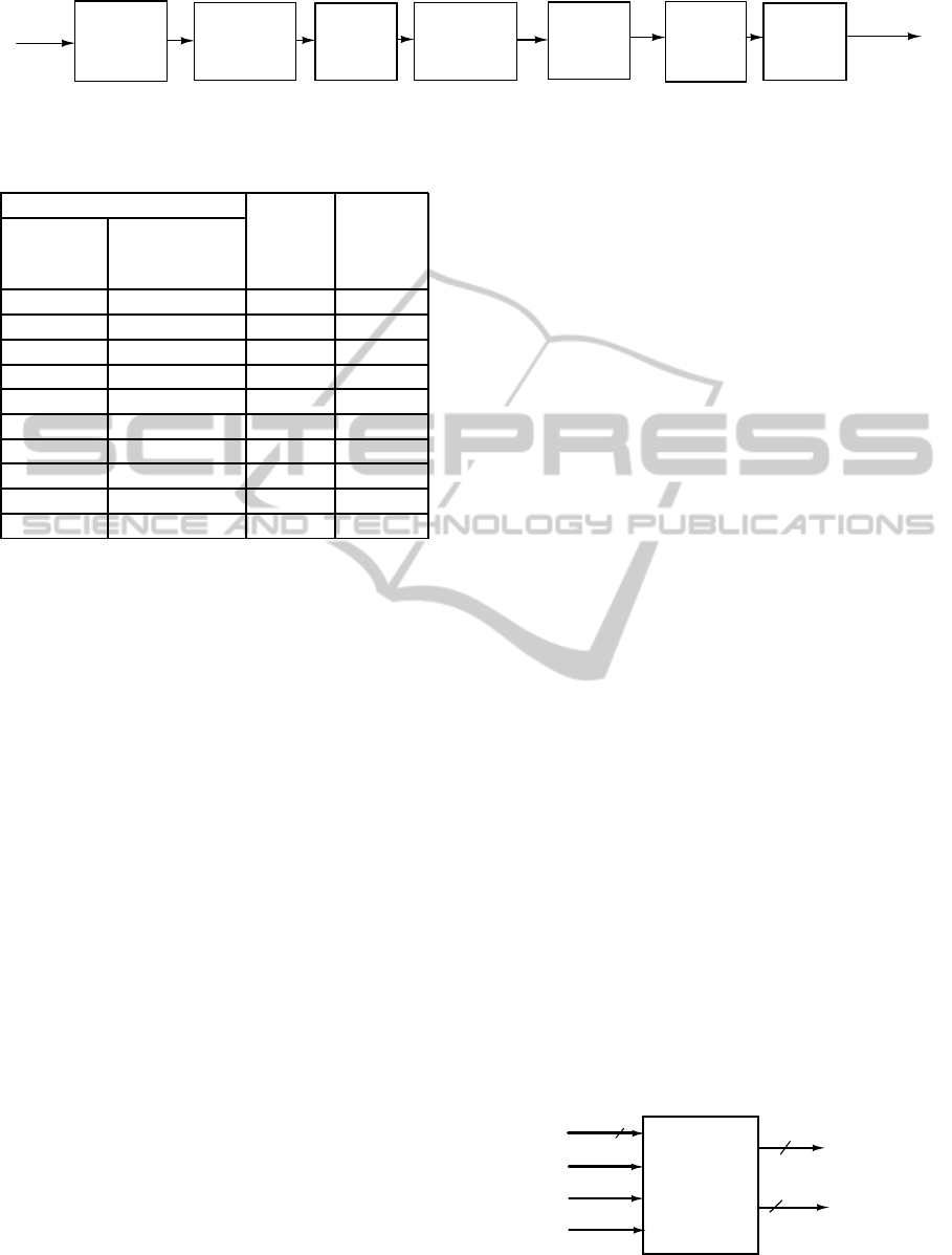
The block diagram of the proposed image compressor
is given in Figure 8. The image compressor takes 8-bit
pixel value from image sensor at every rising edge of
clock (or CLK) signal. The image sensor also sends
row (HSYNC) and frame synchronisation (VSYNC)
to indicate the end of a row and frame respectively.
The image compressor requires computationally sim-
ple methods such as adders, shift registers, counters
etc. The lifting scheme based 1-D wavelet decom-
position of mosaic image is done modifying the ar-
chitecture proposed in (Angelopoulou et al., 2007).
In the modified architecture the first level wavelet de-
composition is done according to the equations given
in (5) and (6). The Golomb-Rice encoder generates
a codeword (CW) of length 32 and codeword index
(CWI). The codeword index holds the index to MSB
of the generated code in the 32-bit CW. No memory
buffer is used in the implementation, only few regis-
ters are employed.
The proposed endoscopic image compression al-
gorithm ( with quantizer q=4 and sub-sampling the
high frequency subband of first level wavelet decom-
position by 4) has been implemented in both MAT-
LAB and VHDL for verification. The image compres-
sor was implemented in MATLAB scripts and VHDL.
The image decompressor is implemented only in
MATLAB. After the functionality of the algorithm
is verified using 120 endoscopic images, the VHDL
code is synthesized and mapped to UMC 130 nm
Faraday high speed (HS) library using Synopsys De-
sign Compiler. After post-synthesis functional verifi-
cation of the algorithm is done using QuestaSim and
MATLAB, the layout is generated using Cadence Soc
Encounter place and route tool. The layout view of
the core of the chip is shown in Figure.9. The pro-
posed image compression algorithm has core size of
0.342 mm x 0.342 mm. The power consumption of
the synthesized design is estimated using Synopsys
PrimeTime PX. The mean power consumption of the
image compressor is 48.4µW when operating at two
frames per second (fps) for encoding 512x512 color
mosaic image. The operating voltage is 1.2V.
The performance comparison of the proposed al-
gorithm with other existing works is given in table
3. We compare our proposed image compressor with
one algorithm which is based on JPEG-LS (Xie et al.,
2006) and two algorithms which are based on DCT
(Lin and Dung, 2011; Turcza and Duplaga, 2013).
All the competing algorithms included in the table
From
image
Image
compressor
8
HSYNC
VSYNC
CLK
DATA
sensor
32
5
CW
CWL
To RF
transmitter
Figure 8: Block diagram of the proposed image compressor
implementation.
ALow-PowerColorMosaicImageCompressorBasedonOptimalCombinationof1-DDiscreteWaveletPacketTransform
andDPCMforWirelessCapsuleEndoscopy
195
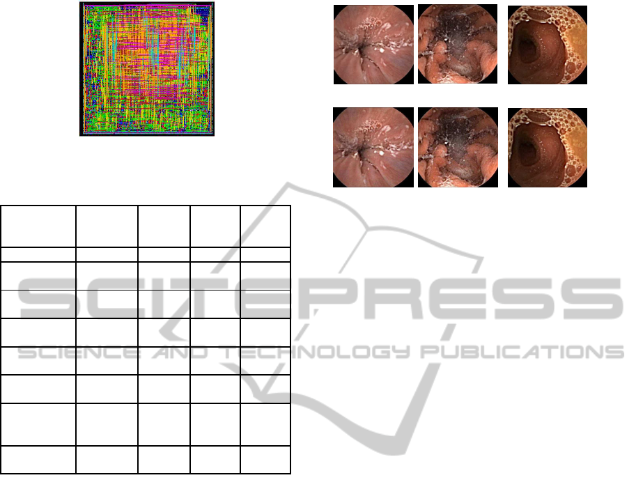
Figure 9: Layout view of the core of the proposed image
compressor implementation.
Table 3: The comparison results with previous works.
(Xie
et al.,
2006)
(Lin
et al.,
2011)
(Turcza
et al.,
2013)
This
work
Methods JPEG-LS DCT DCT DWPT
Compression
Rate (%)
72.7 82.0 91.2 81.31
Overall
PSNR (dB)
46.8 36.2 35.7 39.5
Technology
ASIC
0.18 µm
ASIC
0.18 µm
FPGA
65nm
ASIC
0.13µm
Core
size (µm
2
)
90K gates 318K - 117K
Memory
(byte)
93.81K YES 10.5K 0
Power
consumption
(mW)
1.55
(0.28 fps)
9.17
(2 fps)
7
(7 fps)
0.048
(2 fps)
Supply
Voltage(V)
1.8 1.8 1.2 1.2
were tested with similar images of 512 x 512 reso-
lution. The proposed image compression algorithm
doesn’t require memory and consumes about two or-
ders of less power than other algorithms as shown in
table 3. Our algorithm has also the lowest chip area
as compared to other works. The proposed algorithm
outperforms work in (Xie et al., 2006) in terms of
compression rate. It gives a comparable compression
rate with a work in (Lin and Dung, 2011) but has also
higher image quality. It achieves lower compression
rate than the work in (Turcza and Duplaga, 2013).
However, the work in (Turcza and Duplaga, 2013)
needs about 10.5 KB of memory which consumes
high powerand occupies large silicon area. Generally,
DCT based compression algorithms introduce block-
ing effect due to the quantization of DCT coefficients.
The proposed algorithm doesn’t introduce blocking
effect which is indispensable for precise diagnosis of
medical images. As shown in Figure.10, the proposed
endoscopic image compression algorithm doesn’t in-
troduce noticeable artifacts in the reconstructed im-
age. The proposed endoscopic image compression al-
gorithm meets all the criterion to be a serious candi-
date for WCE.
(a) Cardia (b) Gastric Body (c) Jejunum
(i) 39.38 dB (ii) 39.04 dB (iii) 38.16 dB
Figure 10: Three endoscopic images out of 120 test images
that we have employed in the experimentation. The original
(top) and reconstructed images (bottom) with PSNR value.
5 CONCLUSION
In this paper, we have presented a low-power im-
age compression algorithm for WCE application.
The algorithm uses optimal combination of meth-
ods which are computationally simple. Utilizing
the combination of computationally simple methods
we have achieved a compression rate of 81.3 %.
The compressed image has a high image quality
(39.5 dB) which is greater than the minimum PSNR
(35 dB)(Istepanian et al., 2008; Cosman et al., 1994)
required for accurate medical image diagnosis. The
proposed image compression algorithm has relatively
high compression rate, small chip area, low power
consumption and high image quality which makes it
a good candidate for WCE application.
The future work includes the design of the whole
capsule system and testing its performance in real-
world. The impact of the distortion introduced due to
the image compressor on automatic disease detection
algorithms will be studied.
REFERENCES
Angelopoulou, M. E., Cheung, P. Y. K., Masselos, K., and
Andreopoulos, Y. (2007). Implementation and com-
parison of the 5/3 lifting 2d discrete wavelet transform
computation schedules on fpgas. Springer, Journal of
VLSI Signal Processing.
Bayer, B. E. (1976). Color imaging array.
Chen, X., X., Zhang, L., Li, X., Qi, N., Jiang, H., and
Wang, Z. (2009). A wireless capsule endoscope sys-
tem with low-power controlling and processing asic.
IEEE Transactions on,Biomedical Circuits and Sys-
tems, 3(1):11–22.
BIODEVICES2015-InternationalConferenceonBiomedicalElectronicsandDevices
196

Cheng, C., Liu, Z., Hu, C., and Meng, M. (2010). A novel
wireless capsule endoscope withjpeg compression en-
gine. In 2010 IEEE International Conference on, Au-
tomation and Logistics (ICAL), pages 553–558.
Cosman, P., Gray, R., and Olshen, R. (1994). Evaluating
quality of compressed medical images: Snr, subjective
rating, and diagnostic accuracy. Proceedings of the
IEEE, 82(6):919–932.
Dung, L., Wu, Y., Lai, H., and Weng, P. (2008). A mod-
ified h.264 intra-frame video encoder for capsule en-
doscope. In BioCAS 2008, IEEE Biomedical Circuits
and Systems Conference, pages 61–64.
Gallager, R. and Voorhi, D. V. (1975). Optimal source
codes for geometrically distributed integer alphabets
(corresp.). IEEE Transactions on, Information The-
ory, 21(2):228–230.
Gastrolab (2014). http://gastrolab.net/ (Online avail-
able),last visited 2014.
Getreuer, P. (2011). Color demosaicing with contour sten-
cils. In 17th International Conference on, Digital Sig-
nal Processing (DSP), pages 1–6.
Golomb, S. (1966). Run-length encodings (corresp.). IEEE
Transactions on, Information Theory, 12(3):399–401.
Idden, G., Meron, G., Glukhovsky, A., and Swain, P.
(2000). Wireless capsule endoscopy. Nature.
Istepanian, R., Philip, N., Martini, M., Amso, N., and
Shorvon, P. (2008). Subjective and objective quality
assessment in wireless teleultrasonography imaging.
In EMBS 2008,3 0th Annual International Conference
of the IEEE Engineering in Medicine and Biology So-
ciety.
JPEG (1998). Lossless and near-lossless compression of
continuous-tone still images baseline. JPEG-LS,T.87.
Khan, T. and Wahid, K. (2011a). Low power and low
complexity compressor for video capsule endoscopy.
IEEE Transactions on, Circuits and Systems for Video
Technology, 21(10):1534–1546.
Khan, T. and Wahid, K. (2011b). Subsample-based image
compression for capsule endoscopy. Journal of Real-
Time Image Prococessing, 8(1):5–19.
Korhonen, J. and Junyong, Y. (2012). Peak signal-to-noise
ratio revisited: Is simple beautiful? In Fourth Inter-
national Workshop on, Quality of Multimedia Experi-
ence (QoMEX).
Lin, M. and Dung, L. (2011). A subsample-based low-
power image compressor for capsule gastrointestinal
endoscopy. EURASIP Journal on Advances in Signal
Processing, 2011.
Lin, M., Dung, L., and Weng, P. (2006). An ultra-low-power
image compressor for capsule endoscope. BioMedical
Engineering OnLine, 5(14).
Moglia, A., Menciassi, A., and Dario, P. (2008). Recent
patents on wireless capsule endoscopy. Recent Patents
on Biomedical Engineering.
Pattanaik, S., Mahapatra, K., and Panda, G. (2006). A
novel lossless image compression algorithm using
arithmetic modulo operation. In IEEE Conference on
Cybernetics and Intelligent Systems, pages 1–5.
Rice, R. F. (1991). Some practical universal noiseless cod-
ing techniques - part iii. Tech. Rep.JPL-91-3, Jet
Propulsion Laboratory.
Sweldens, W. (1995). Lifting scheme: A new philosophy
in biorthogonal wavelet constructions. In proc. SPIE,
volume 2569.
Turcza, P. and Duplaga, M. (2011). Low power fpga-based
image processing core for wireless capsule endoscopy.
Sensors and Actuators A: Physical, 172(2):552–560.
Turcza, P. and Duplaga, M. (2013). Hardware-efficient low-
power image processing system for wireless capsule
endoscopy. IEEE Journal of Biomedical and Health
Informatics, 17(6):1046–1056.
Wahid, K., Ko, S., and Teng, D. (2008). Efficient hardware
implementation of an image compressor for wire-
less capsule endoscopy applications. In IJCNN’08,
IEEE International Joint Conference on Neural Net-
works (IEEE World Congress on Computational In-
telligence).
Xie, X., Li, G., Chen, X., Li, X., and Wang, Z. (2006). A
low-power digital ic design inside the wireless endo-
scopic capsule. IEEE Journal of Solid-State Circuits,
41(11):2390–2400.
Xie, X., Li, G., and Wang, Z. (2007). A near-lossless image
compression algorithm suitable for hardware design
in wireless endoscopy system. EURASIP Journal on
Advances in Signal Processing.
Zhang, N. and Wu, X. (2004). Lossless compression of
color mosaic images. In ICIP ’04, International Con-
ference on Image Processing.
ALow-PowerColorMosaicImageCompressorBasedonOptimalCombinationof1-DDiscreteWaveletPacketTransform
andDPCMforWirelessCapsuleEndoscopy
197
