Development of a Multispectral Gastroendoscope to Improve the
Detection of Precancerous Lesions in Digestive Gastroendoscopy
Sergio Ernesto Martinez Herrera
1
, Yannick Benezeth
2
, Matthieu Boffety
3
, François Goudail
3
,
Dominique Lamarque
1
, Jean-François Emile
1
and Franck Marzani
2
1
Université de Versailles Saint Quentin en Yvelines, Versailles, France
2
Le2i,Université de Bourgogne, Dijon, France
3
Institut d’Optique Graduate School, Palaiseau, France
1 STAGE OF THE RESEARCH
The actual stage of research is the beginning of the
second year of the PhD thesis. The duration of the
thesis is three years. The first year has begun with
the state of the art involving two main parts. The
first one focused on medical aspects related to the
development and staging of stomach cancer. The
second part was oriented to the actual technology
and image processing techniques which are used to
help in the diagnosis of malignancies in the stomach.
The review of the state of the art mainly focused on
the study of multispectral imaging. An overview of
the state of the art is presented in section 4. Then, a
multispectral endoscope prototype has been
developed; this system is described in section 5. It is
based on the use of a filter wheel to modify the
regular white light of a gastroendoscopic system.
Afterwards, a set of image pre-processing techniques
has been developed to improve the usability of the
multispectral images obtained by the prototype.
These techniques are also described in section 5.
Finally, in section 6 are presented the future work
and the expected outcome.
2 OUTLINE OF OBJECTIVES
In order to improve the diagnosis during
gastroendoscopy, practitioners need additional
information from the tissue characteristics in a non-
invasive, efficient and accurate way.
The actual technology used to perform
gastroendoscopy is mainly based on the visual
exploration under white light; an illustration is
presented in figure 1a. Unfortunately, it is often
difficult to visualize malignancies in the tissue with
this technology. Practitioners with all degrees of
experience, including those with many years of
practice mention this situation.
The diagnosis of gastric pathologies is performed
based on biopsy acquisition and its histological
analysis, which is the microscopic evaluation of
tissue. This is considered to be the most reliable
technique for diagnosis. Unfortunately, the
collection is difficult because in many cases there
are no macroscopic differences between healthy and
wounded tissue. In consequence, practitioners
usually collect biopsies randomly. This situation
leads to the acquisition of tissue without any
pathology, which produces undesirable false
negative diagnosis.
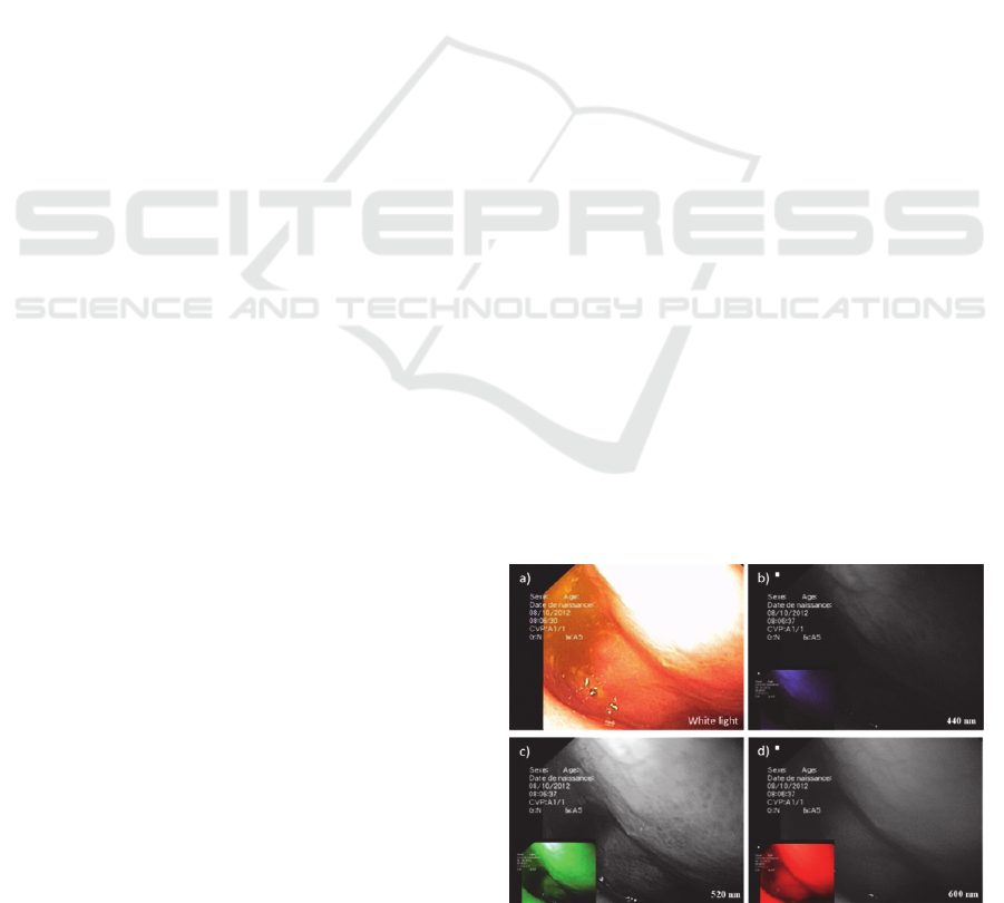
Figure 1: a) Colour image under white light. b), c), d)
Monoband images which highlight different features.
Considering that the gastric pathologies are
related to modifications in the properties of the
tissue, there is an important need to measure these
variations. Based on previously successful
applications, we believe that multispectral imaging
can help in the identification of early stages of
15
Martinez Herrera S., Benezeth Y., Boffety M., Goudail F., Lamarque D., Emile J. and Marzani F..
Development of a Multispectral Gastroendoscope to Improve the Detection of Precancerous Lesions in Digestive Gastroendoscopy.
Copyright
c
2014 SCITEPRESS (Science and Technology Publications, Lda.)

gastric cancer, even if these lesions present subtle
colour and morphological differences in comparison
with healthy tissues using conventional white light
illumination.
In summary, there are two main objectives in this
PhD thesis. The first one is the design and
development of a prototype capable to acquire
multispectral images of the stomach during
gastroendoscopy. The system must be compatible
with the actual systems used in gastroendoscopy.
The second objective is to propose tools and
methods to identify cancerous tissue at an early
stage. This second part is not presented here. We
present in the remainder of the document, the
proposed acquisition system and the image pre-
processing techniques implemented.
3 RESEARCH PROBLEM
The first challenge of the PhD is the acquisition of
multispectral images during gastroendoscopy. Some
recent works have focused on the analysis of the
reflectance from gastric tissue using spectroscopy
(Bashkatov et al., 2007). On the other hand, other
recent works were focused on the analysis of
multispectral images of gastric tissue from ex-vivo
samples (Galeano et al., 2012); (Kiyotoki et al.,
2013). To the best of our knowledge, there is no
work on the analysis of pre-cancerous lesions using
in-vivo multispectral images of the stomach tissue
acquired during gastroendoscopy. Consequently, the
first research problem to solve is the development of
a system to acquire the data, which is not a trivial
task due to the inherent constrains from the working
environment.
The second problem to solve is the registration
between successive monoband images. Due to the
configuration of the acquisition system, the
monoband images that compose the multispectral
images are acquired sequentially and present a
shifting. It is necessary to register the monoband
images to form the multispectral image. This
registration is highly complicated because many
assumptions, upon which the majority of the state of
the art techniques rely, are not respected for our
data. These methods are usually based either on the
use of anatomical references, texture features or the
underlying assumptions to the use of optical flow,
where there is a small displacement of a constant
intensity between two images. In our particular case,
the images obtained are not compliant with these
fundamental assumptions, for instance the gastric
tissue is highly homogeneous with subtle texture in
some cases, it is in constant movement (non-rigid
deformations) and the environment is moist (large
variations in photometric properties). Moreover,
monoband images of the same area at two different
wavelengths present strong differences. In figure 1 is
shown an example of these difficulties with 3
monoband images from a gastric multispectral
image.
The two above problems are detailed in this
document. We can also mention the difficulty to
extract information from these images. The first
problem will be given by the quality of the images.
The methods to detect pre-cancerous lesions should
be sufficiently robust to operate on noisy images.
Then, the methods should be fast enough to identify
in real time tissue which is more likely to develop
cancer, in order to help the practitioner during the
gastroendoscopy. These issues will be a major part
of the work during the second and third year of the
PhD thesis.
Before introducing the acquisition system and
the image processing techniques, we present in the
following section the state of the art.
4 STATE OF THE ART
Nowadays, the majority of gastroendoscopy imaging
devices provide colour images acquired under white
light. Some systems have been developed that
increase the visualization of the lesions from a
macroscopically point of view. These systems can
be classified in two main categories. The first one
takes advantage of an external agent, for instance a
dye is used to highlight specific features of the
lesions. The main technique in this category is
chromoendoscopy (Kida et al., 2003).
The second class of systems increases the
spectral resolution of the images in order to enhance
the visualization of the tissue. The Narrow band
Imaging (NBI proposed by Olympus) or Multi Band
Imaging (FICE proposed by Fuji) belongs to this
category. For these systems, a false colour image is
formed with 2 or 3 monoband images at a specific
wavelength (Wong Kee Song et al., 2008
). These
techniques can be considered as a virtual version of
chromoendoscopy.
Even if they are limited to a few bands, these
techniques show the potential of multispectral
imaging for gastroendoscopy. A multispectral image
is formed by monoband images taken at different
wavelength. This technique has an important
advantage since it provides spatial and spectral
information. This situation leads to use image and
VISIGRAPP2014-DoctoralConsortium
16
signal processing algorithms for data treatment.
There are different examples of successful
implementations of this technique oriented to
medical applications. For instance, it has been used
to increase the proportion of anomalies found in
skin, but also to characterize and delimitate lesions
(Tomatis et al., 2005
)
. Recent approaches have
showed that it is possible to retrieve biological
parameters from the tissue under controlled
acquisition conditions (Jolivot et al., 2011).
Multispectral imaging presents some
disadvantages, for instance the amount of memory
required for a single image (width x height x total of
wavelengths). In consequence, the computational
cost increases significantly. Furthermore, the images
are acquired typically sequentially; this can be
problematic in case of non-static scenarios.
After reviewing the actual technologies to detect
pre-cancerous lesions, as well as the advantages and
disadvantages of multispectral imaging, we
introduce in the following section the acquisition
prototype and the algorithms used.
5 METHODOLOGY
In this section we describe the development of the
multispectral prototype, as well as the pre-
processing image techniques implemented.
5.1 Prototype of the Acquisition System
Multispectral imaging acquires information in two
spatial dimensions (width and height) and one
spectral dimension (wavelength). The acquisition is
performed in most of the cases through two
dimensions at the time, creating two options (Simon
et al., 2013); (Grahn and Geladi, 2007).
The first one acquires one spatial and the spectral
dimension; this option demands some kind of
motion to scan the other spatial dimension.
The second one acquires the spatial dimensions
whereas the remaining spectral dimension is
scanned. In practice, this option can take advantage
of a CCD camera to acquire monoband images at
different wavelengths. Therefore, this configuration
is selected over the other due to its compatibility to
use the camera from the current gastroendoscopes.
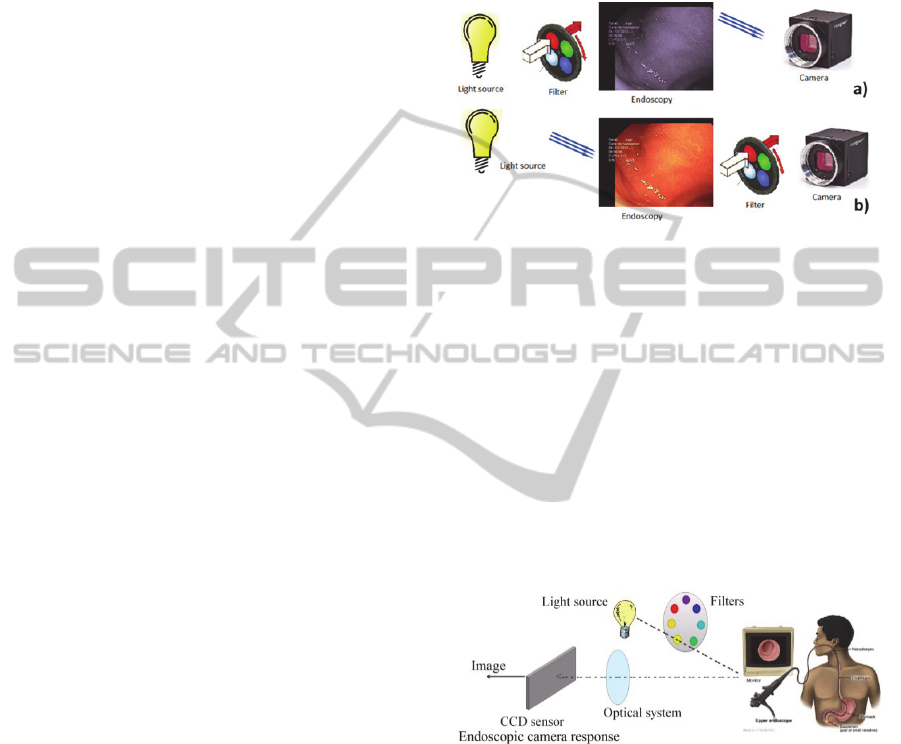
Physically, there are two main options to scan
the spectral dimension as is presented in figure 2.
The first option is to illuminate the scene with a
series of specific wavelengths. This can be achieved
for instance by using a tunable light source or
filtering the light from a single light source. This last
option is commonly used in practice through a filter
wheel.
The second option is to filter the incoming light
from the scene to the sensor, which can also be
achieved by placing a filter wheel in front of the
camera or by using a multispectral camera.
Figure 2: Techniques to acquire multispectral images. a)
Single wavelength illuminates the scene. b) Filtering the
light arriving to the camera.
For reasons of simplicity and compatibility with
the actual gastroendoscopic systems, we decided to
modify the source of light of the regular
gastroendoscopes. The final configuration is
presented in figure 3, where the source of light is a
Xenon lamp, filtered by a filter wheel. This light is
transmitted through the gastroendoscope (Olympus
Exera II) to illuminate the stomach. Then, the
camera from the gastroendoscope is used to capture
the image which is finally transmitted to a computer
connected to the gastroendoscopic station.
Figure 3: Multispectral acquisition prototype.
The filter wheel includes six filters in the visible
range from 440 to 640 nm with an equidistant
spectral separation. This option was selected over
the others due to the wide range of available filters
that facilitates the customization. Moreover the cost
is reasonable and the light power allows strong input
ranges in comparison with the wavelength generator
light source. The output of this system is a video
sequence at 25fps with a resolution of 640x480
pixels, from which we can extract the multispectral
images.
The video is first deinterlaced using the widely
known algorithm of Yadif (Hegenbart et al., 2013).
DevelopmentofaMultispectralGastroendoscopetoImprovetheDetectionofPrecancerousLesionsinDigestive
Gastroendoscopy
17
Then, the sequences of monoband images that
formed the multispectral images are extracted from
the video. The speed of the filter wheel is configured
in order to obtain 4 monoband images from each
wavelength. The first and fourth are the transition
between two filters and are discarded. The second
and third images can be used to generate the
multispectral image; in our application, we use the
third image.
The moist environment of the stomach produces
areas of specular reflection in the images, these
regions are easily removed using a threshold.
The acquisition time of a multispectral image is
approximately one second. Because the stomach is
in constant movement, the monoband images
acquired sequentially present a shifting. This shifting
can be reduced increasing the acquisition speed, but
it is limited to the speed of the filter wheel and the
frame rate of the gastroendoscopic camera. In the
following section is addressed this shifting issue.
5.2 Image Registration
This stage is crucial for the image analysis, since it
allows the superposition of the monoband images.
The first step is a pre-processing step in order to
enhance the contrast information. The algorithm of
contrast limited adaptive histogram equalization is
used in each monoband image.
Then, an affine model is used to model the
transformation between consecutive monoband
images. The parameters of the transformation are
computed using the hierarchical motion-based
estimation (Bergen et al., 1992). The transformation
matrix has six degrees of freedom, which covers the
relative small variations caused in the image by the
movement of the endoscope and the tissue during
the acquisition.
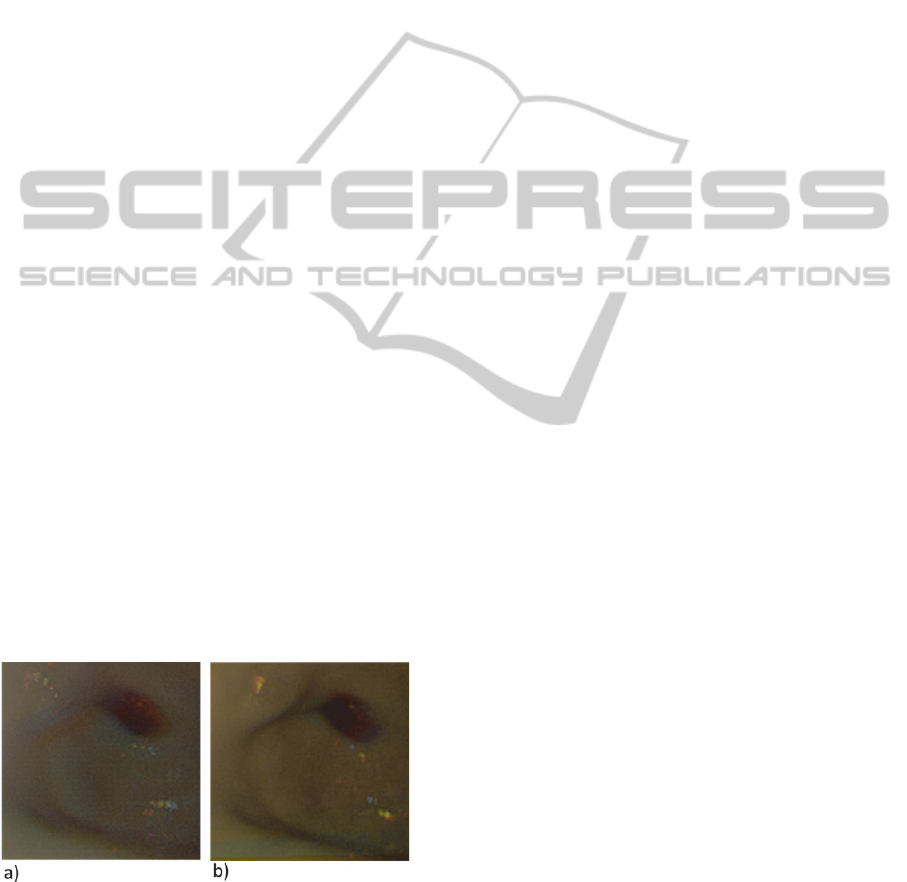
Figure 4: Virtual white light image computed from a)
original monoband images and b) registered monoband
images.
In order to minimize the cumulative error, we
visualize the six monoband images as a sequence,
where the center image is used as a common image
for two subsequences.
The fourth monoband image is assumed to be the
center image and functions as reference for the
registration of the other images. Then, to explain the
procedure, we use the smaller subsequence as an
example (images fourth, fifth and sixth); the fifth
monoband image is registered to the reference
image. Then, the sixth image is registered to the fifth
image producing a temporal image; later on, we
apply to this image the transformation before
estimated to register the fifth image with the
reference image. If there where further images, this
procedure can be iteratively repeated until all the
images from the subsequence are registered. Then,
the same procedure is applied to the other
subsequence.
Finally, when all the transformations are known,
they are applied to the original data to produce a
new set of six monoband images. In order to
highlight the advantages of the registration, figure 4
shows two images whose simulate an endoscopic
image acquired under white light. It is clear that the
figure 4b generated from the registered images is
sharper in comparison with 4a.
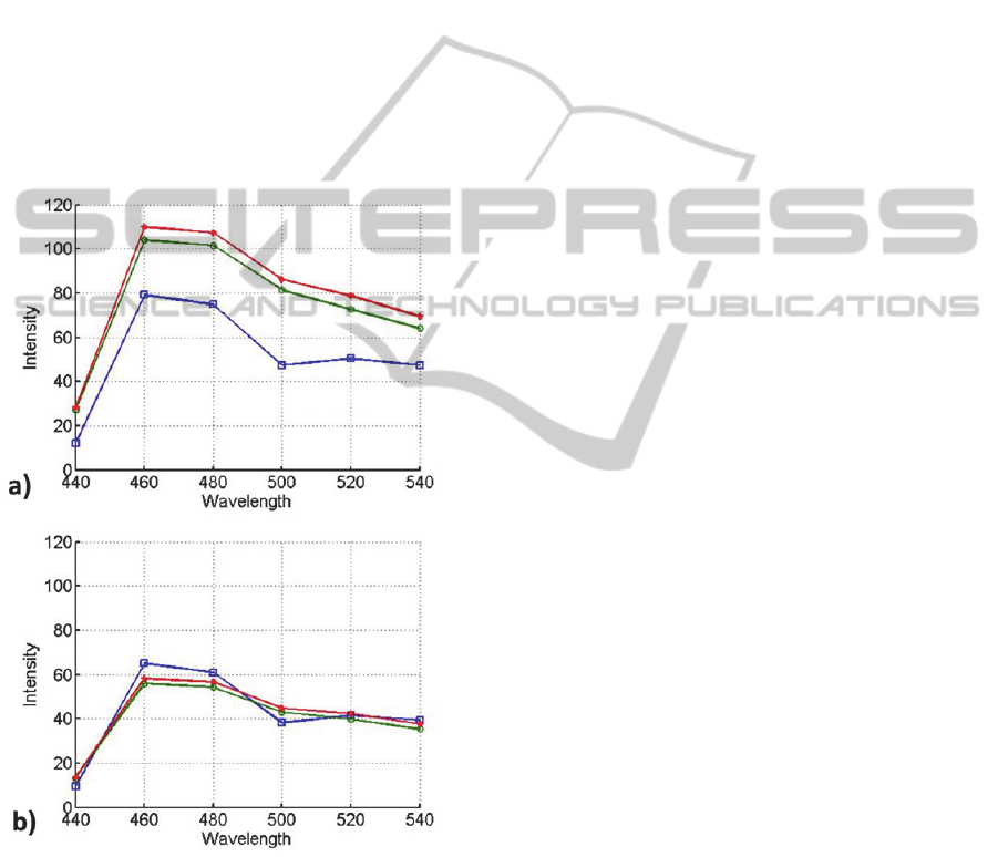
5.3 Normalization
The fact that the distance and the orientation
between the tissue and the camera are not constant,
has an important impact in the amplitude of the
estimated spectrum as is shown in figure 5a.
We believe that the shape of the spectrum is
more significant than the amplitude to differentiate
precancerous lesions; therefore, a normalization step
is clearly necessary in order to accurately identify
malignancies in gastric tissue. In this sense, the Area
Under the Curve (AUC) is selected as a spectral
normalizer function. Figure 5b presents the spectra
after normalization, where the spectral shape
remains, facilitating its comparison. This
characteristic is highly desired in the input data for
classification algorithms.
The multispectral images acquired with the
prototype and the described treatment produce
promising spectra. These findings lead the path to
the second and third year of PhD in order to propose
methods to identify pre-cancerous lesions at an early
stage.
6 EXPECTED OUTCOME
The three years of PhD thesis are expected to
VISIGRAPP2014-DoctoralConsortium
18
produce two main results.
The first one is a multispectral acquisition
system for gastroendoscopy. This system is being
designed to be compatible with the actual acquisition
systems used in gastroendoscopy.
The acquired multispectral image during
gastroendoscopy leads to the second outcome, which
is the identification of cancerous lesions at an early
stage. The research is oriented to the proposal and
development of tools and methods oriented to
identify pre-cancerous lesions. These methods are
expected to be robust to noise due to the acquisition
conditions and fast enough, in order to recognize in
real time the tissue, which is more likely to develop
cancer. These algorithms will be a major part of the
work during the second and third year of the PhD
thesis.
Figure 5: Spectrum from healthy tissue, a) original
spectrum, b) normalized spectrum.
REFERENCES
Bashkatov, A. N., Genina, E. A., Kochubey, V. I.,
Gavrilova, A. A., Kapralov, S. V., Grishaev, V. A.,
Tuchin V. V., 2007. Optical properties of human
stomach mucosa in the spectral range from 400 to
2000 nm: Prognosis for gastroenterology. In Medical
Laser Application, 22(2), p95-104.
Bergen, J. R., Anandan, P., Hanna, K. J., Hingorani, R.,
1992. Hierarchical model-based motion estimation. In
ECCV’92, 588, p237-252.
Galeano, J., Jolivot, R., Benezeth, Y., Marzani, F., Emile,
J.-F., Lamarque, D., 2012. Analysis of Multispectral
Images of Excised Colon Tissue Samples Based on
Genetic Algorithms. In int. conf. on Signal Image
Technology & Internet Based Systems (SITIS), 25-29
Nov, Naples, Italy, pp. 833-838.
Grahn, H. & Geladi, P., 2007. Techniques and
Applications of Hyperspectral Image Analysis, West
Sussex: Wiley. P1-13.
Jolivot, R., Vabres, P., Marzani, F., 2011. Reconstruction
of hyperspectral cutaneous data from an artificial
neural network-based multispectral imaging system. In
Computerized Medical Imaging and Graphics, 35(2),
p85-88.
Hegenbart, S., Uhl, A., Wimmer, G., Vecsei, A., 2013. On
the effects of de-interlacing on the classification
accuracy of interlaced endoscopic videos with
indication for celiac disease. In Computer-Based
Medical Systems (CBMS), 2013 IEEE 26th
International Symposium, 20-22 June, Porto, Portugal,
pp. 137-142.
Kida M., Kobayashi K., Saigenji K., 2003. Routine
chromoendoscopy for gastrointestinal diseases:
indications revised. In Endoscopy, 35(7), p590-596.
Kiyotoki, S., Nishikawa, J., Okamoto, T., Hamabe, K.,
Saito, M., Goto, A., Fujita, Y., Hamamoto, Y.,
Takeuchi, Y., Satori, S., Sakaida, I., 2013. New
method for detection of gastric cancer by
hyperspectral imaging: a pilot study. In Journal of
Biomedical Optics, 18(2), p26010.
Simon C., Mansouric, A., Marzani, F., Booch, F., 2013.
Integration of 3D and multispectral data for cultural
heritage applications: survey and perspectives. In
Image and Vision Computing, 31(1), p91-102.
Tomatis, S., Carrara, M., Bono A., 2005. Automated
melanoma detection with a novel multispectral
imaging system: results of a prospective study. In
Physics in Medicine and Biology, 50(8), p1675-1687.
Wong Kee Song, L. M., Adler, D. G., Conway J. D.,
Diehl, D. L., Farraye, F. A., Kantsevoy, S.V., Kwon,
R., Mamula, P., Rodriguez, B., Shah, R. J., Tierney,
W. M., 2008. Narrow band imaging and multiband
imaging. In Gastrointestinal Endoscopy, 67(4), p581-
589.
DevelopmentofaMultispectralGastroendoscopetoImprovetheDetectionofPrecancerousLesionsinDigestive
Gastroendoscopy
19
