
EEG Motor Imagery Classification of Upper Limb Movements
Maria Claudia F. de Castro, Jo
˜
ao Pedro de O. P. Galhianne and Esther Luna Colombini
Electrical Engineering Department, Centro Universit
´
ario da FEI, S
˜
ao Bernardo do Campo, Brazil
Keywords:
EEG,
Band-power Extraction, Pattern Recognition, Linear Discriminant Analysis (LDA).
Abstract:
C EEG channel data are usually used when building systems that aim at distinguishing among right and left
hand movements. Few alternatives use multichannel systems when bigger sets of motor imagery are subject
to classification and more inputs are required. In this context, this work proposes the use of 8 EEG channels
(F,C,P, and O), disposed in a non-conventional set up, to classify up to 4 motor imagery of the upper limbs
through a Linear Discriminant Analysis classifier. A spatial feature selection, prior to classification, is applied
in order to improve the classification accuracy. For the many channel combinations tested, results suggest
that, in addition to the motor areas, other brain areas should be considered. For the proposed system, the best
classification accuracy was achieved when distinguishing between left arm and left hand (89.74%) and using
only the electrodes in F areas. For the right versus left hand a 71.80% rate was obtained, with electrodes either
in P and O areas or in F and P areas. To discriminate between arms and hands, independently of the body side,
the best score was 83.33%, for F and P channels, whereas for right and left limbs the best score was 66.02%,
with only P channels. The best classification accuracy for the 4 movement problem achieved 50.00%, using
all electrodes.
1 INTRODUCTION
Brain Computer Interfaces (BCI) are communication
systems that use the electrical brain activity as input
of a system that will translate them into a control sig-
nal, for an external device, that represents the sub-
ject’s wish. Originally, this technology was developed
for people with severe motor disabilities and common
applications include spelling devices, wheelchair, and
neuromotor prostheses control. However, nowadays,
entertainment applications for healthy users are gain-
ing space, as applications for games and virtual reality
interfaces (Hoffmann et al., 2007; Veen, 2009; Millan
et al., 2010). BCI systems can be classified into two
major categories depending on the signal that they
use. Some are based on Endogenous Potentials, such
as those used in imagined movement that are volun-
tarily generated by the user, and others are based on
Exogenous Potentials that are externally induced by
an stimuli (Veen, 2009).
The brain activity occurs in many regions of the
brain, either on the cortex, basal ganglia, cerebellum,
and thalamus, changing its oscillatory frequency ac-
cording to the mental and physical states of the sub-
ject. The main bands or rhythms typically observable
are: delta (1 - 4 Hz), theta (4 - 8 Hz), alpha (8 - 12 Hz),
Beta (12 -28 Hz), and Gama above 28 Hz (Hoffmann
et al., 2007; Hema et al., 2010).
The field of motor imagery has shown a predom-
inant interest in the right and left hand movements
using EEG from C3, Cz, and C4 channels. (Xu
and Song, 2008) achieved 90%− 92% accuracy using
Discrete Wavelets Transforms (DWT) and Autore-
gressive (AR) Model features with a Linear Discrimi-
nant Analysis (LDA) classifier, whereas (Hema et al.,
2009) applied an Elman Neural Network over EEG 4
band power features, achieving an average classifica-
tion of 72%. (Huang and Wu, 2010) used the average
energy of the C3 and C4 channels, a Wavelet Package
Transform, and the Quadratic Discriminant Analysis
classifier achieving a maximum rate of 88.71%. (Ku-
mar and Fumitoshi, 2010) proposed the use of a Rel-
ative Spectral Power as feature, applying it over the 5
EEG frequency bands. The classification rate using a
LDA classifier was 76.43%, whereas (Dolezal et al.,
2011), using the Power Spectral Density (PSD) with
a Support Vector Machine (SVM), achieved 75% of
classification.
With a 14-electrode set up, located around the mo-
tor area, the best results achieved by (Higashi et al.,
2009), using correlation coefficients based on Rhyth-
mic Component Extraction with a LDA classifier,
ranged from 74.9% to 83%. Performing a spatial
feature selection from a 64-electrode set up, (Xiao
Claudia F. Castro M., Pedro de O. P. Galhianne J. and Luna Colombini E..
EEG Motor Imagery Classification of Upper Limb Movements.
DOI: 10.5220/0004235003140317
In Proceedings of the International Conference on Bio-inspired Systems and Signal Processing (BIOSIGNALS-2013), pages 314-317
ISBN: 978-989-8565-36-5
Copyright
c
2013 SCITEPRESS (Science and Technology Publications, Lda.)

et al., 2009) discriminated between two types of mo-
tor imagery among a set of four (right and left hand,
foot, and tongue) using Energy Entropy of the Short-
term Fourier Transform as feature. Comparing the
performance of a Linear Discriminant classifier, a
Back-Propagation Neural Network, and a SVM, the
best accuracy was achieved by the Linear Discrim-
inant classifier, with average classifications between
82.4% − 88%. With a set of 29-electrodes and us-
ing Independent Component Analysis prior to DWT
to obtain the features, and Bhattacharyya distance ma-
trices and scalp plots to classify, (Morash et al., 2008)
reached an average classification around 35% to dis-
tinguish among the same four motor imagery classes.
It can be noticed that there is a prevalence of using
only C3 and C4 channels to distinguish between two
motor imagery, mostly restricted to the right and left
hand movements, with a few alternatives using mul-
tichannel systems aiming at classifying a bigger set
of motor imagery. In this context, this work proposes
the use of 8 EEG channels (F,C,P, and O), disposed in
a non-conventional set up, to classify up to 4 motor
imagery of the upper limbs with a LDA classifier. A
spatial feature selection is applied, prior to classifica-
tion, in order to verify the best electrode combination
to improve classification performance.
2 MATERIALS AND METHODS
During the experiment, an able body subject was
seated on a comfortable arm chair, with the body re-
laxed. Six series of 5 repetitions of 4 random com-
mand sequences were given: close right hand, close
left hand, flex right arm, flex left arm, those which
should be imagined. EEG was recorded at 1000 Hz
using a Bioamplifier plus PowerLab 16/30 configura-
tion from AdInstruments, according to the approved
protocol (COEP - USJT - No.088/2011).
The signal, from each of the 8-channel set up, was
acquired transversally from electrodes Fz, Cz, Pz, Oz
to electrodes F3, F4, C3, C4, P3, P4, O1, and O2
(Figure 1). A segment of 2.5 s of each execution was
selected and the Spectral Power Magnitude Averages
in different frequency bands were computed as fea-
tures, as follows: alpha (8 - 12 Hz), Beta1 (12 - 16
Hz), Beta2 (16 - 20 Hz), Beta3 (20 - 28 Hz). For
the Gamma band, the effect of different configura-
tions were investigated: (1) 28 - 32 Hz; (2) 28 - 64
Hz; (3) 28 - 100 Hz; (4) Gamma1 (28 - 32 Hz), and
Gamma2 (32 - 64 Hz); (5) Gamma1 (28 - 32 Hz),
Gamma2 (32 - 64 Hz), and Gamma3 (64 - 100 Hz). A
spatial feature selection was also performed aiming to
find efficient electrode combinations that carry useful
and discriminative information.
Figure 1: Electrode channel set up.
A classifier based on Linear Discriminant Analy-
sis (LDA) was used in order to distinguish different
groups of data. Experiments were performed to dis-
criminate each of the motor imagery: right hand ver-
sus left hand, right arm versus left arm, right arm ver-
sus right hand, left arm versus left hand, hands versus
arms, and right limb versus left limb. LDA is a well
known technique based on linear combinations of the
ratio between two covariances: between-class scatter
matrix and the within-class scatter matrix. It usually
results in a reliable classification accuracy, requiring a
few number of samples and a low computational cost
if compared to other classification methods such as
Neural Networks.
3 RESULTS AND DISCUSSION
The classification accuracies achieved for each fre-
quency band when varying the boundaries of the
Gamma frequency band are shown in Figure 2. In
general, the impact of this variation was low, below
5%, specially when aiming at discriminating among
the four movements, right arm and right hand, left arm
and left hand, and between right and left limbs. Nev-
ertheless, the increase of the upper band boundary (28
- 64 Hz, 28 - 100 Hz) or the use of the total frequency
band (28 - 32 Hz, 32 - 64 Hz, 64 - 100 Hz) resulted
in a diminished classification accuracy to distinguish
between right and left hand or right arm and right
hand. The increase of the upper boundary had also
a negative effect into the classification rate to differ-
entiate between right and left arm. However, this sit-
uation generated the best discrimination rate between
left arm and left hand or arms versus hands. Although
small, the effect of the gamma band configuration is
different for each experiment. This suggests an inves-
tigation a priori and an appropriate use depending on
the ultimate goal.
The classification rates achieved by the applica-
tion of the LDA can be shown in Figures 3 and 4.
Figure 3 shows the classification accuracies reached
EEGMotorImageryClassificationofUpperLimbMovements
315
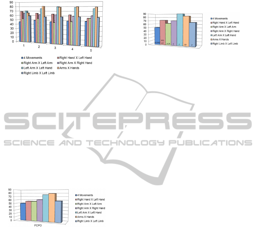
Figure 2: Classification accuracies for each frequency band
varying the Gamma frequency boundaries ( 1) 28 - 32 Hz;
2) 28 - 64 Hz; 3) 28 - 100 Hz; 4) Gamma1 (28 - 32 Hz), and
Gamma2 (32 - 64 Hz); 5) Gamma1 (28 - 32 Hz), Gamma2
(32 - 64 Hz), and Gamma3 (64 - 100 Hz).
considering all channels. The best average classifi-
cation was 78.85%, obtained when distinguishing be-
tween motor imagery related to hands and arms, inde-
pendently of the body side, followed by a 75.64% of
classification for left arm versus left hand. The classi-
fication accuracy when distinguishing between right
and left hands, a common problem presented in the
literature, was 56.43%, the same value achieved to
distinguish between right and left arms, whereas the
4 movements problem reached 50.00% accuracy.
Figure 3: Classification accuracies using a LDA classifier
over the F, C, P, and O channels.
The results for the spatial feature selection,
showed in Figure 4, improved all classification accu-
racies except for the 4 Movement problem, which is
the most complex among those presented. The classi-
fication accuracy to distinguish between left arm and
left hand movement imagination achieved 89.74%,
using only the electrodes in the F areas, whereas for
the right versus left hands a 71.80% rate was obtained
with electrodes either in P and O areas or in F and
P areas. To distinguish between arms and hands,
independently of the body side, the best score was
83.33%, considering only F and P channels. For the
discrimination between right and left limbs, the best
score was 66.02% with only P channels. It is impor-
tant to mention that these two last experiments were
performed with groups assembled with mixed data:
there was no equivalent movement imagination for
both limbs.
Figure 4: Best classification accuracies after spatial feature
selection.
In the predominant protocol for motor imagery,
the BCI systems use only C3, Cz, and C4 signals to
distinguish right versus left hands (Hema et al., 2009;
Xu and Song, 2008; Xiao et al., 2009; Huang and Wu,
2010; Kumar and Fumitoshi, 2010; Dolezal et al.,
2011). This is justified by the fact that when the sub-
ject performs an upper limb motor imagery the con-
sidered waves in the contra-lateral electrode concen-
trates more energy, while in the same side the energy
is suppressed (Xiao et al., 2009).
Despite the fact that this work has used a non-
conventional electrode set up, the spatial feature
selection process suggests that other brain regions
should be considered. Most of the experiments with
better classification rates have not used the electrodes
in C areas after applying the spatial feature selection.
Specifically, the best average classification accuracy
achieved to distinguish between right and left hands
used electrodes in F and P areas or in P and O areas.
To distinguish between right arm and right hand or
left arm and left hand, the best accuracy was achieved
using only F channels. When dealing with arms ver-
sus hands classification, electrodes in F and P areas
demonstrated the best results, and for right versus left
limbs, only P channels were used to achieve a similar
performance. The frontal cortex (F area) is related to
activities of planning movements, the parietal cortex
(P area) is an area of association for proprioceptive in-
formation also related to movement control, whereas
the visual cortex (O area) could be used in order to
visualized the execution of the movement.
Another point that must be considered is that, ac-
cording to (Veen, 2009), the comparison among BCI
systems suggests that there is a trade-off between
speed and accuracy: slower systems that consider
a long period of data demonstrate higher accuracies
than faster ones. This can be related to the number
of features samples that are available to the classifier,
supplying it with more useful information about the
motor imagery. Another point refers to the period of
time that the subject has to learn how to use motor
imagery. The use of BCI in real applications, during
device control, provides continuous feedback to the
subject regarding the action that is implemented. This
BIOSIGNALS2013-InternationalConferenceonBio-inspiredSystemsandSignalProcessing
316

process enables the subject to learn how to better use
the motor imagery control. Some researchers consid-
ering a feedback stage and this training can provide a
performance improvement (Veen, 2009).
In this work no feedback was provided to the sub-
ject and a 2.5 s data period was considered, that com-
paratively to others (Xu and Song, 2008; Kumar and
Fumitoshi, 2010; Huang and Wu, 2010; Dolezal et al.,
2011) is a short period. Nevertheless, the classifica-
tion rates were consistent or even higher than those
achieved by other systems.
On the other hand, the lower accuracies achieved
for the classification between right and left arm, right
and left limbs, and also to distinguish the four motor
imagery set up, situations that were not found in the
literature for comparison, need further investigation.
Maybe a higher period of time and other electrodes
could be considered to provide more information to
the classifier. Other features and classifiers might be
tested in order to evaluate their performances under
this environment set up.
4 CONCLUSIONS
This work presented a motor imagery classification
system that uses a non-conventional electrode set up
and a spatial feature selection aiming at distinguish-
ing up to four upper limb motor imagery. The results
suggest that in addition to the motor areas (C3 and
C4) other brain areas should be considered. New sets
of experiments were proposed to classify between left
arm and left hand movement imagination and to dis-
criminate between arms and hands, resulting in high
classification accuracy. Furthermore, the classifica-
tion of 4 upper limb motor imagery was evaluated
and, for that, the results have shown that further im-
provements, such as the use of more electrodes, the
increase of the data period, and the use of other fea-
tures and classifiers are required. Finally, in order to
generalize the results, experiments with more subjects
are necessary.
ACKNOWLEDGEMENTS
The authors would like to thank FEI, CNPq and
FAPESP for supporting.
REFERENCES
Dolezal, J., Cerny, V., and Stastny, J. (2011). Constructing
a Brain-Computer Interface. In International Confer-
ence on Applied Electronics, pages 1–4.
Hema, C. R., Paulraj, M. P., Yaacob, S., Adom, A. H., and
Nagarajan, R. (2009). EEG Motor Imagery Classifi-
cation of Hand Movements for a Brain Machine In-
terface. Biomedical Soft Computing and Human Sci-
ences, 14(2):49–56.
Hema, C. R., Paulraj, M. P., Yaacob, S., Adom, A. H., and
Nagarajan, R. (2010). An Analysis of the Effect of
EEG Frequency Bands on the Classification of Mo-
tor Imagery Signals. Biomedical Soft Computing and
Human Sciences, 16(1):121–126.
Higashi, H., Tanaka, T., and Funase, A. (2009). Clas-
sification of single trial EEG during imagined hand
movement by rhythmic component extraction. In 31st
Annual International Conference of the IEEE EMBS,
pages 2482–5.
Hoffmann, U., Vesin, J.-M., and Ebrahimi, T. (2007). Re-
cent advances in brain-computer interfaces. In IEEE
International Workshop on Multimedia Signal Pro-
cessing, pages 1–8, Chania, Greece. IEEE.
Huang, S. and Wu, X. (2010). Feature extraction and clas-
sification of EEG for imagery movement based on
mu/beta rhythms. In 3rd International Conference on
Biomedical Engineering and Informatics, volume 2,
pages 891–894. IEEE.
Kumar, M. and Fumitoshi, M. (2010). Relative Spectral
Power ( RSP ) and Temporal RSP as Features for
Movement Imagery EEG Classification with Linear
Discriminant Analysis. In SICE Annual Conference,
pages 439–448, Taipei, Taiwan.
Millan, J. D. R., Rupp, R., Muller-Putz, G. R., Murray-
Smith, R., Giugliemma, C., Tangermann, M., Vidau-
rre, C., Cincotti, F., Kubler, A., Leeb, R., Neuper, C.,
Muller, K. R., and Mattia, D. (2010). Combining
Brain-Computer Interfaces and Assistive Technolo-
gies: State-of-the-Art and Challenges. Frontiers in
Neuroscience, 4:161–193.
Morash, V., Bai, O., Furlani, S., Lin, P., and Hallett, M.
(2008). Classifying EEG signals preceding right hand,
left hand, tongue, and right foot movements and motor
imageries. Clinical Neurophysiology, 119(11):2570–
8.
Veen, E. R. G. V. D. (2009). Survey of state-of-the-art
eeg-based bci systems as reliable computer interface
mechanisms. In 11th Twente Student Conference on
IT, pages 1–7. University of Twente.
Xiao, D., Mu, Z., and Hu, J. (2009). A Linear Discrimi-
nation Method Used in Motor Imagery EEG Classifi-
cation. In Fifth International Conference on Natural
Computation, pages 94–98. IEEE.
Xu, B.-G. and Song, A.-G. (2008). Pattern recognition of
motor imagery EEG using wavelet transform. Journal
of Biomedical Science and Engineering, 1(May):64–
67.
EEGMotorImageryClassificationofUpperLimbMovements
317
