
Differences of Functional Connectivity Brain Network in Emotional
Judgment
Mehran Amadlou
1
, Kazuko Hiyoshi-Taniguchi
2
, Jordi Solé-Casals
3
, Hironori Fukuyama
4
,
Andrzej Cichocki
2,Ϯ
and François-Benoît Vialatte
1,2,Ϯ
1
Laboratoire SIGMA, ESPCI ParisTech, Paris, France
2
LABSP, Riken BSI, Wako-Shi, Japan
3
Digital Technologies Group. University of Vic, Vic, Spain
4
Human Brain Research Centres, Kyoto University Graduate of Medicine, Kyoto, Japan
Keywords: EEG, Emotion, Neurodynamics, Synchrony.
Abstract: Using combined emotional stimuli, combining photos of faces and recording of voices, we investigated the
neural dynamics of emotional judgment using scalp EEG recordings. Stimuli could be either combioned in a
congruent, or a non-congruent way. As many evidences show the major role of alpha in emotional
processing, the alpha band was subjected to be analyzed. Analysis was performed by computing the
synchronization of the EEGs and the conditions congruent vs. non-congruent were compared using
statistical tools. The obtained results demonstrate that scalp EEG ccould be used as a tool to investigate the
neural dynamics of emotional valence and discriminate various emotions (angry, happy and neutral stimuli).
1 INTRODUCTION
Neural synchrony of neural assemblies is thought to
be correlated with cognitive functions and mental
representation. Despite years of investigations, much
further work is required to explore the various
functions of oscillations and neural synchrony
(Uhlhaas et al., 2009). This is especially the case for
affective cognition, which is a recent topic of
interest in neuroscience (see Duncan and Barrett for
a review). Judgment is important for decision
making, and involves both cognitive and infra-
cognitive processes. In social cognition, judging the
emotion of another human being is important to
interpret communications. For instance, patients
with emotional judgment disorders, such as patients
suffering from major depression (Grimm et al.,
2006), can have serious social impairments. Our
purpose in this manuscript is to investigate the
neural synchrony of human emotional judgments.
A huge literature emphasizes the role of sub-
cortical areas in emotion processing. However, these
areas do not work independently one from another,
Ϯ
AC and FBV have equal contribution and should be considered
as co-last authors of the present manuscript.
and consequently emotion processing necessarily
involves large-scale networks of neural assemblies
(see e.g. Tsuchiya and Adolfs, 2007).
What would happen if subjects were exposed to
contradictory visual and auditory stimuli? Such
contradiction is termed as a “McGurk effect”
(McGurk and MacDonald, 1976) – the visual and
auditory stimuli do not carry the same message.
Subjects confronted to these emotional stimuli, and
asked to provide feedbacks on their internal
perceptions while their neural activities are recorded,
are confronted to the difficulty of binding
contradictory emotional features.
The purpose of our study was to induce a
controlled perturbation in the emotional system of
the brain by multi-modal stimuli, and to control if
such stimuli could induce reproducible changes in
EEG signal. We use a combination of photos and
voices with congruent or non-congruent emotional
valence. As the synchronization and functional
connectivity plays a major role in flowing
information among brain regions and then for
information processing, we analyze the EEG data
using the functional connectivity, with the goal of
finding the differences of brain dynamics during
judgment in the congruent and non-congruent
276
Amadlou M., Hiyoshi-Taniguchi K., Solé-Casals J., Fukuyama H., Cichocki A. and Vialatte F..
Differences of Functional Connectivity Brain Network in Emotional Judgment.
DOI: 10.5220/0004194002760279
In Proceedings of the International Conference on Bio-inspired Systems and Signal Processing (BIOSIGNALS-2013), pages 276-279
ISBN: 978-989-8565-36-5
Copyright
c
2013 SCITEPRESS (Science and Technology Publications, Lda.)
emotional conditions.
2 METHOD
2.1 Participants
The data were recorded in RIKEN Brain Science
Institute (RIKEN BSI), Tokyo, Japan. 12 young
healthy adults (10 females) were recruited with ages
ranged from 21 to 24 with mean of 21.9 years. The
participants had no history of any
neurological/psychiatric disorders.
As assessed through the Edinburgh handedness
test, all participants were right handed. The Positive
and Negative Affect Schedule (Watson et al., 1988)
was collected for each subject before and after the
experiment, and no subject displayed unusual
PANAS scores (which would have been indicative
of mood disorders).
All participants signed an informed consent
form, and the experiment complied with the Riken
BSI’s ethic review board guidelines.
2.2 Emotional Task
We exposed these subjects to combined audio-visual
stimuli. Stimuli were presented for 2 sec, the
subjects was asked to answer afterwards within a 3
sec window, and then had 5 sec of rest (one trial =
10 sec). The audio-visual stimuli (see Fig. 1) were
composed using simultaneous combinations of
auditory and visual stimuli with three emotional
valences (Angry - A, Happy – H, Neutral - N), either
congruent (e.g. H x H) or non-congruent (e.g. H x
A). Audio stimuli consisted of voice recordings of
the Japanese word ‘arigato’ (thank you) pronounced
with the three different intonations (A, H, and N).
Visual stimuli consisted of faces of women
expressing the same emotional valences, taken from
the JACfee and JACNeuf Japanese-Caucasian photo
databases (Biehl et al., 1997).
The emotional task included 180 stimuli
presenting in a pre-decided random order, so that
two consecutive emotions were always different, and
so that the same number of trials occurred for all
possible pairs of stimuli. For all trials, the task was
to judge if the percept was angry or happy – by
pressing a button.
2.3 EEG Recordings
The EEGs were recorded during the emotional tasks.
They were collected with a 32-channel Biosemi
EEG system with active electrodes in a shielded
room. Sampling rate was fixed at 1024 Hz, notch
filter at 50 Hz and analog band-pass filter between
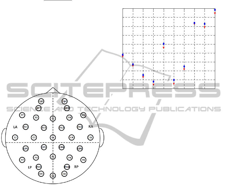
0.5 and 100 Hz. Fig. 2 shows positions of the
electrodes.
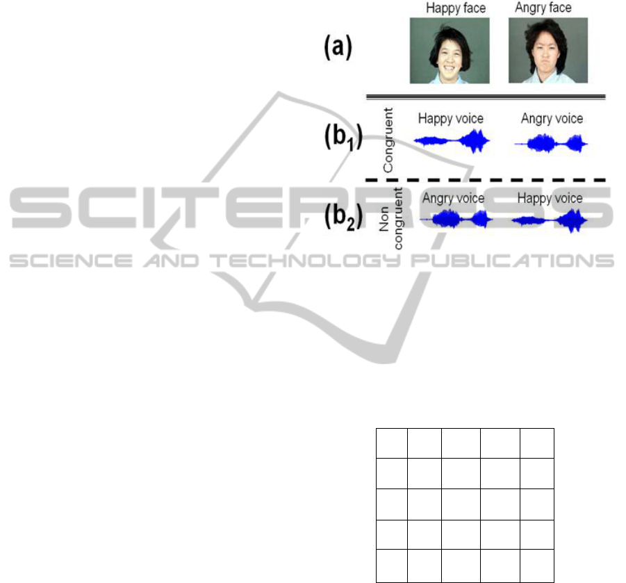
Figure 1: McGurk effect. Visual stimuli (a) are combined
with audio stimuli (b). Subjects will expect congruent
stimuli (b1), where visual and auditory clues are
concordant (e.g. happy face and happy voice). Non-
congruent stimuli (b2), where visual and auditory clues are
discordant (e.g. happy face and angry voice), will induce
distortions in either the visual or auditory perception (this
distortion is termed as a “McGurk effect”).
Table 1: p-values of the inter and intra regions in
discrimination of congruent and non-congruent conditions.
LA RA LP RP
LA 0.33 0.55 0.31 0.14
RA 0.55 0.02
*
0.04
*
0.19
LP 0.31 0.04
*
0.50 0.18
RP 0.14 0.19 0.18 0.10
*: p-value less than 0.05.
2.4 Data Analysis
Fig. 2 shows the topography used in this study,
consisting four regions: Left Anterior (LA), Right
Anterior (RA), Left Posterior (LP), and Right
Posterior (RP). The alpha frequency (8-12 Hz) EEGs
were extracted by applying Butterworth band-pass
filter. Then using the conventional cross-correlation
function, the functional connectivity between each
pair-channel was computed in the alpha band,
DifferencesofFunctionalConnectivityBrainNetworkinEmotionalJudgment
277
according to the following formula:
cov[ , ]
[,]| |
XY
XY
SXY
(1)
Where X and Y are signals of two channels, |x|
indicates absolute value of x,
],cov[ yx is the
covariance of x and y, and
x
is standard deviation
of x.
Then by averaging the correlation coefficients
over the channels within and between the regions,
the intra- and inter- connectivity of the four regions
(respectively) were computed in the alpha band. The
Mann-Whitney statistical test was used to compare
the differences of the obtained synchronization
values between the congruent and non-congruent
conditions.
Figure 2: Illustration of the brain topography used in this
study, which contains: Left Anterior (LA), Right Anterior
(RA), Left Posterior (LP), and Right Posterior (RP).
3 RESULTS
Fig. 3 shows the mean synchronization values of
inter- and intra- regions in congruent (red triangles)
and non-congruent (blue circles) cognitions. The x-
axis shows the pair regions in the studied
topography and the y-axis shows synchronization
values. It shows all synchronization values in the
non-congruent condition are more than those in the
congruent condition.
Table I presents the p-values obtained by the
Mann-Whitney test for distinguishing the inter- and
intra- regional synchronization values between the
two conditions. The significant p-values (less than
0.05) are related to the right anterior – right anterior
centimeter.
Figure 3: Mean values of inter- and intra- regional
synchronizations in congruent (red triangles) and non-
congruent (blue circles) emotional judgments. The x-axis
shows the pair regions in the studied topography and the
y-axis shows synchronization values.
4 CONCLUSIONS
In this study the differences of brain connectivity in
emotional judgment between congruent and non-
congruent emotional conditions was studied. To the
best knowledge of the authors the current paper
presented the first study on analysis of functional
connectivity in emotional judgment.
It was shown the alpha synchronization in the
overall brain in the non-congruent condition is
higher than that in the congruent condition.
Judgment in the non-congruent condition is more
difficult, compared with the congruent condition,
and therefore the higher alpha synchronization in the
non-congruent condition may be related to the
greater demanding and more effort of the brain for
judging emotions.
The obtained significant p-values between the
conditions in the right anterior – right anterior and
right anterior – left posterior connectivity shows the
ability of the alpha synchronization (in the
associated regions) for discrimination of congruent
and non-congruent conditions. Therefore the alpha
synchronization may have a good potential for
diagnosis of the disorders with deficient emotional
judgments and also it may be useful for their
treatment using EEG neurofeedback training.
LA - LA LA - RA LA - LP LA - RP RA - RA RA - LP RA - RP LP - LP LP - RP RP - RP
0.35
0.4
0.45
0.5
0.55
0.6
0.65
0.7
0.75
INTRA- and INETR- REGIONS
SYNCHRONIZATION VALUE
BIOSIGNALS2013-InternationalConferenceonBio-inspiredSystemsandSignalProcessing
278

ACKNOWLEDGEMENTS
Many thanks go to the International
Neuroinformatics Coordinating Facility (INCF) for
the travel grant provided to support this project. This
work has also been partially supported by the
University of Vic to Dr. Jordi Solé-Casals under the
grant R0904.
REFERENCES
Biehl, M., Matsumoto, D., Ekman, P., Hearn, V., Heider,
K., Kudoh, T., Veronica, T. Matsumoto and Ekman's
Japanese and Caucasian Facial Expressions of
Emotion (JACFEE): Reliability Data and Cross-
National Differences. Journal of Nonvernal Behavior,
21(1):3-21, 2008.
Duncan, S., Barrett, L.F. Affect is a form of cognition: A
neurobiological analysis. Cognition and Emotion,
21:1184-1211, 2007.
Grimm, S., Schmidt, C.F., Bermpohl, F., Heinzel, A.,
Dahlem, Y., Wyss, M. Segregated neural
representation of distinct emotion dimensions in the
prefrontal cortex—an fMRI study. NeuroImage,
30:325-340, 2006.
McGurk, H., MacDonald, J. Hearing lips and seeing
voices. Nature, 264(5588):746-748, 1976.
Tsuchiya, N., Adolphs, R. Emotion and consciousness,
Trends Cogn Sci, 11(4):158-167, 2007.
Uhlhaas, P.J., Pipa, G., Lima, B., Melloni, L.,
Neuenschwander, S., Nikolić, D., Singer, W. Neural
synchrony in cortical networks: history, concept and
current status. Front Integr Neurosci.3:17, 2009.
Watson, D., Clark, L.A., Tellegen, A. Development and
validation of brief measures of positive and negative
affect: The PANAS Scales. Journal of Personality and
Social Psychology, 47:1063-1070, 1988.
DifferencesofFunctionalConnectivityBrainNetworkinEmotionalJudgment
279
