
SELECTIVELY BONDED POLYMERIC CHECK VALVE FOR
THE RELIABLE REGULATION OF INTRAOCULAR PRESSURE
Seongmin Im, Byungphil Mun, Jaeyong An, Jongchan Choi
School of Mechatronics, Gwangju Institute of Science and Technology, Gwangju, Korea
Sung Yang
1,2,3
, Jonghyun Lee
1,2,3
1
School of Mechatronics,
2
Graduate Program of Medical System Engineering
3
Department of Nanobio Materials and Electronics, Gwangju Institute of Science and Technology, Gwangju, Korea
Keywords: Glaucoma drainage device, Polymer, Check Valve, Selectively bonding, Intraocular Pressure, FEA, MEMS.
Abstract: A glaucoma drainage device for the regulation of intraocular pressure is presented. The device consists of
three biocompatible polymer layers: the cover (top layer), the diaphragm (intermediate layer), and the
baseplate with a cannular (bottom layer). Finite element analyses (FEA) were performed to find optimal
design parameters for the targeted cracking pressure: the diameter of orifice, the thickness of intermediate
layer, the width of the channel, and the overlapped length of the orifice over valve seat. Top and bottom
layers were made by polydimethylsiloxane (PDMS) replica molding process, and an intermediate thin layer
with through-holes was fabricated by PDMS fill-in molding method and hydrophobic treatment. The
overlapped area of the bottom layer was coated with Cr and Au by using PDMS as a shadow mask. Metal
layers are not to be bonded between the bottom and the intermediate layers so that the device showed the
enhanced reliability in operation and the higher yield in production. Oxygen plasma treatment was
performed for irreversible bonds between separate three PDMS layers. The experimental cracking pressure
of the fabricated valve was 2.50 kPa, which is very close to the target value (2.67 kPa). The experiments
showed that the proposed polymer check valve can regulate the pressure of the aqueous humour, fluid in an
anterior chamber, within the normal intraocular pressure range (15~20mmHg) with a high repeatability.
1 INTRODUCTION
In microfluidic systems, valves are one of the most
important components to control the flow of the
systems. One of the most important characteristics
of microvalves is reliability, and high reliability
makes microfluidic systems successful. Check
valves are passive microvalves which allow
unidirectional flow. In micro check valve, reliability
can be defined as the consistency of cracking
pressure, flow rate, and diode-like characteristic.
Check valves can be used for flow regulation, on/off
switch, and sealing of liquid/gas/vacuum (Kwang W
Oh et al., 2006). Glaucoma drainage devices and
drug delivery are representative applications of
micro check valve for bionics. Currently, the reliable
fabrication and operation are the key issues in the
micro check valve (Ronalee Lo et al., 2009).
Though prior micro check valves have several
advantages: simple fabrication, polymer and cheap,
there is a stiction problem in the bonding process of
polymer layers (Nooli Jeon et al.,2002). To detach
the overlapped area between valve seat and valve
orifice, air or water should be introduced quickly
during bonding process before polymer layers are
irreversibly bonded. If the overlapped area is not
completely detached, cracking pressure and flow
rate of the fabricated device will vary seriously
during operation as shown in Fig. 1.
Glaucoma is the second leading cause of
blindness in the world, and it is estimated that 66.8
million people are suffering from glaucoma
(Quigley HA., 1996). Glaucoma is theoretically
defined as a progressive optic neuropathy as a result
of elevation of intraocular pressure (IOP) above the
physiological level of individuals (Shiose Y.,1990).
Glaucoma drainage devices (GDDs) have the
potential to regulate flow reliably and to maintain
IOP below 20mmHg.
234
Im S., An J., Choi J., Mun B., Yang S. and Lee J..
SELECTIVELY BONDED POLYMERIC CHECK VALVE FOR THE RELIABLE REGULATION OF INTRAOCULAR PRESSURE.
DOI: 10.5220/0003167302340237
In Proceedings of the International Conference on Biomedical Electronics and Devices (BIODEVICES-2011), pages 234-237
ISBN: 978-989-8425-37-9
Copyright
c
2011 SCITEPRESS (Science and Technology Publications, Lda.)

Figure 1: A PDMS diaphragm valve and stiction problem; (a) Schematic drawing of a PDMS check valve, (b) Illustration of
a normal valve under forward pressure, and (c) Illustration of a valve with stiction problem.
This paper describes the advancements of the
existing micro check valve through the selective
bonding of PDMS layers: the high consistency of
cracking pressure, flow rate, and diode-like
characteristic (Seongmin Im et al., 2010). Also, it
will be confirmed that the proposed micro check
valve has a great potential as a component of a
glaucoma drainage device.
2 DESIGN AND OPERATION
2.1 Design
Fig. 2 shows the micro check valve with two layers,
and each layer is made of PDMS. There are four
design parameters for the targeted cracking pressure:
the diameter of hole, the thickness of membrane, the
width of the channel, and the overlapped length of
the hole over valve seat. The thickness of membrane
is the most critical factor for the cracking pressure.
The top layer is thin membrane which has a hole
(valve orifice) in it. The thickness of the membrane
is 122um, and the diameter of the hole is 350um.
The bottom layer is micro channel, and defines the
fluidic channel and valve seat. The dimensions of
the channel are 100um width and 100um height, and
the diameter of the cylindrical post is 450um. Cr and
Au are deposited on the valve seat with 20nm
respectively. These thin Cr/Au layers are introduced
to prevent the stiction problem between the valve
orifice and the valve seat during bonding process.
A glaucoma drainage device integrates the cover
(top layer), the diaphragm (intermediate layer), and
baseplate with a cannular (bottom layer) as shown in
Fig. 3. The base plate is a square, 5mm each side.
The cannula dimensions are 15mm length, 1mm
width, and 900um height, and internal microchannel
is 100um width and 100um height. The square-
shaped valve cover is equipped with the cylindrical
post array (450um in diameter and the 80um in
height). The valve cover is bonded on the
Figure 2: Illustration of a PDMS diaphragm valve with
Cr/Au layer on the valve seat for selective bonding of
PDMS layers.
intermediate layer after flipping down to prevent
conjunctiva from directly contacting with valve
orifice so that the stable valve operation of the micro
check valve will be secured in terms of cracking
pressure and flow rate after the implantation.
Figure 3: Illustration of a glaucoma drainage device. The
device consists of the cover (top layer), the diaphragm
(intermediate layer), and the baseplate with a cannular
(bottom layer). The cover is bonded on the diaphragm
after flipping down.
2.2 Operation Principle
The proposed micro check valve is normally-closed
allowing one directional flow. When the internal
pressure is higher than external pressure, the valve
opens, and the valve remains closed when the
external pressure is higher than internal pressure.
After the implantation of a GDD, the end of the
SELECTIVELY BONDED POLYMERIC CHECK VALVE FOR THE RELIABLE REGULATION OF INTRAOCULAR
PRESSURE
235
cannula is located in the anterior chamber of the eye
ball. When the IOP is higher than the cracking
pressure of a GDD, the valve drains the surplus
aqueous humour to regulate the pressure of the
anterior chamber.
3 FABRICATION
The fabrication of the bottom layer started with spin
coating of SU-8 on the silicon wafer. The SU-8 in
100um height on the silicon wafer was patterned,
and it served as a mold. PDMS was poured on the
mold, and was cured at 90
℃ for 1h. The PDMS
replica was separated from the SU-8 mold, and the
shadow mask was aligned on the PDMS replica. Cr
and Au are deposited on the valve seat for the
selective bonding between the cover and the
diaphragm.
The fabrication of the diaphragm (intermediate
layer) with a through-hole started with making a
PDMS replica. The PDMS replica has a post 350um
in diameter and 122um in height. The hydrophobic
treatment using CH4 and He was performed on the
surface of the PDMS replica. The PDMS replica was
put on a silicon wafer, and uncured PDMS was filled
in the space between the PDMS replica and silicon
wafer. The uncured PDMS layer was cured at 90℃
for 1h, and the diaphragm was separated from the
molds (Jongchan Choi et al., 2010). The cover of the
device was simply made using soft-lithography.
Oxygen plasma treatment enables the surface
modification of PDMS layers and irreversible bonds
each layer. An aligner was used to assemble three
layers, and the valve orifice and the valve seat were
selectively bonded.
Figure 4: Fabrication sequence; A CH
4
/He plasma surface
treatment of a PDMS mold was performed to make a
diaphragm with a through-hole. Soft-lithography process
was used to fabricate the bottom layer.
Figure 5: Micrographs of the check valve (Top view); (a)
the bottom layer after sputtering of Cr and Au, (b) a
diaphragm with a through-hole, and (c) a micro check
valve after bonding the diaphragm and the bottom layer.
4 EXPERIMENT AND RESULTS
The performance of the fabricated check valve was
tested by using a hydrostatic pressure and analytical
balance. After connecting the micro check valve
with a pressurized water source, the valve was
placed on the analytical balance. In order to
experimentally find the cracking pressure of the
valve, the flow rates of three devices were recorded
with respect to the input pressure. The data were
attained for 3 min at high pressure (≥ 3000 Pa) and
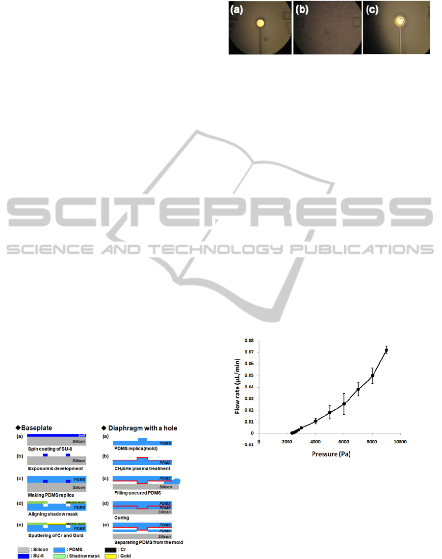
for 10 min at low pressure (< 3000 Pa). From the Fig
6, the mean value and standard deviation of the
cracking pressure was found to be 2444 ± 87.76 Pa
and, which is very close to the target value (2666 Pa)
for the glaucoma drainage device. When reverse
pressures over 10 kPa was applied, the leakage was
not occurred.
Figure 6: Outlet flow rate versus input pressure. The
cracking pressure is 2444 ± 87.76 Pa (n=3). The steady
state outlet flow rates were 0.025 ± 0.009 μL/min (n=3, at
6000 Pa). When the reverse pressure over 10 kPa was
applied, the leakage was not observed.
Fig. 7 shows the experimental result of the
repeatability test. The flow rates in the steady state
were nearly 0 μL/min at 2350 Pa (slightly lower than
cracking pressure) and 0.0081 μL/min ± 2% at 4000
Pa (sufficiently higher than desirable intraocular
pressure). The negligible variance in the
repeatability test indicates that the reliability of the
valve is considerably enhanced, and the selectively
BIODEVICES 2011 - International Conference on Biomedical Electronics and Devices
236

bonded diaphragm valve has great potential as a
component of a glaucoma drainage device.
For the future work, the fabricated glaucoma
drainage device will be implanted in a rabbit’s eye to
find the in-vivo cracking pressure and to monitor the
postoperative fibrosis. Furthermore, the
biocompatibility of the device will be examined
through in-vivo experiments. Even though the body
of the device is made of an approved biocompatible
material, Cr/Au layers of the device should be
checked of biocompatibility.
Figure 7: Repeatability of the outlet flow rate. The steady
state flow rates were 0 μL/min (n=7, at 2350 Pa) and
0.0081 μL/min±2% (n=7, at 4000 Pa).
ACKNOWLEDGEMENTS
This work was partially supported by the World
Class University (WCU) program at GIST through a
grant provided by the Ministry of Education,
Science and Technology (MEST) of Korea (Project
No. R31-2008-000-10026-0)
REFERENCES
Kwang W. Oh, Chong H. Ahn, 2006, “A Review of
Microvalves”, J. Micromech. Microeng, Vol. 16, pp.
13-39
Ronalee Lo, Po-Ying Li, Saloomeh Saati, Rajat N.
Agrawal, Mark S. Humayun, Ellis Meng, 2009, “A
Passive MEMS Drug Delivery Pump for Treatment of
Ocular Diseases”, Biomedical Microdevices, DOI
10.1007/s10544-009-9313-9
Noo Li Jeon, Daniel T. Chiu, Christopher J. Wargo,
Hongkai Wu, Insung S. Choi, Janelle R. Anderson,
George M. Whitesides, 2002 "Design and Fabrication
of Integrated Passive Valves and Pumps for Flexible
Polymer 3-Dimensional Microfluidic Systems",
Biomedical Microdevices, 4:2,117-121
Quigley H. A., 1996, Number of people with glaucoma
worldwide., Br J Ophthalmol; 80:389
–
393
Shiose Y., 1990, Intraocular pressure: new perspectives.,
Surv Ophthalmol;34:413
–
435
Seongmin Im, Byungphil Mun, Jaeyong An, Jongchan
Choi, Sung Yang, and Jong-Hyun Lee, 2010,
“Selectively bonded polymeric Check Valve for the
Regulation of Intraocular Pressure”, KSME
Conference on Dynamics and Control, Gwangju,
pp.286
Jong Chan Choi, Sang Youl Yoon, Sung Yang, 2010,
“Simple PDMS double molding method for PDMS
membrane with micro through-hole via CH4/He
plasma surface treatment”, The12th Korea MEMS
Conference, Busan, pp. 373
SELECTIVELY BONDED POLYMERIC CHECK VALVE FOR THE RELIABLE REGULATION OF INTRAOCULAR
PRESSURE
237
