
COGNITIVE REASONING IN INTELLIGENT MEDICAL
INFORMATION SYSTEMS
Visual Data Perception Algorithms in Medical Decision Support Systems
Marek R. Ogiela
1
, Ryszard Tadeusiewicz
1
, Lidia Ogiela
2
AGH University of Science and Technology
1
Institute of Automatics,
2
Department of Company Management
Al. Mickiewicza 30, PL-30-059 Kraków, Poland
Keywords: Intelligent systems, image understanding, pattern classification, medical imaging, artificial intelligence
Abstract: This paper presents new approach in application of picture languages for cognitive analysis and reasoning of
selected medical visualization. It will be shown new opportunities for applying these methods to undertake
tasks of the automatic understanding of image semantics in intelligent medical information or computer-
aided diagnosis systems. These systems are applied in various tasks supporting decisions taken in the wide
area of health care and medical imaging. The possibility of obtaining the information about semantic
content of the medical images may contribute considerably to the creation of new intelligent cognitive
medical systems. This article shows that structural techniques of artificial intelligence may be applied in the
case of tasks related to automatic classification and machine perception of semantic pattern content in order
to determine the medical meaning of the images. In the paper, we describe some examples presenting ways
of applying such techniques in the creation of cognitive vision systems for selected classes of medical
images.
1 INTRODUCTION
In the foundations of image understanding there are
many algorithms and AI approaches to the task of
intelligent visual data perception and analysis.
Among them one is the most important enabling to
make a deeper semantic and cognitive analysis.
These are picture languages consisting formal
grammars for pictorial pattern analysis as well as
languages of shape features description allowing
multidimensional pattern classification. In this paper
will be presented the way of application of such
formalisms to the task of understanding of medical
visual data, especially in the intelligent medical
information systems. We try to show how the tasks
of automatic understanding of medical data may be
done using cognitive analysis approach, which allow
to make a semantic perception of analyzed
visualization. Generally, the perception of an image
requires a deeper analysis aimed at the determination
of significant semantic features (Albus, 2001). Such
features
enable a further semantic image
interpretation or a semantically oriented indexation
in databases (especially when objects are retrieved
from various diagnostic examinations or determine
different disease entities). A proper semantic
interpretation of the data being analyzed is very
important because in the case of medical images
often happens that the same illnesses are visualized
in various forms of images that are registered and
processed (Fig.1). This is the main reason why
attempts have been made to create a system that
would automatically find the message (i.e. the
content) carried by analyzed medical images.
Due to the fact that the number of combinations
of features that characterize images is not limited, it
can be assumed that perception may refer
the image
to potentially unlimited number of classes. This may
be achieved by cognitive analysis, in which
specified languages of image description must be
used.
The general approach in the cognitive analysis is
the initial interpretation of images and specification
of important features. The proper selection of such
features is conducted by means of image pre-
processing. Next features are subsequently described
with the use of a picture language generated by an
appropriately defined attributed grammar. Properties
described in this way can be later reproduced in the
222
R. Ogiela M., Tadeusiewicz R. and Ogiela L. (2004).
COGNITIVE REASONING IN INTELLIGENT MEDICAL INFORMATION SYSTEMS - Visual Data Perception Algorithms in Medical Decision Support
Systems.
In Proceedings of the Sixth International Conference on Enterprise Information Systems, pages 222-225
DOI: 10.5220/0002603402220225
Copyright
c
SciTePress
course of structural reasoning conducted by the
prepared information system.
The main advantage of this approach is its
possibility to interpret the meaning of a much bigger
class of images than the ones, which were used for
the writing of the formal language. This results from
the fact that the used grammar rules generalise the
descriptions introduced and allow one to interpret
new cases, previously not defined.
For such interpretation of the mentioned
structures and for a verification of lesion
advancement level, a graph grammar (Ogiela, 2003),
and an attributed grammar have been proposed.
Before coming to the cognitive interpretation of
the changes, it is necessary to preserve the sequence
of preliminary operations, which are included in the
image pre-processing. The goal of this analysis is to
obtain new representation in the form of width
graphs, which show the pathological changes
occurring in these structures.
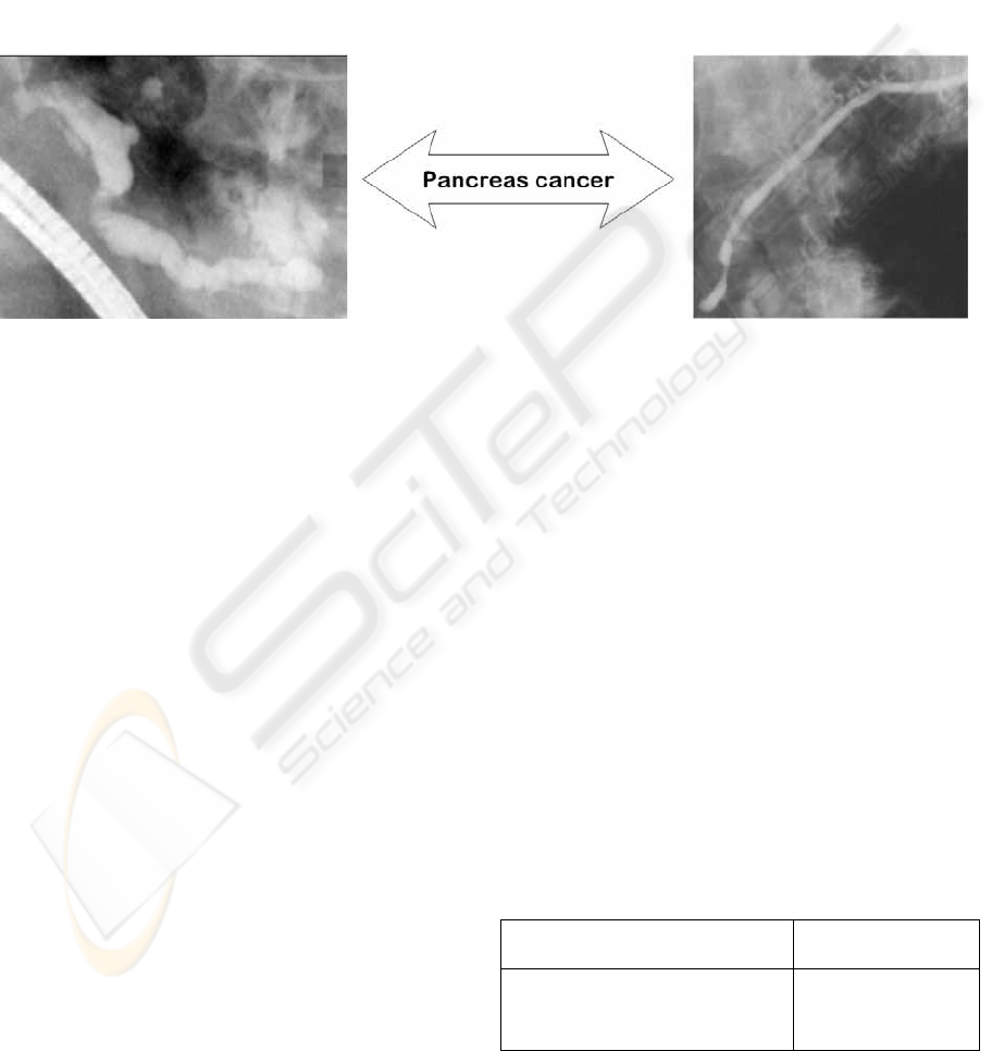
Figure 1: Two images carrying the same message. In both cases, the symptoms of pancreas cancer are visible
During the initial analysis of visualisations, the
following operations are executed: segmentation,
skeletonization, and the application of a
straightening transformation to transform the
contour of the analysed structure into two-
dimensional graph, which shows a profile of a
straightened organ. The graphs obtained in such way
are the starting point in the classification of
morphological features by using context-free
grammars. In order to define primary components on
the obtained width graphs as terminal symbols
describing these components, an algorithm of linear
approximation was used. As a result of
approximation the sequences of terminal symbols
for every graph was received, which constitute an
input to syntax analysers and semantic classifiers.
2 PICTURE GRAMMAR FOR
COGNITIVE ANALYSIS
A possibility to conduct cognitive analysis will be
presented on the examples of patterns received
during the diagnostic examinations of renal pelvis,
and coronary arteries.
2.1 Coronary Image Interpretation
Analysis of coronary arteries is extremely important
from the point of view of correct diagnosis of
myocardial ischaemia states caused by coronary
atheromatosis sclerosis lesions resulting in stenoses
of artery lumen, which in consequence lead to
myocardial ischaemia disease. This disease can take
the form of either stable or unstable angina pectoris
or myocardial infraction (Khan, 1996).
The following attributed grammar has been
proposed to diagnose various types of stenosis
shapes:
V
N
= {SYMPTOM, U, H, D}
V
T
= {h, u, d} for h∈[-10°, 10°], u∈(10°, 90°),
d∈(-10°, -90°)
STS = STENOSIS
SP:
STENOSIS → D H U
STENOSIS → D U | D H
Lesion = Stenosis
H → H h | h
D → D d | d
U → U u | u
w
sym
= w
sym
+ w
h
h
sym
= h
sym
+ h
h
...
COGNITIVE REASONING IN INTELLIGENT MEDICAL INFORMATION SYSTEMS - Visual Data Perception
Algorithms in Medical Decision Support Systems
223

This grammar allows to detect different forms of
coronary artery stenosis, which may characterize the
different disease units (angina pectoris or infarct).
Using attributes permits to calculate the numerical
parameters and semantic information of detected
lesions, which allows to characterize the degree of
lesion development.
The simplicity of this grammar results mainly
from the big generation capacity of context-free
grammars, understood mainly as possibilities to
describe complex shapes by means of a small
number of introductory rules, that is grammar
productions.
2.2 Renal Pelvis Cognitive Analysis
In the case of analysis of renal radiograms, the main
task is to recognise local stenoses or dilations of
upper segments of urinary tracts and attempt to
define the correct morphology of renal pelvis and
renal calyxes. Lesions in those structures can
suggest the occurrence of renal calculi or deposits,
which causing ureter artresia can lead to diseases
such as acute extrarenal uraemia or hydronephrosis.
An analysis of the correct morphology of ureter
lumen will be conducted with the use of context-free
attributed grammar.
Diagnosing morphological lesions in the form of
ureter stenosis or dilations has been conducted with
the use of the following attributed grammar:
V
N
= {LESION, STENOSIS, DILATATION, HOR,
SLOPE_UP, SLOPE_DOWN}
V
T
= {h, v, nv} for h∈[-8°, 8°], su∈(8°, 180°),
sd∈(-8°, -180°)
STS = LESION
SP:
LESION → STENOSIS
STENOSIS → SLOPE_DOWN HOR
SLOPE_UP
STENOSIS → SLOPE_DOWN
SLOPE_UP
STENOSIS → SLOPE_DOWN HOR
Lesion = Stenosis
LESION → DILATATION
DILATATION → SLOPE_UP HOR
SLOPE_DOWN
DILATATION → SLOPE_UP
SLOPE_DOWN
DILATATION → SLOPE_UP HOR
Lesion =
Dilatation
HOR → HOR h | h
SLOPE_DOWN → SLOPE_DOWN
sd | sd
SLOPE_UP → SLOPE_UP su | su
w
sym
= w
sym
+ w
h
;
h
sym
= h
sym
+ h
h
...
3 SELECTED RESULTS
As a result of cognitive analysis using linguistic
approach it is possible to understand pathogenesis of
the deformations viewed on x-ray images of the
organs under consideration, what means the
possibility of recognize some kind of diseases even
on images absolutely not similar one to other.
Presented approach is applicable even if no
templates of healthy and pathological organs at all or
if number of recognized classes goes to infinity. In
particularly applications of the presented grammars
deliver almost complete information concerning the
visual morphological irregularities of investigated
organs. An analysis of the morphological changes
was carried out based on a set containing few dozens
of images. The efficiency of gaining recognition of
information with semantic character, in all cases
exceeded the threshold of 93%. In Fig. 2 are
presented examples, which show the description of
the changes in ureter ducts, and coronary arteries.
The results obtained owing to the application of
the characterized methods, confirm the immense
opportunities offered by syntactic methods in the
cognitive analysis of medical visualizations showing
dangerous pathological lesions.
4 CONCLUSION
Development of the intelligent information systems
and techniques of visual data semantics analysis
made possible to understand the medical meaning of
any images coming from diagnostic research.
However, the full automatic analysis and
interpretation of such data is still a real problem,
advanced techniques of artificial intelligence must
be applied to enable the creation of systems that can
both recognize and understand visual data (Ogiela
2003).
Thus the aim of the presented techniques was to
show an innovative concept of the application of
structural pattern analysis in the creation of
cognitive information systems.
ICEIS 2004 - ARTIFICIAL INTELLIGENCE AND DECISION SUPPORT SYSTEMS
224
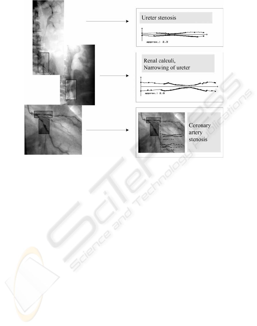
Figure 2: Results of disease symptom interpretation in intelligent diagnosis support system
Such systems are able to understanding and
determining the semantic meaning of medical
images of certain classes. It is worth mentioning that
machine perception using such methods may lead to
an automatic interpretation of medical images in the
way it is done by a specialist. It may enable the
determination of not only crucial changes but also
the consequences of existing irregularities and
finally the optimal directions and methods of
conducting a suitable therapy. Automatic
understanding of the image content can have
numerous further applications for example such
information can be used to monitor therapeutic
processes or to forecast disease development as well
as the patient’s future state of health.
ACKNOWLEDGEMENT
This work was supported by the AGH University of
Science and Technology under Grant No.
10.10.120.39.
REFERENCES
Albus, J.S., Meystel, A.M., 2001. Engineering of Mind:
An Introduction to the Science of Intelligent Systems.
John Wiley & Sons.
Khan, M.G., 1996. Heart Disease Diagnosis and Therapy.
Williams & Wilkins, Baltimore.
Ogiela, M.R., Tadeusiewicz, R., New approach for
cognitive analysis and understanding of medical
patterns and visualizations. Proceedings of SPIE, vol.
5203 Andrew G. Tescher (eds.), SPIE, Bellingham
WA, 2003, pp.615-622.
Ogiela, M.R., Tadeusiewicz, R., Cognitive Vision Systems
in Medical Applications. Lecture Notes in Artificial
Intelligence, 2871 (2003) 116-123.
Ogiela, M.R., Tadeusiewicz, R., Artificial Intelligence
structural imaging techniques in visual pattern analysis
and medical data understanding. Pattern Recognition,
36 (2003) 2441-2452.
Sonka, M., Fitzpatrick, J.M. (eds.), 2000. Handbook of
Medical Imaging: Vol. 2- Medical image processing
and analysis. SPIE PRESS, Bellingham WA.
COGNITIVE REASONING IN INTELLIGENT MEDICAL INFORMATION SYSTEMS - Visual Data Perception
Algorithms in Medical Decision Support Systems
225
