
Fourier Ptychography Microscopy Resolution Improvement
Employing Refocusing
Arban Uka
a
, Gent Imeraj
b
, Bjorna Qesaraku and Besmir Shehu
Department of Computer Engineering, Epoka University, Tirane, Albania
Keywords: Microscopy, Fourier Ptychography, Refocusing, Microfluidic Chambers.
Abstract: Fourier ptychography is a computational imaging technique that is used to overcome the physical limitations
in determining the spatial resolution of an optical system by combining a large number of low resolution
images. The low resolution images are acquired using programmed illumination from an array of light sources,
thus enabling the scanning of the k-space, which is the reciprocal space of the real domain. The use of this
approach would improve the resolution when biological samples from patients are analyzed in
multiparametric chambers. When an off-center light source is used at an oblique incidence angle, the optical
path length changes thus defocusing the expected image. The common depth of focus of microscopes is a few
micrometers and when a chamber of thickness from 0.5-1.0 cm is used, an adjustment of the focusing is
needed. Here in this work, we report Fourier ptychography using LED illumination and an improved image
quality is acquired when refocusing is implemented.
1 INTRODUCTION
Acquisition of high resolution images is essential in
medical imaging and this is facilitated by an optimal
combination of high-end optical systems and
computational imaging. Costly machineries often
have bench-top design that are sturdy and need the
associating components to adapt to their design. The
development of the experimental instruments has
enabled the measurement of several vital parameters
of patients in the same setup. In this case, in order to
use a small amount of diagnostic specimen (saliva,
blood etc) microfluidic platforms are used
(Chmayssem et al., 2021). To monitor the health state
of cells under different stress conditions microscopy
is employed. To increase the resolution of microscope
intuitive modifications have been applied in the
illumination such as replacing the light source by a
LED array (Zheng et al., 2011) and then later this
LED array enabled illumination is used to scan the
reciprocal space (k-space) of the light propagation.
This technique has led to the development of Fourier
ptychography that enables the increase of the
resolution of the acquired images in both phase and
amplitude (Zheng et al., 2013). This technique can
a
https://orcid.org/0000-0003-0037-0207
b
https://orcid.org/0000-0002-7877-3906
overcome the physical limitations of the microscope
as it can increase the depth of focus, thus enabling a
broader range of focused sample. Tian et al. (2014)
implemented multiplexed illumination to reduce the
number of images to be acquired thus optimizing the
runtime of the experiment and the time needed to run
the reconstruction algorithms when acquiring the
final image. This latter contribution would prove
valuable in case when one is using microfluidic
platforms and monitoring is conducted by
microscopes. If a biological sample is undergoing
some change because of some induced stress, then
one would have to acquire all the images and then
analyse them in a short time. The work reported so far
in the literature has been the proof of principle and is
applied on bare samples that are attached on
microscope glass slides. Even when applied on
simple glass slides in a multiplexed mode, some of
the images could diverge out of focus as the depth of
focus is limited. This happens as the optical path
length in air surrounding the sample (on the side of
the incoming light illumination and on the side of the
outgoing modified light past the sample) is different
when different LEDs are turned on. In this case the
optical path length is comparable to the geometric
Uka, A., Imeraj, G., Qesaraku, B. and Shehu, B.
Fourier Ptychography Microscopy Resolution Improvement Employing Refocusing.
DOI: 10.5220/0010915500003123
In Proceedings of the 15th International Joint Conference on Biomedical Engineering Systems and Technologies (BIOSTEC 2022) - Volume 1: BIODEVICES, pages 191-195
ISBN: 978-989-758-552-4; ISSN: 2184-4305
Copyright
c
2022 by SCITEPRESS – Science and Technology Publications, Lda. All rights reserved
191

path length. When microfluidic chambers are used,
their larger than one index of refraction (comparing
to air) leads to an increase of the difference between
the optical path length and the geometric path length.
Considering that a single LED source results in a
normal incidence to the sample, all the other
secondary LED illumination sources have an oblique
incident angle and this brings to the attention the need
to refocus the sample. At the same time one may
consider that the microfluidic chambers do not have a
perfect width across the volume as few micrometers
of difference in thickness may be inevitable. This
constitutes another source of the undesired
defocusing. At the same time, complex fluids that are
used in the microfluidic channels may exhibit varying
index of refraction as a result of mixing with different
densities of biological samples and this will then
change the optical path length. All these arguments
reminds one on the need of careful refocusing before
each image acquisition. Here in this work we compare
the performance of Fourier ptychography
reconstruction as the focus control is implemented
each time we acquire an image. We observe that a
refocusing improves the quality of the reconstructed
image.
2 THEORY AND
EXPERIMENTAL DESIGN
The main limitation in medical imaging using optical
microscopy is the spatial resolution. Under the visible
light spectrum, it is not possible to observe images of
objects smaller than half of the wavelength of the
incident light source (0.4 to 0.7 μm). Furthermore, in
live cell examination, unsuccessful results may be
obtained because often contrast is low. Staining with
selective dyes, which are used to increase the
contrast, may modify the sample and introduce other
structural features that are not present in the
specimen. With all the associating challenges, there is
a continuous increase in the number of patients being
examined using microscopic techniques rather than
other diagnosis methods, as they are consistently
accurate and of low cost with the latter being essential
in resource-limited settings. An increase in the
demand for digital microscopes in the current
coronavirus pandemic has been observed. Some of
the greatest innovators that operate in the market of
digital portable microscopes include Carl Zeiss AG,
Olympus Corporation, Keyence Corporation, Nikon
Corporation, Leica Microsystems etc. These
benchtop commercial microscopes can be used to
implement Fourier ptychography.
2.1 Theory and Mathematical
Apparatus
Fourier ptychography is an excellent example of how
one can apply algorithms in both the real (x-space)
and the reciprocal domains (k-space) to improve the
spatial resolution (micrometer scale). The
information in the reciprocal domain (with the unit of
𝑚
) is indirectly collected as the k vector - denoting
the direction of the light illumination to the camera
sensor – can be selected for each image by turning on
LED one by one. It uses objective lens with small
numerical aperture and is able to increase the field of
view. This method requires no manual scanning of the
specimen plane, since a LED array is used as an
illumination source. So instead of using a narrow
beam of light to illuminate the sample, different angle
illuminations are provided by programming the LED
array to turn on individual LEDs without having to
move any part of the physical system.
2.2 Experimental Design
The major construct is the combination of the
programmable LED array, sample, light gathering
system, and the CMOS camera. As mentioned earlier
LED array is placed sufficiently below the sample so
that the illumination is considered to be spatially
coherent. In order to achieve this, the sample must be
lifted at a fixed distance of few centimeters above the
fixed LED matrix. This is done by means of metric
adapter plates fixed on the metric breadboard using a
90° metric adapter that glues them together. 32x32
LED array will be used with a spacing of 4 mm
between neighboring LEDs. This spacing is
convenient to cover the sample area and to clearly
define the changes and shifts in each data
measurement. The structure of the experimental setup
is shown in Figure 1. A magnifying lens (model
number 378-805-3) with a relatively small numerical
aperture is used with. It is a 50x magnifying lens with
infinity correction.
This means that the image is directly passed to the
camera sensor without being diffracted and modified
inside the path from objective to the sensor of the
camera. In order to perform the bright field refocusing
that will correct for geometrical shifts and phase
aberrations that degrade the resolution of the final
image, the focus will be adjusted manually as
BIODEVICES 2022 - 15th International Conference on Biomedical Electronics and Devices
192
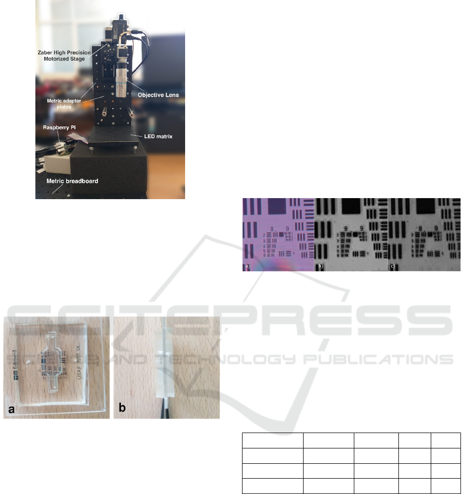
Figure 1: Experimental setup used in this work
proposed by using a step-controlled motor with high
precision. The camera together with the lens is
mounted to the Zaber motor (Zaber Technologies Inc.
with a resolution of 0.0476 m) that is responsible for
controlling the up and down movements of the
sample each time the illumination angle hence the
change of focus occurs.
Figure 2: Microfluidic chamber material with width of 5
mm. a) Front view of the chamber enclosing USAF 1951 b)
side view of the microfluidic chamber.
Its own software controls this motor while the LEDs
are turned on sequentially by using a raspberry pi and
a python script that identifies how the LEDs are
turned on and the time slots for each of them being
turned on. To acquire the images a CMOS sensor
camera is used. It imposes a lot of advantages
compared to CCD cameras where the most important
one was the faster data acquisition rate. These
cameras have lower cost compared to other cameras
that may be used in modern digital microscopes. The
USAF 1951 that facilitates a quantitative analysis is
enclosed inside a double-sided microfluidic chamber
as shown in Figure 2. The use of the chamber
increased the working distance by 1 mm.
3 RESULTS
The output of the algorithm is a reconstructed image
with higher resolution, one image per each dataset
composed of 25 images. The details become more
significant at the end of the preset number of
iterations. The image with and without focus for each
dataset are compared with each other by means of
algorithms that calculate the contrast and the
sharpness of each output. Beginning with the first and
second dataset ‘5x5 LEDs with USAF 1951 with and
without refocusing’, the synthesized numerical
aperture of Fourier Ptychography is 0.9363 and the
reconstructed image is displayed below in Figure 3
together with the central raw image with maximum
supposed intensity.
Figure 3: a) Original image captured with central LED, b)
Reconstructed image without applying focus adjustment c)
Reconstructed image with focus adjustment.
Computational power is used to determine the
image quality of both reconstructed images with the
refocus method and without. The measured
parameters are Brightness, Contrast, Sharpness, PIQE
(Perception based Image Quality Evaluator) and
NIQE (Natural Image Quality Evaluator). The
respective values are displayed in Table 1:
Table 1: Image quality measurement parameters.
Brightness Sharpness PIQE NIQE
Original Image 127.90 3.39 6.18 3.71
Rec. Img NR 102.56 8.04 26.83 6.19
Rec. Img WR 86.60 7.45 26.15 5.95
The other datasets produced similar results, but
since they were turned into grayscale during the
reconstruction process, it was more difficult to
observe the differences caused by effect of refocusing
the image. Another important aspect of FPM is the
effect of thickness, or the consideration of the third
dimension of the sample. Since Fourier algorithm
takes into account the slightest detail of the image, the
angular illumination will cause some aberrations to
the obtained data. The effect of such thickness was
discussed in Chapter 4, and the experiment results are
also analyzed. Theoretically the glass specimen
Fourier Ptychography Microscopy Resolution Improvement Employing Refocusing
193
imposed an error to the data acquisition process of
nearly 1 percent, which can or cannot be negligible.
This depends in the application of FPM which is
about slightest details in minute length scale. In case
of micro fluidic chamber of thickness nearly 4 mm
per each side, the error was very significant when
compared to the same sample without the chambers.
The thickness effect completely distorted the signal
of the image, which was reconstructed with a not very
pleasant resolution. So, this must also be taken into
consideration when using different types of samples
with varying thickness.
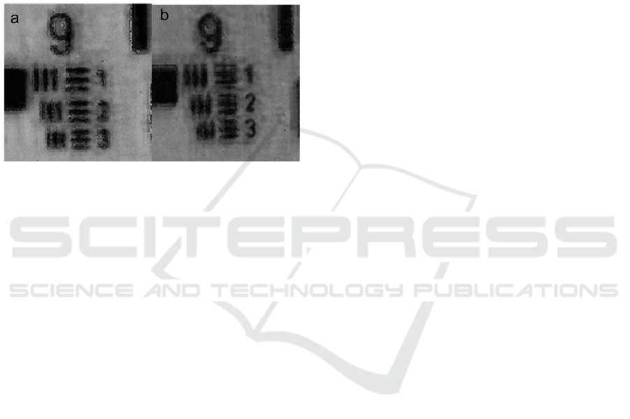
Figure 4: Reconstructed image a) without refocus, b) with
refocus.
4 CONCLUSIONS
In this work we proposed a different approach to the
increment of reconstructed image resolution, by
adjusting the focus manually using a geometrical
approach to solve the problem. The relevant concepts
to the theoretical approach of the proposed solution
were also explained. Theoretically the work proves to
be successful, relying also in the computational
power to resolve the adjustment of image resolution.
When estimating the performance of an experiment
that is conducted for the first time, there may be a lot
of space for improvement. However, assuming the
aberrations arising from moderate conditions in
which the experiment was realized, the results proved
the point even though not much significant
improvement was made in the resulting images. In the
reconstruction process, much less raw images are
used, preventing the computer from drowning in
unnecessary data of low to none important
information of the phase and amplitude (including
darkfield images as well). Reducing the number of
images to 25 was not based on any logical fact rather
than experience in image reconstruction. The
illuminating source was enough to produce
electromagnetic waves with oblique incidence,
however in practice illumination is not coherent,
which leads to images having reduced spatial
coherence. The NA was increased from the synthetic
NA produced in the algorithm of FP and the image
resolution was increased up to 4 times. This could be
further improved by increasing the number of
captured images which comes with expense in the
computational run time and large number of iterations
required.
The effect of refocusing became much clear
during the data analysis process, where the refocused
raw data managed to produce an image of a higher
quality and resolution. The manual adjustment was
made step by step in order to carefully observe the
change in the shape and 3D effect of each captured
image. Even though it took a few minutes of
adjustment for 25 images, the pattern of motion can
be translated into an algorithm and run by the Zaber
Console Software in order to perform the active
refocusing in an automated way. Another important
aspect that was discussed in the fourth chapter was
the thickness of the sample. According to the
theoretical analysis, it imposed no notable error for
the glass sample. However, this was not the case for
the practical aspect, where the resolution was
decreased significantly.
The purpose of this study was to introduce a
different path towards the automatization of the
microscope with the help of Artificial Intelligence.
The corrections that are made physically before the
data acquisition reduce the computational time by
performing just as well, not even better. Active
focusing is in its early stages and still requires the
expertise of biologists and scientists that can train the
AI in the appropriate way.
ACKNOWLEDGEMENTS
This project has received funding from the European
Union’s Horizon 2020 research and innovation
program under grant agreement No 760921
(PANBioRA).
REFERENCES
Chmayssem, A., Verplanck, N., Tanase, C. E., Costa, G.,
Monsalve-Grijalba, K., Amigues, S., ... & Mailley, P.
(2021). Development of a multiparametric (bio) sensing
platform for continuous monitoring of stress
metabolites. Talanta, 229, 122275.
Zheng, G., Kolner, C., & Yang, C. (2011). Microscopy
refocusing and dark-field imaging by using a simple
LED array. Optics letters, 36(20), 3987-3989.
BIODEVICES 2022 - 15th International Conference on Biomedical Electronics and Devices
194

Zheng, G., Horstmeyer, R., & Yang, C. (2013). Wide-field,
high-resolution Fourier ptychographic microscopy.
Nature photonics, 7(9), 739-745.
Tian, L., Li, X., Ramchandran, K., & Waller, L. (2014).
Multiplexed coded illumination for Fourier Ptycho-
graphy with an LED array microscope. Biomedical
optics express, 5(7), 2376-2389.
Kellman, M., Bostan, E., Chen, M., & Waller, L. (2019,
May). Data-driven design for fourier ptychographic
microscopy. In 2019 IEEE International Conference on
Computational Photography (ICCP) (pp. 1-8). IEEE.
Konda, P. C., Taylor, J. M., & Harvey, A. R. (2015,
September). High-resolution microscopy with low-
resolution objectives: correcting phase aberrations in
Fourier ptychography. In Optical Systems Design 2015:
Computational Optics (Vol. 9630, p. 96300X).
International Society for Optics and Photonics.
Lee, D., Ryu, S., Kim, U., Jung, D., & Joo, C. (2015).
Color-coded LED microscopy for multi-contrast and
quantitative phase-gradient imaging. Biomedical optics
express, 6(12), 4912-4922.
Zuo, C., Sun, J., & Chen, Q. (2016). Adaptive step-size
strategy for noise-robust Fourier ptychographic
microscopy. Optics express, 24(18), 20724-20744.
Zheng, G., Horstmeyer, R., & Yang, C. (2013). Wide-field,
high-resolution Fourier ptychographic microscopy.
Nature photonics, 7(9), 739-745.
Tian, L., & Waller, L. (2015). 3D intensity and phase
imaging from light field measurements in an LED array
microscope. optica, 2(2), 104-111.
Huang, W., Pan, S., Zhou, Q., Liao, M., Zhang, C., Tang,
Q… & Peng, X. (2020, October). Positional
misalignment correction for Fourier ptychographic
microscopy based on intensity distribution. In
Advanced Optical Imaging Technologies III (Vol.
11549, p. 115490D). International Society for Optics
and Photonics.
Bian, L., Suo, J., Dai, Q., & Chen, F. (2017). Fourier
ptychography for high space-bandwidth product
microscopy. Advanced Optical Technologies, 6(6),
449-457.
Claveau, R., Manescu, P., Elmi, M., Pawar, V., Shaw, M.,
& Fernandez-Reyes, D. (2020). Digital refocusing and
extended depth of field reconstruction in Fourier
ptychographic microscopy. Biomedical optics express,
11(1), 215-226.
Dong, S., Horstmeyer, R., Shiradkar, R., Guo, K., Ou, X.,
Bian, Z., ... & Zheng, G. (2014). Aperture-scanning
Fourier ptychography for 3D refocusing and super-
resolution macroscopic imaging. Optics express,
22(11), 13586-13599.
Konda, P. C., Loetgering, L., Zhou, K. C., Xu, S., Harvey,
A. R., & Horstmeyer, R. (2020). Fourier ptychography:
current applications and future promises. Optics
express, 28(7), 9603-9630.
Zheng, G., Kolner, C., & Yang, C. (2011). Microscopy
refocusing and dark-field imaging by using a simple
LED array. Optics letters, 36(20), 3987-3989.
Dong, S., Liao, J., Guo, K., Bian, L., Suo, J., & Zheng, G.
(2015). Resolution doubling with a reduced number of
image acquisitions. Biomedical optics express, 6(8),
2946-2952.
Dai, X., Konda, P. C., Xu, S., & Horstmeyer, R. (2021,
March). Polarization and phase imaging using an LED
array microscope. In Polarized Light and Optical
Angular Momentum for Biomedical Diagnostics (Vol.
11646, p. 116460U). International Society for Optics
and Photonics.
Kellman, M., Chen, M., Phillips, Z. F., Lustig, M., &
Waller, L. (2018). Motion-resolved quantitative phase
imaging. Biomedical optics express, 9(11), 5456-5466.
Bian, L., Suo, J., Situ, G., Zheng, G., Chen, F., & Dai, Q.
(2014). Content adaptive illumination for Fourier
ptychography. Optics letters, 39(23), 6648-6651.
Williams, A., Chung, J., Yang, C., & Cote, R. J. (2017).
Fourier ptychographic microscopy for rapid, high-
resolution imaging of circulating tumor cells enriched
by microfiltration. In Circulating Tumor Cells (pp. 107-
117). Humana Press, New York, NY.
Fourier Ptychography Microscopy Resolution Improvement Employing Refocusing
195
