
A Supervised Quantification of the Color Names Characterizing the
Visual Component Color in the ABCD Dermatological Criteria for a
Further Melanoma Inspection
Jinen Daghrir
1,2
, Lotfi Tlig
2
, Moez Bouchouicha
3
, Noureddine Litaiem
4
, Faten Zeglaoui
4
and Mounir Sayadi
2
1
Universit ´e de Sousse, ISITCom, 4011, Hammam Sousse, Tunisia
2
Universit ´e de Tunis, ENSIT, Laboratory SIME, Tunisia
3
Aix Marseille Univ, Universit ´e de Toulon, CNRS, LIS, Toulon, France
4
University of Tunis El-Manar, Faculty of Medicine of Tunis, Department of Dermatology,
Charles Nicolle Hospital, Tunis, Tunisia
Keywords:
Melanoma Inspection, Medical Imaging, Color Name Extraction, Machine Learning, Computer Vision,
Image Processing.
Abstract:
Digital imaging is widely used for creating automated systems for medical purposes such as the diagnosis
of certain kinds of diseases. One typical use of these computer vision diagnosis systems in dermatology is
the inspection of melanoma skin cancer, which is one of the most fatal skin cancer. For the early detection
of melanoma, a lot of systems have been proposed. Most of them use some visual features through image
processing methods, such as color processing and border and texture inspection. Color variation is a good clue
to differentiate melanoma and benign lesions. Thus, it is important to process skin lesion images to extract
the various colors. The paper presents a new method that extracts the different color names from a skin lesion
in a supervised way based on observed skin condition types. These features can ensure accurate melanoma
detection with other types of features. To demonstrate the effectiveness of our suggested representation, we
construct a prediction system for inspecting the malignancy of skin lesions. The experimental results show a
consistent improvement in the prediction performance against other color representations.
1 INTRODUCTION
Skin cancer is the uncontrolled growth of abnormal
skin cells. It is caused by unrepaired DNA dam-
age that activates mutations or genetic defects, which
stimulate the skin cells to rapidly multiply and form
malignant tumors. Among the three main types of
skin cancer, two of them are frequently diagnosed,
which are Basal and Squamous cell carcinoma. These
are considered non-melanoma skin cancer and not
life-threatening (Khazaei et al., 2019). However,
melanomas, which is the deadliest form of skin can-
cer, are less common but they represent the most
fatal cancer since they can quickly spread to other
parts of the body. A melanoma arises through a
malignant transformation of melanocytes which are
derived from the neural crest neoplasia (Dimitriou
et al., 2018) causing about 60,000 cancer deaths in
2018 (Khazaei et al., 2019). It represents as 0.7% of
all cancer deaths. The incidence rate from 1973 to
2009 shows a rise in the number of cases (Heinzer-
ling and Eigentler, 2021) which is particularly wor-
rying. A particular interest in creating automated
systems for melanoma inspection has been the chal-
lenge of the healthcare management community. It
is now crucial to use supportive imaging to identify
melanomas at an early stage when the odds of cur-
ing it are completely high, thereby reducing mortal-
ity (Khazaei et al., 2019).
Computer-Aided Diagnosis (CAD), has been de-
signed to improve and facilitate a quick and accu-
rate diagnostic process based on strategies invented
by physicians. One widely used clinical clue is
the ABCDE signs, which is a useful indicator for
melanoma. The ABCD rule (Stolz et al., 1991) of
dermatoscopy, based on multivariate analysis of only
four criteria was introduced by Stolz et al. and ex-
panded to ABCDE in 2004. The rule represents an
analytical method for the evaluation of melanocytic
Daghrir, J., Tlig, L., Bouchouicha, M., Litaiem, N., Zeglaoui, F. and Sayadi, M.
A Supervised Quantification of the Color Names Characterizing the Visual Component Color in the ABCD Dermatological Criteria for a Further Melanoma Inspection.
DOI: 10.5220/0010865300003188
In Proceedings of the 8th International Conference on Information and Communication Technologies for Ageing Well and e-Health (ICT4AWE 2022), pages 147-154
ISBN: 978-989-758-566-1; ISSN: 2184-4984
Copyright
c
2022 by SCITEPRESS – Science and Technology Publications, Lda. All rights reserved
147
lesions that clinicians and the general public can uti-
lize to help detect melanoma (Abbasi et al., 2004).
Melanoma often manifests some or all of the ABCDE
features, namely asymmetry (A), border irregularity
(B), color variability (C), diameter greater than 6 mm
(D), and evolution (E) or change in the color or size
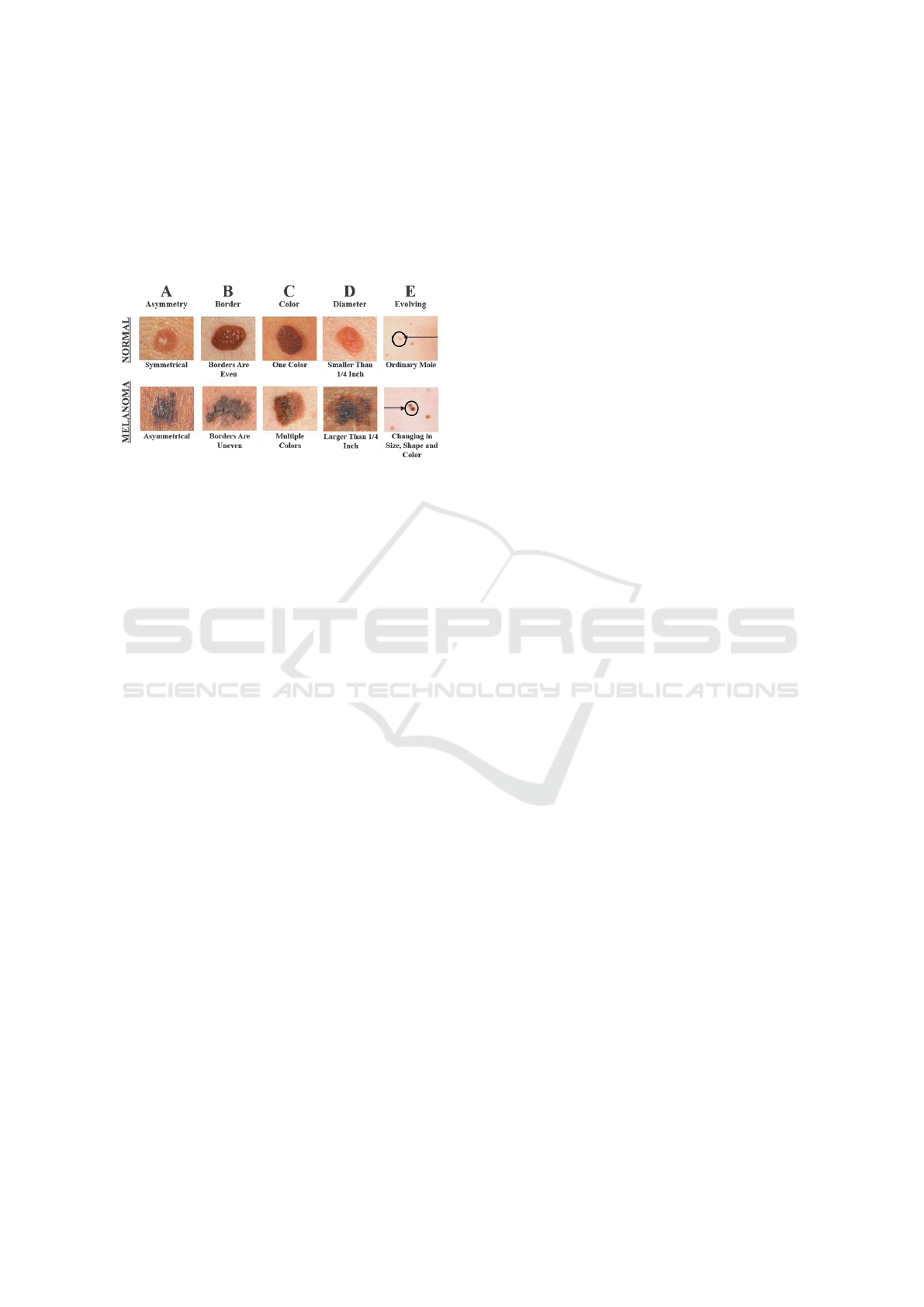
(see Fig. 1). The color variability means that the col-
Figure 1: ABCDEs of detecting melanoma: Aspects and
differences between benign and malignant skin lesions.
ors of a skin lesion are not uniform. Since most be-
nign lesions mainly contains one color, often a single
shade of brown, having a variety of colors is a warn-
ing signal as given by Fig. 1. Melanoma can con-
tain different shades of brown, black, red, white, or
blue colors. Due to the sensitivity of the ABCDE rule,
physicians tend to use other new features to recognize
melanoma. The blue-black color is one of these fea-
tures. It is defined by the presence of a combination
of blue and black pigmented areas involving at least
10% of the lesion surface (Argenziano et al., 2011).
While some melanomas begin within an atypical
mole, though it can be hard to observe the different
colors in an atypical mole. However, it may be ame-
lanotic, not having any of the skin pigment that typ-
ically turns a mole brown or black. Thus, defining
the different colors in a skin lesion is not a straight-
forward process. Accordingly, it becomes harder to
recognize the melanoma. Even dermatologists will
not be able to examine it by the naked eye which can
lead them to remove a portion of a mole for exami-
nation in a lab, which can delay diagnosis. The auto-
matic inspection of melanomas is composed of a vari-
ety of steps including preprocessing, extraction of the
region of interest, post-processing, and lesion inspec-
tion, which are the various steps of a classical pattern
recognition system, including image acquisition, im-
age processing, segmentation, characterization, and
classification of the lesion in question (Maglogiannis
and Doukas, 2009a).
One important step is skin lesion characterization,
which consists in extracting a set of relevant and dis-
criminative primitives that can describe precisely a
skin lesion. These characteristics must ensure non-
redundancy, relevance, discrimination, and robustness
to noise.
The ABCD rule is widely used in automated com-
puter diagnosis systems, which investigates the asym-
metry, the border, the color, and the diameter or differ-
ential structures (Maglogiannis and Doukas, 2009a).
Other features can be employed like the seven-point
checklist (Spalding, 1993) which contains three ma-
jor aspects ( change in size, shape, and color) and
four minor aspects (diameter, inflammation, crusting
or bleeding, and sensory change). These criteria can
be quantitatively determined by the change of the tex-
ture, color, and structure of the skin lesion. A lot
of studies have been introduced to examine the color
characteristics inside a lesion and to define the num-
ber of colors. Skin diseases are restricted to only six
defined colors, for which the color name features are
extracted to achieve accurate inspection under any il-
lumination condition.
A lesion may include light brown, dark brown,
black, red, white, and slate blue (Maglogiannis and
Doukas, 2009b). Nevertheless, the lesion can be ame-
lanotic in some cases. Thus, these color names be-
come worthless for melanoma inspection, and other
degrees of colors should be examined. Besides, the
human perception of color is very complex, as men
and women can differently describe a color. Women
can distinguish even the tiniest differences between
two colors, contrarily to men who see them identi-
cal. Hence, learning about the different color shades
which can differentiate that a malignant and benign
lesion is not easy, Hence, learning about the different
color shades which can differentiate that a malignant
and benign lesion is not easy, so our main contribution
is to build a machine learning-based method to extract
the most relevant color names. This color representa-
tion will be used then for skin lesion classification.
Our aim is to design a low-dimensional representa-
tion that can efficiently detect the different colors in a
skin lesion, more specifically the black and blue color
which has been proved that it is an accurate clue for
melanoma inspection.
This paper is organized as follows. In the next
section, the most color features used in the literature
for melanoma inspection are reported. After that, we
present the way to define the color names using ma-
chine learning and use them to classify skin lesions.
Then, we introduce the conducted experiments and
the results evaluating the classification process. So
the conclusions and future work are drawn in the last
section.
ICT4AWE 2022 - 8th International Conference on Information and Communication Technologies for Ageing Well and e-Health
148

2 RELATED WORK
Classifying skin lesions addresses the problem of
defining skin lesions as malignant or benign. The
classification process is led by some visual clues that
characterize a skin lesion. These features differentiat-
ing malignant and benign lesions should be quantified
and should have a high probability of being true clas-
sified. More essentially, when detecting melanoma,
the decrease in false negatives (misclassified malig-
nant lesions) is critical.
Examining the shape, color and texture have been
the consideration of many researchers. The color
of a lesion is considered as a crucial criterion for
melanoma inspection. This is because the blue-black
rule has also proved to be a good practice in the di-
agnosis process (Argenziano et al., 2011) in addition
to the fact that the ABCD rule defines the most used
and accurate clue for dermatologists. The color fea-
tures have been examined on different color represen-
tations, such as Red, Green, Blue (RGB), HSV (Hue,
Saturation, Value) or the spherical coordinate LAB
average and variance responses for pixels. The color
inconsistency is quantified by calculating the mini-
mum, maximum, average and standard deviations for
each channel (Menzies et al., 2005; Maglogiannis and
Zafiropoulos, 2004a; Maglogiannis and Zafiropoulos,
2004b). In (Manousaki et al., 2006), the authors used
the color texture for determining the nature of skin le-
sions by measuring the lacunarity in the distribution
of colors. Furthermore, in (Yang et al., 2018), the au-
thors have tested the color SIFT (Abdel-Hakim and
Farag, 2006), which examines the color texture. An-
other interesting yet simple method is to examine the
color variegation in a lesion by calculating the vari-
ance of the local average color (Zhang et al., 2003).
Examining the uniformness of the tumor color, it can
be quantified by comparing the colors inside a le-
sion and the healthy skin colors as in (Claridge et al.,
2003). In (Yang et al., 2018), Yang et al. put forward
clinical skin lesion diagnosis using a representation
inspired by dermatologists, where the color is intro-
duced by two representations, defining the different
color names and the continuous color values of le-
sions, which indicates for each pixel the probability
of belonging to a color bin.
3 PROPOSED METHOD
As a CAD system, many frameworks based on image
processing have been proposed and have proved their
efficiency in melanoma inspection. In the literature,
a large variety of classification methods have been
adopted: KNN, SVM, ANN, etc. (Magalhaes et al.,
2021; Melbin and Raj, 2021a). In the last decades,
regarding the evolution of Convolutional Neural Net-
works (CNN), CAD systems have become more and
more oriented into the implementation of semantic
techniques called Deep Learning (DL) (Gonzalez-
Diaz, 2018; Saeed and Zeebaree, 2021). When us-
ing DL, the low-level features discriminating malig-
nant and benign lesions are automatically extracted.
This representation has shown a limitation in some
cases. Thus, it is important to extract hand-crafted
features that have been used and proved by dermatolo-
gists for the diagnosis process. Generally, these prim-
itives represent only the ABCD rule that describes the
color, the border and the texture to find some dif-
ferential structures (Daghrir et al., 2020). The color
of the lesion is still a crucial criterion for diagnosing
melanomas. It represents a variety of colors. More-
over, dermatologists have proposed other important
rules for diagnosing melanomas like the blue-black
rule (Argenziano et al., 2011) and the ugly duck-
ling (Grob and Bonerandi, 1998). The blue-black
rule is defined as the presence of the blue and black
color in a lesion surface (Daghrir et al., 2020). Thus,
extracting color features is extremely important in
melanoma inspection. For instance, some systems
represent the color by defining the Color Name (CN)
features which are linguistic color labels representing
different colors in a skin lesion. As discussed above,
we can notice that the color is with a high sensitivity
in the whole melanoma CAD system. In this work, we
suggest a new color feature extraction method.First
we determine the different color names of skin le-
sions. Second,we search is a selection step the most
pertinent color names that ensure accurate melanoma
detection. The overall implementation of the extrac-
tion process is given in (see Fig. 2). To demonstrate
the effectiveness of our proposed method, we test the
selected features on a set of skin lesion images (see
Fig. 3).
3.1 Supervised Color Name Extraction
Skin lesions may contain many different colors re-
garding the healthy skin color, the kind of the skin dis-
ease, the stage of the disease, etc. Thus, it is important
to choose which colors are more likely to be present
in a skin lesion. Melanomas may mainly include six
colors. These colors can be presented with different
degrees. For an effective color name representation,
we extract the dominant six colors from a set of im-
ages using the K-means algorithm. Centroids for the
algorithm initially depend on the six considered col-
ors: light brown, dark brown, black, red, white, and
A Supervised Quantification of the Color Names Characterizing the Visual Component Color in the ABCD Dermatological Criteria for a
Further Melanoma Inspection
149
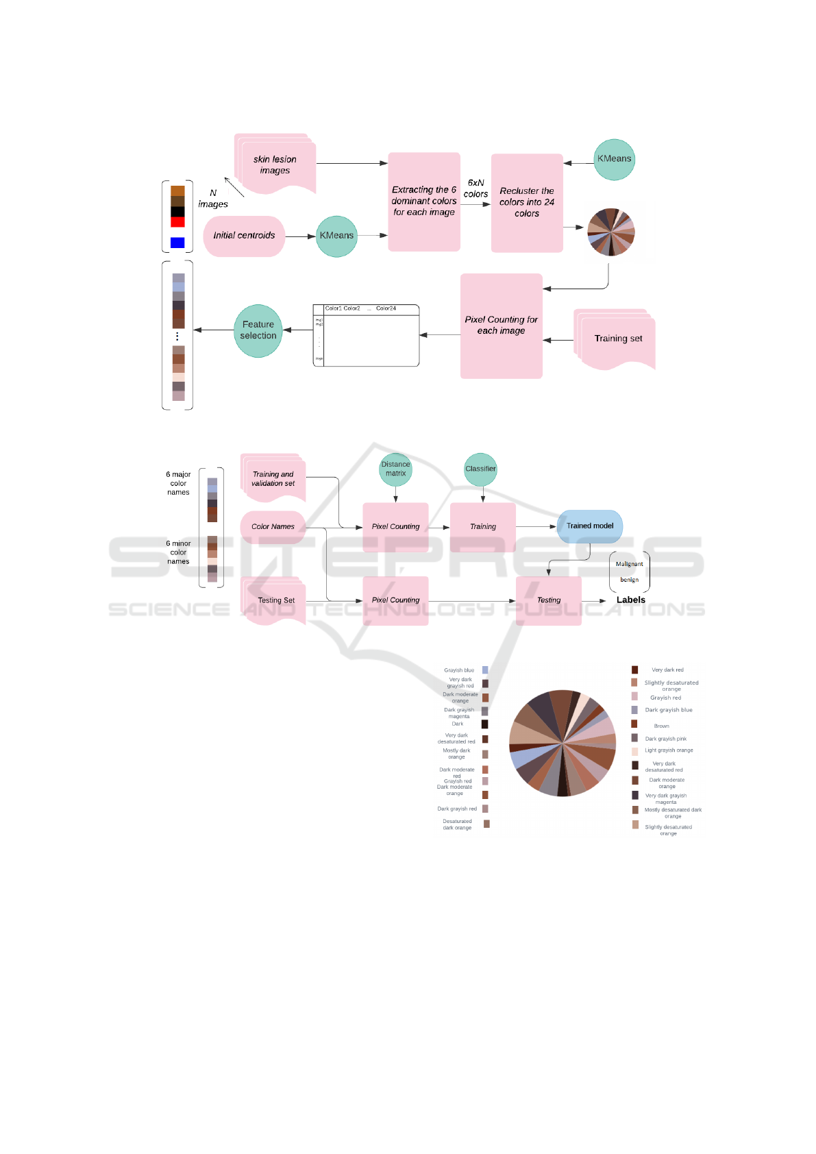
Figure 2: Layout architecture of method of extracting color-names.
Figure 3: Layout architecture of evaluating extracted color names.
slate blue. K-means iteratively minimizes the intra-
class and maximizes the inter-class distances to create
a final partition of image pixels into six groups. Each
input pixel is characterized by three intensities in the
RGB color space. When convergence is reached, six
centroids will be assigned for each color. For more
details about K-means clustering, please see (Melbin
and Raj, 2021b). Gathering all the extracted colors
will serve then to do clustering one more time to fi-
nally extract the 24 major colors, which are displayed
in the pie chart shown in Fig. 4. We assume that for
each color four instances are adopted. After identify-
ing, the major 24 colors associated with skin lesions,
the process of preserving only those which guaran-
tee an effective representation of lesion color names
is done using feature selection, which is a trend in
a lot of machine learning systems. We use the in-
finite Feature Selection (inf-FS) (Roffo et al., 2015)
which is a feature selection method that performs a
ranking step in an unsupervised way, followed by a
cross-validation strategy to select only the most repre-
Figure 4: The most dominant 24 colors extracted from a set
of images.
sentative features. Ranking individually the relevancy
of features is done utilizing class labels: malignant or
benign. On the other hand, using a distance matrix,
we define how the 24 various color values frequently
occur in the lesion (see Fig 3). In the processed im-
age, every pixel value is assigned to the nearest color
ICT4AWE 2022 - 8th International Conference on Information and Communication Technologies for Ageing Well and e-Health
150

name regarding the distance of its intensity. Finally,
by counting the pixels of each color name and apply-
ing the inf-FS procedure, the suggested process gen-
erates a ranked features that refer to their character-
ization relevance. The six major colors found after
applying the feature selection, affirm the efficiency of
the black-blue clue proposed by physicians in inspect-
ing the malignancy of skin lesions. The slight blue
and black colors are highly ranked (see figure3).
3.2 Classifying Skin Lesion using Color
Name Features
After extracting the most relevant color names, the
skin lesion classification is implemented. The most
attractive feature of this process is to evaluate the im-
portance of using only the best-ranked color names.
Generally, using them would be more accurate in clas-
sifying skin lesions. However, counting the pixels for
the best color names using the distance matrix will
create an unfair distribution since some pixels have
different yet distant colors regarding the best color
names. These pixels will be assigned anyway with
the nearest color name, so it is crucial to disturb the
pixels partitioning by using alternative color names.
It would be more appropriate to use the six worst
color names for that purpose. For the classification
task, we have use the K-Nearest Neighbor (KNN) al-
gorithm (Coomans and Massart, 1982). This method
demonstrates a high classification accuracy especially
when using a low-dimensional representation.
4 EXPERIMENTS AND RESULTS
The experiments are conducted using the ISIC2017
(International Skin Imaging Collaboration)
dataset (Codella et al., 2018). The images are
split into three sets, the training set which is used for
ranking the color names and for training the KNN,
and the validation and testing set which are used to
evaluate the performance of our color name extrac-
tion method. We have also used the SD-198 (Sun
et al., 2016), which contains 198 skin diseases rep-
resented by 6,584 images. The authors provide two
split strategies. We have used the fifty split, which
contains 3,292 training and 3,292 testing images. The
SD-198 dataset was trained in (Yang et al., 2018) by
using different low-level features describing the three
visual components: texture, color, and border. We
compare our proposed color name extraction method
with the other color features used in the literature.
To demonstrate the performance of any classification
task, the accuracy is basically introduced, which is
the fraction of correctly classified points and the total
number of points.
Accuracy =
T P + T N
T P + T N + FP + FN
(1)
where TP = True positive, FP = False positive, TN =
True negative and FN = False negative.
However, in some cases, accuracy does not really
indicate the relevancy of the system. For example
when evaluating a binary classifier, one class having
positive labels can appear in the validation set more
than the other one having negative labels. Thus, to
overcome this class imbalance problem, sensitivity,
specificity and balanced accuracy are defined to ef-
ficiently evaluate the classification task. The sensitiv-
ity known also as recall, measures the proportion of
actual positives that are correctly identified.
Sensitivity =
T P
T P + FN
(2)
Nevertheless, specificity measures the proportion of
actual negatives that are correctly classified.
Speci f icity =
T N
T N + FP
(3)
As a consequence, balanced accuracy, which is the
arithmetic mean of both sensitivity and specificity
metrics will properly introduce the pertinence of ma-
lignancy inspection.
Balanced accuracy =
sensitivity + speci f icity
2
(4)
In the experiments, we first find the best color names
introducing the best representative features and we
use the validation data set to gain insights into what
the optimal hyperparameter K is. Therefore, we sug-
gest three scenarios to demonstrate the performance
of the extracted color names using the validation data.
The first, called Scenario A, illustrates the use of the
six major and six worst color names. The second
named Scenario B, is defined as the use of only the
best color names. On the other hand, we use all the
24 color names for scenario C.
Figure 5: Balanced accuracy using different scenarios with
various values of K on validation set.
A Supervised Quantification of the Color Names Characterizing the Visual Component Color in the ABCD Dermatological Criteria for a
Further Melanoma Inspection
151

Table 1: Balanced accuracy using different scenarios applied on testing set.
Scenario A Scenario B Scenario C
Accuracy(%) 77.7 77 62.7
Sensitivity(%) 17.4 11.6 26.7
Specificity(%) 87.7 88.1 68.8
Balanced accuracy(%) 52.6 49.8 47.7
Generally, when the number of classes is odd, hy-
perparameter K must be even. Thus, by generating
the model on different even values of K and checking
their performance, we will understand which number
of neighbors should be considered in the testing pro-
cess. In Fig. 5, we can say that the classifier does a
little better when using a small number of K with a
small number of color names. However, for the third
scenario, when the number of Ks is higher, balanced
accuracy is higher than when applying the other sce-
narios. The experiments show also that when using
the proposed color name combination the balanced
accuracy is higher than using the other scenarios.
Now, we have test the performance of the KNN
using the three scenarios as well, with K=3 (Table 1).
Based on balanced accuracy, it is obvious that sce-
nario A achieve the best performance. Finally, the
disturbing strategy reports the best classification per-
formances so far referring to the other scenarios. It
is evident that adding further color names is generally
useful. With the three scenarios, balanced accuracy
is somewhat minimal, probably because we train the
classifier with an unbalanced dataset having a limited
number of features.
We compare the extracted color names with other
several hand-crafted color features used in the state of
the art. Table 2 reports the prediction performance of
using various representations using KNN with k=3.
As we have a limited and unbalanced dataset, we use
the 5-Fold Cross Validation strategy to report how
well the representations are working. Thus, five accu-
racies as well as their mean using the colorSIFT, CH
(Color Histogram), and our proposed method (SCN)
are reported. It is obvious that all the color representa-
tions succeeded in accurately diagnosing melanoma.
Nevertheless, the best representation is the proposed
one, which reaches about 75% accuracy with only 12
features, unlike the other used representations.
Convolutional Neural Networks (CNNs) have
been widely used in the literature to automati-
cally characterize skin lesions, for their notable
performance. Therefore, in a previous research
work(Daghrir et al., 2020), we have used a CNN fol-
lowed by a fully-connected layer with a softmax ac-
tivation function to classify malignant and benign le-
sions on the ISIC2017 dataset. The extracted features
achieve an 85.5% accuracy, about 8% greater com-
pared to the achievement of our proposed features
(77.7% accuracy in Table 1). This result shows that
our proposed method can reflect the comprehensive
medical information relating to the black-blue feature.
As a result, when using other different features de-
scribing also the color, texture, and shape of the le-
sions, our suggested color name features will be more
relevant.
We compare also our proposed color name extrac-
tion using the SD-198 dataset. Thus, the whole pro-
cess was tested using 198 skin diseases. A set of the
most present 24 colors and their ranks in identifying
the type of the disease are extracted. In table 3, we
report the accuracy of using different color features
proposed and used in (Yang et al., 2018), as well as
the color-based features extracted using our proposed
method. It is shown that our method does not perform
well, it achieves only 5.58% accuracy. It fails in rec-
ognizing the different skin diseases, mainly because
of the variability and the specificity of the skin dis-
eases. In (Yang et al., 2018), the authors also have
compared the classification performance using deep
features derived from fine-tuned CNNs. Mainly, a
CNN achieves an accuracy of 53.35% in classifying
the 198 skin diseases, which is incomparable with the
use of our proposed color-based features.
Although, the use of more than 12 color names
(all the 24 extracted color names) slightly improves
the accuracy to 5.99% using KNN. This can some-
how prove the efficiency of our proposed method in
precisely identifying melanoma since it manifests the
presence of a very limited number of colors( gener-
ally 6 colors). However, 198 skin diseases might be
characterized by a huge number of color names.
Thus, the huge number of skin diseases in a
dataset, limits the performance of our method, as it is
shown in Table 3, 5.58% against 52.6% accuracy for
recognizing 198 skin diseases, compared to the use of
the ISIC2017.
5 CONCLUSION
In this paper, we have presented a new method of
color name extraction in a supervised way using fea-
ture selection to rank the extracted color names. The
application of the extracted color names has been
ICT4AWE 2022 - 8th International Conference on Information and Communication Technologies for Ageing Well and e-Health
152
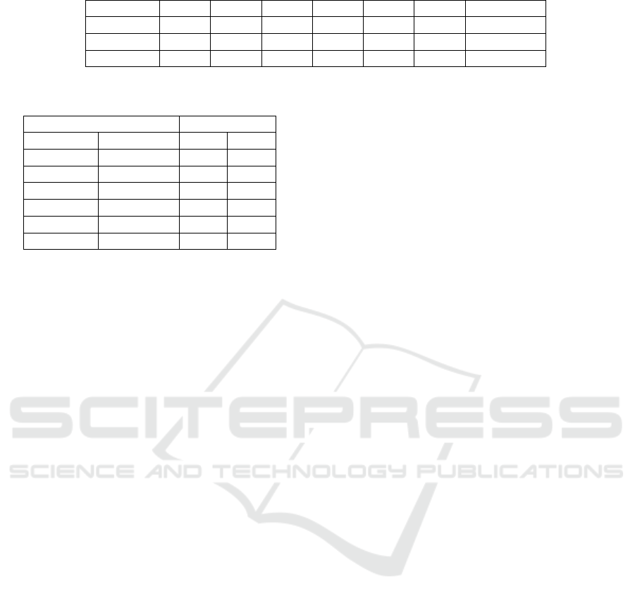
Table 2: Accuracy using different validation Folds with different color representations on the ISIC2017 dataset.
Accuracy Fold1 Fold2 Fold3 Fold4 Fold5 Mean Dimension
SCN 0.78 0.76 0.77 0.78 0.67 0.75 12
CH 0.71 0.78 0.75 0.77 0.63 0.72 255
colorSIFT 0.69 0.71 0.75 0.71 0.62 0.69 10000
Table 3: Accuracy using different representations and clas-
sifiers on the SD-198 dataset.
Accuracy
Features Dimension KNN SVM
CH 256 12.33 4.19
CN 21000 20.03 20.23
colorSIFT 21000 21.29 22.51
CN-L 21000 42.50 38.91
CCV-L 21000 42.80 40.13
SCN 12 5.58 4.73
proved utilizing a classifier with three different par-
titions. These color names are mainly extracted to
classify skin lesions for more accurate inspection of
melanomas, which are considered as the most fa-
tal skin cancer. The proposed method has shown a
notable performance for diagnosing melanomas. A
comparison of different handcrafted features is pre-
sented as well, which proves the efficiency of our
color name features against the state-of-the-art color
representations. Accordingly, using only our pro-
posed color-based features shows a promising result
compared to automatically extracted features using
deep learning. However, our proposed representation
method shows a limitation when using a benchmark
dataset that contains several skin conditions. Thus,
these color names can be further used with other
hand-crafted features and more sophisticated machine
learning models to inspect melanomas to ameliorate
the diagnosis process. Fuzzy features of color names
could be also introduced in future work.
REFERENCES
Abbasi, N. R., Shaw, H. M., Rigel, D. S., Friedman, R. J.,
McCarthy, W. H., Osman, I., Kopf, A. W., and Polsky,
D. (2004). Early diagnosis of cutaneous melanoma:
revisiting the abcd criteria. Jama, 292(22):2771–
2776.
Abdel-Hakim, A. E. and Farag, A. A. (2006). Csift: A sift
descriptor with color invariant characteristics. In 2006
IEEE computer society conference on computer vision
and pattern recognition (CVPR’06), volume 2, pages
1978–1983. Ieee.
Argenziano, G., Longo, C., Cameron, A., Cavicchini, S.,
Gourhant, J.-Y., Lallas, A., McColl, I., Rosendahl, C.,
Thomas, L., Tiodorovic-Zivkovic, D., et al. (2011).
Blue-black rule: a simple dermoscopic clue to recog-
nize pigmented nodular melanoma. British Journal of
Dermatology, 165(6):1251–1255.
Claridge, E., Cotton, S., Hall, P., and Moncrieff, M. (2003).
From colour to tissue histology: physics-based inter-
pretation of images of pigmented skin lesions. Medi-
cal Image Analysis, 7(4):489–502.
Codella, N. C., Gutman, D., Celebi, M. E., Helba, B.,
Marchetti, M. A., Dusza, S. W., Kalloo, A., Liopy-
ris, K., Mishra, N., Kittler, H., et al. (2018). Skin
lesion analysis toward melanoma detection: A chal-
lenge at the 2017 international symposium on biomed-
ical imaging (isbi), hosted by the international skin
imaging collaboration (isic). In 2018 IEEE 15th in-
ternational symposium on biomedical imaging (ISBI
2018), pages 168–172. IEEE.
Coomans, D. and Massart, D. L. (1982). Alternative k-
nearest neighbour rules in supervised pattern recogni-
tion: Part 1. k-nearest neighbour classification by us-
ing alternative voting rules. Analytica Chimica Acta,
136:15–27.
Daghrir, J., Tlig, L., Bouchouicha, M., and Sayadi, M.
(2020). Melanoma skin cancer detection using deep
learning and classical machine learning techniques: A
hybrid approach. In 2020 5th International Confer-
ence on Advanced Technologies for Signal and Image
Processing (ATSIP), pages 1–5. IEEE.
Dimitriou, F., Krattinger, R., Ramelyte, E., Barysch, M. J.,
Micaletto, S., Dummer, R., and Goldinger, S. M.
(2018). The world of melanoma: epidemiologic, ge-
netic, and anatomic differences of melanoma across
the globe. Current oncology reports, 20(11):1–9.
Gonzalez-Diaz, I. (2018). Dermaknet: Incorporating the
knowledge of dermatologists to convolutional neural
networks for skin lesion diagnosis. IEEE journal of
biomedical and health informatics, 23(2):547–559.
Grob, J. and Bonerandi, J. (1998). The ‘ugly duckling’sign:
identification of the common characteristics of nevi
in an individual as a basis for melanoma screening.
Archives of dermatology, 134(1):103–104.
Heinzerling, L. and Eigentler, T. K. (2021). Skin cancer
in childhood and adolescents: Treatment and implica-
tions for the long-term follow-up. In Late Treatment
Effects and Cancer Survivor Care in the Young, pages
349–355. Springer.
Khazaei, Z., Ghorat, F., Jarrahi, A., Adineh, H., Sohrabi-
vafa, M., and Goodarzi, E. (2019). Global incidence
and mortality of skin cancer by histological subtype
and its relationship with the human development in-
dex (hdi); an ecology study in 2018. World Cancer
Res J, 6(2):e13.
Magalhaes, C., Tavares, J. M. R., Mendes, J., and Vardasca,
R. (2021). Comparison of machine learning strategies
for infrared thermography of skin cancer. Biomedical
Signal Processing and Control, 69:102872.
A Supervised Quantification of the Color Names Characterizing the Visual Component Color in the ABCD Dermatological Criteria for a
Further Melanoma Inspection
153

Maglogiannis, I. and Doukas, C. N. (2009a). Overview
of advanced computer vision systems for skin lesions
characterization. IEEE transactions on information
technology in biomedicine, 13(5):721–733.
Maglogiannis, I. and Doukas, C. N. (2009b). Overview
of advanced computer vision systems for skin lesions
characterization. IEEE transactions on information
technology in biomedicine, 13(5):721–733.
Maglogiannis, I. G. and Zafiropoulos, E. P. (2004a). Char-
acterization of digital medical images utilizing sup-
port vector machines. BMC Medical Informatics and
Decision Making, 4(1):1–9.
Maglogiannis, I. G. and Zafiropoulos, E. P. (2004b). Char-
acterization of digital medical images utilizing sup-
port vector machines. BMC Medical Informatics and
Decision Making, 4(1):1–9.
Manousaki, A. G., Manios, A. G., Tsompanaki, E. I., and
Tosca, A. D. (2006). Use of color texture in determin-
ing the nature of melanocytic skin lesions—a qualita-
tive and quantitative approach. Computers in biology
and medicine, 36(4):419–427.
Melbin, K. and Raj, Y. J. V. (2021a). Integration of modi-
fied abcd features and support vector machine for skin
lesion types classification. Multimedia Tools and Ap-
plications, 80(6):8909–8929.
Melbin, K. and Raj, Y. J. V. (2021b). Integration of modi-
fied abcd features and support vector machine for skin
lesion types classification. Multimedia Tools and Ap-
plications, 80(6):8909–8929.
Menzies, S. W., Bischof, L., Talbot, H., Gutenev, A.,
Avramidis, M., Wong, L., Lo, S. K., Mackellar, G.,
Skladnev, V., McCarthy, W., et al. (2005). The per-
formance of solarscan: an automated dermoscopy im-
age analysis instrument for the diagnosis of primary
melanoma. Archives of dermatology, 141(11):1388–
1396.
Roffo, G., Melzi, S., and Cristani, M. (2015). Infinite fea-
ture selection. In Proceedings of the IEEE Interna-
tional Conference on Computer Vision, pages 4202–
4210.
Saeed, J. and Zeebaree, S. (2021). Skin lesion classification
based on deep convolutional neural networks archi-
tectures. Journal of Applied Science and Technology
Trends, 2(01):41–51.
Spalding, J. (1993). The doctor with an inherited defect
of colour vision: effect on clinical skills. The British
Journal of General Practice, 43(366):32.
Stolz, W., H
¨
olzel, D., Riemann, A., Abmayr, W., Prze-
tak, C., Bilek, P., Landthaler, M., and Braun-Falco, O.
(1991). Multivariate analysis of criteria given by der-
matoscopy for the recognition of melanocytic lesions.
In Book of Abstracts, Fiftieth Meeting of the American
Academy of Dermatology, Dallas, Tex: Dec, pages 7–
12.
Sun, X., Yang, J., Sun, M., and Wang, K. (2016). A
benchmark for automatic visual classification of clin-
ical skin disease images. In European Conference on
Computer Vision, pages 206–222. Springer.
Yang, J., Sun, X., Liang, J., and Rosin, P. L. (2018). Clinical
skin lesion diagnosis using representations inspired by
dermatologist criteria. In Proceedings of the IEEE
Conference on Computer Vision and Pattern Recog-
nition, pages 1258–1266.
Zhang, Z., Moss, R. H., and Stoecker, W. V. (2003). Neu-
ral networks skin tumor diagnostic system. In Inter-
national Conference on Neural Networks and Signal
Processing, 2003. Proceedings of the 2003, volume 1,
pages 191–192. IEEE.
ICT4AWE 2022 - 8th International Conference on Information and Communication Technologies for Ageing Well and e-Health
154
