
A Successful Treatment of Pemphigus Foliaceus Patient with
Systemic Corticosteroid
Widyaningsih Oentari
1*
, Irma D. Roesyanto-Mahadi
1
1
Department of Dermatology and Venereology, Faculty of Medicine,
Universitas Sumatera Utara, Haji Adam Malik General Hospital, Medan
Keywords: Pemphigus foliaceus, diagnosis, treatment
Abstract: Pemphigus is a group of IgG-related autoimmune disease of skin and mucous membrane. It cause
acantholysis with blisters and erosion as the clinical manifestation. There are three main groups of
pemphigus, which is vulgaris, foliaceus, and paraneoplastic. Female 68 years old, came with chief complain
of blisters that were spreading on her face, body, and both extremities since 1 year ago. From
dermatological examination, we found multiple bullae and erosions sized lenticular to plaque with
erythematous base and multiple hyperpigmented maculae with crusts on the face, body and both extremities.
Biopsy was taken from newly formed vesicle and histopathological examination supported characteristics of
pemphigus foliaceus. The patient was treated with NaCl 0,9% compress for 15 minutes every 4-6 hours,
fusidic acid cream twice a day, 32 mg methylprednisolone every morning, and clarithromycin 500 mg once
a day. After control, the patients showed improvement in her skin lesions.
1 INTRODUCTION
Pemphigus is a group of IgG-related autoimmune
disease of skin and mucous membrane resulting
acantholysis with clinical manifestation of blisters
and erosion (Payne et al, 2012; Kasperkiewicz et al,
2017). Pemphigus can be classified into three main
groups, such as vulgaris, foliaceus, and
paraneoplastic (Kasperkiewicz et al, 2017). In
pemphigus vulgaris (PV), the blisters can be found
on the suprabasal layer, whereas in pemphigus
foliaceus (PF) on granular layer (Payne et al, 2012;
Kasperkiewicz et al, 2017). Patients with
paraneoplastic pemphigus usually have neoplasm
associated with lymphoid tissues and caused by
combination of autoimmune humoral and cellular
reaction (Kasperkiewicz et al, 2017). Both PV and
PF have their variants. Variants of PV usually
appear locally, such as pemphigus vegetans of
Hallopeau and pemphigus vegetans of Neumann.
While, PF that appear locally is pemphigus
eritematosa (Payne et al, 2012).
Epidemiology of pemphigus varies throughout
the world (Payne et al, 2012; Kasperkiewicz et al,
2017). The incidence is quite rare, which is 2-10
cases per one million population in some areas of the
world and the prevalence is 0,1-0,7 per 100.000
population (Dimarco, 2016). Pemphigus vulgaris is
a subtype that often found in Europe, United States
and Japan, especially in women aged 50-60 years
old. Pemphigus foliaceus is less common compare to
PV and commonly found as endemic disease in
South America and North Africa (Kasperkiewics et
al, 2017; Pollmann et al, 2018). The age of onset of
PF is 40 to 60 years old. However, the endemic form
of PF can appear in second or third decades (James
et al, 2011). Paraneoplastic pemphigus is less
common that PV and PF, usually can be found in
adult aged 45 to 70 years old (Kasperkiewicz et al,
2017). Pemphigus seldom found in children (Payne
et al, 2012). The different onset of age is related to
genetic, hormonal, and environmental factors
(Kasperkiewicz et al, 2017).
In addition to anamnesis and physical
examination, the diagnosis of pemphigus can be
supported by histopathology and serology
examination. Direct immunofluorescence
examination can also be done to detect IgG antibody
on the surface of keratinocytes. Also, measurement
of IgG antibody titers to desmoglein can be done
through enzyme-linked immunosorbant assay
(ELISA) (Dimarco, 2016; Pollmann et al, 2018).
Through this case report, we would like to present
patient with pemphigus foliaceus.
Oentari, W. and Mahadi, I.
A Successful Treatment of Pemphigus Foliaceus Patient with Systemic Corticosteroid.
DOI: 10.5220/0009990804250429
In Proceedings of the 2nd International Conference on Tropical Medicine and Infectious Disease (ICTROMI 2019), pages 425-429
ISBN: 978-989-758-469-5
Copyright
c
2020 by SCITEPRESS – Science and Technology Publications, Lda. All rights reserved
425
2 CASE
The patient, female 68 years old, came with chief
complain of blisters that were spreading on her face,
body, and both extremities since 1 year ago. At first,
the blisters were only found on her left hand, then
after a couple of months it started spreading to her
legs and body. One month ago, the blisters are also
appeared on her face. The blisters especially appear
after being exposed to friction and scratch, easily
ruptured, leaving painful erosion. There were no
blisters on her mouth, eyes, or genital area. The
patient denied any drug allergy and similar lesion in
family. She also denied any other medical condition
or consumes any medication.
Physical examination generally was within
normal limits. From dermatological examination, we
found multiple bullae and erosions sized lenticular to
plaque with erythematous base and multiple
hyperpigmented maculae with crusts on the face,
body and both extremities. We then carried out
punch biopsy on patient’s bulla for histopathology
examination. According to several examination that
was done in the patients, we have differential
diagnoses of pemphigus vulgaris, pemphigus
foliaceus, and bullous pemphigoid. The patient was
treated with NaCl 0,9% compress for 15 minutes
every 4-6 hours, fusidic acid cream twice a day, 32
mg methylprednisolone every morning, and
clarithromycin 500 mg once a day.
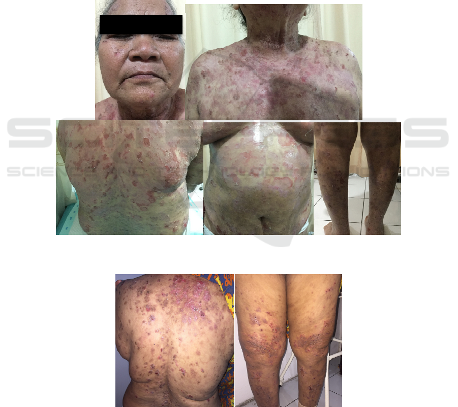
Figure 1. In generalized area, there were multiple bullae and erosions sized lenticular to plaque with erythematous base and
hyperpigmented maculae with crust
Figure 2. Multiple papule in lenticular to plaque sized with erosion, hyperpigmented maculae with crusts in her back, chest,
and lower extremities
ICTROMI 2019 - The 2nd International Conference on Tropical Medicine and Infectious Disease
426

Histopathological examination showed stratified
squamous epithelial cell with hyperkeratosis and
thickening on stratum granulosum and acanthosis.
There are subcorneal blister surrounded by
lymphocyte infiltrate. It was concluded that the
diagnosis was pemphigus foliaceus. After control,
the patients showed improvement in her skin lesions.
3 DISCUSSION
Pemphigus is caused by autoantibody, especially
IgG
4
, against its antigen, which is desmoglein, a
trans membrane glycoprotein of desmosome.
Desmoglein is part of cell’s adhesion molecule
cadherin. (Payne et al, 2012; Pollmann et al, 2018).
Pemphigus is mainly caused by antibodies to
desmoglein 1 (Dsg1, 160-kDa) in PF, desmoglein 3
(Dsg3, 130-kDa) in PV that predominantly in
mucosal membrane, or both in muco-cutaneous PV.
(Payne et al, 2012;Dimarco, 20 et al,2016);Hammers
et al, 2016) Dsg1 and 3 are found in varying
amounts in the skin and mucous membrane. Dsg1 is
found more in upper layer of epidermis, while Dsg3
is found in lower layer of epidermis. This causes
different clinical manifestation in PV and PF.
(Dimarco, 2016; Pollmann et al, 2018;Ruocco et
al,2013). There are two main types of PF, which is
idiopathic PF, that are found universally and appears
sporadically, and fogo selvagem (FS) which is an
endemic form of PF associated with several
geographic areas.(James et al,2011)
IgG autoantibody of PV and PF bound NH
2
domain of Dsg ectodomain. This domain is the same
area as the area that plays a role in desmoglein
intercellular adhesion, which directly caused
acantholysis. In addition, studies on keratinocytes
show that loss of intercellular adhesion due to
autoantibodies causes desmoglein internalization
and degradation. (Payne et al, 2012; Pollmann et al,
2018). Pathogenic antibodies to desmoglein tend to
bind to matured desmoglein on keratinocytes.
(Pollmann et al, 2018). In PF, pathogenic IgG binds
to Dsg1, causes phosphorylation of p-38 mitogen-
activated protein kinase (MPAK) thus encouraging
apoptosis in keratinocytes.(James et al,2011)
Clinical manifestation of PF and PV can
resemble one another, which is superficial blisters
that rupture easily causing erosions. (Pollmann et al,
2018;James et al,2011) Patients usually complain
about pain and burning sensation on their skin
lesions. (Payne et al, 2012;James et al,2011) It can
be located on chest, face, scalp, upper back and
traumatized area. (Pollmann et al, 2018) Skin lesions
in PV can be found on the entire surface of the skin,
but rarely on palms and feet. There are several clues
that can help us distinguish PF from PV. Skin
lesions of PF are initially found locally and spread
on seborrheic location. This condition can expand to
entire body and can be aggravated by ultraviolet
light. (Payne et al, 2012) Bullae in PF is more prone
to rupture than PV, therefore the clinical
manifestation of PF usually are small erosions with
crusts. (Hammers et al, 2016) Also, PF rarely
involves the mucous membrane. (Dimarco, 2016)
Both V and PF show a positive Nikolsky sign.
(Hammers et al, 2016) Initially in our patient, skin
lesions appeared on her left hand, which lasted for
several weeks, then spreads slowly throughout her
body. There are no similar lesions that were reported
on mucous area, therefore we considered PF as one
of differential diagnoses. However, given the similar
clinical manifestation of PV and PF, we still could
not eliminate PV as differential diagnosis.
Another differential diagnosis from this patient is
bullous pemphigoid. It usually found in adult age
more than 60 years old and caused by IgG
autoantibody against antigen in dermo-epidermal
junction causing subepidermal blister. Skin lesions
that are usually found are tense skin blisters on
normal or erythematous skin on flexors, lower thighs
and abdomen. Sometimes, skin lesions can be found
in the mucous membrane. Nikolsky sign is negative
in patients with bullous pemphigoid. (Culton et
al,2012)
Our patient complained about easily rupture
blisters with erosions that appeared after friction.
Therefore, based on anamnesis and physical
examination, we concluded that bullous pemphigoid
could be excluded from differential diagnoses.
The diagnosis of pemphigus should be supported
by histopathology and laboratory tests. Biopsy
samples are usually taken from newly formed
vesicle or edge of blister. Moreover, direct
immunofluorescent should be carried out by taking
biopsy at least 1 cm from blisters or inflamed skin.
Enzyme-linked immunosorbent assay (ELISA) is
also useful to measure IgG antibody titers against
desmoglein in patient’s serum. (Dimarco, 2016;
Pollmann et al, 2018). In this patient, we taken
biopsy sample from newly formed vesicle from her
back.
Histopathologically, pemphigus shows loss of
intraepidermic cell adhesion. (Pollmann et al, 2018).
While in PV, histopathological examination shows
suprabasal blister with acantholysis with “row of
tombstones” that sign of bad prognosis. In PF,
acantholysis can be found right under stratum
corneum and in stratum granulosum. Other finding
A Successful Treatment of Pemphigus Foliaceus Patient with Systemic Corticosteroid
427

include subcorneal pustules contains neutrophil.
(Payne et al, 2012; Hammers et al, 2016) Old PF
lesion usually shows papillomatosis, acanthosis,
hyperkeratosis, parakeratosis and follicular plug.
Increase pigment formation can also be found in
melanocytes of basal layer and there is capillary
dilatation in papilla dermis. There are also infiltrates
consist of neutrophils, eosinophils, and lymphocytes.
(James et al,2011) Histopathological characteristics
of PF is often difficult to differentiate with bullous
impetigo or staphylococcal scalded skin syndrome.
(Payne et al, 2012) According to this informations,
we concluded that histopathological examination of
this patient support diagnosis of pemphigus
foliaceus. Differential diagnosis of bullous
pemphigoid could be eliminated because usually the
blisters are located in subepidermis with eosinophils,
neutrophils and monosit infiltrate in superficial
dermis.(Culton et al,2012)
Immunofluorescence examination of pemphigus
showed IgG autoantibody on the surface of
keratinocytes. In patients with PV and PF, direct and
indirect immunofluorescence can give similar
findings, which is IgG on epidermal cell surface
with netlike intraepidermal staining pattern (Payne
et al, 2012; Pollmann et al, 2018). Biopsy samples
for direct immunofluorescence can be taken from
perilesion area and stored on Michel’s transport
media before examination. While in indirect
immunofluorescence, patient’s serum was incubated
with tissues obtained from esophagus of monkey,
human skin, or bladder epithelium from mice or
rabbit.(Pollmann et al, 2018). We could not perform
this examination on this patient because it is not
available in our healthcare facilities.
Serological examination such as ELISA can be
used to identify and monitor IgG antibody serum
level in pemphigus. According to the type of
autoantigen, such as Dsg1 and Dsg3, we can identify
different type of pemphigus in patients (Pollmann et
al, 2018). Unfortunately, this modalities also not yet
available and routinely tested at our healthcare
facilities so that this examination is not carried out in
this patient.
There has been no FDA-approved therapy for
pemphigus. (Payne et al, 2012) General
recommendation for treatment of pemphigus remain
inconclusive because of various research designs
and different outcomes from previous studies.
(Pollmann et al, 2018;Singh et al,2011) Initial
therapy for pemphigus is high dose of systemic
corticosteroid equivalent 0,5-1,5 mg/kg/day of
prednisone with adjuvant immunosuppressive
medication. (Pollmann et al, 2018;Singh et
al,2011;Hertl et al,2015) Patients that unresponsive
to initial therapy or have contraindication for high
dose of corticosteroid can be given second line
therapy, which is intravenous immunoglobulin 2
g/kg/cycle. Monoclonal antibody of anti CD20 is
also useful in patients with refractory pemphigus
(Pollmann et al, 2018 (Pollmann et al, 2018;Singh et
al,2011) Pemphigus foliaceus with local skin
eruption can be treated with topical corticosteroid.
However, if the disease is active and widespread, we
can use similar therapy as pemphigus vulgaris(Payne
et al, 2012) Our patient were given NaCl 0,9%
compress for 15 minutes every 4-6 hours, fucidic
acid cream twice a day, methylprednisolone 32 mg
every morning, and clarithromycin 500 mg once a
day. After control, there were improvements in
patient complaints.
4 CONCLUSION
Pemphigus is group of autoimmune disease
associated with IgG to desmosome of stratified
squamous epithelial skin and mucous membrane,
causing acantholysis with clinical manifestation of
easily rupture blisters and erosions. We reported 68
years old female patient with blisters on her face,
body and extremities since 1 year ago. Differential
diagnosis includes pemphigus vulgaris, pemphigus
foliaceus, and bullous pemphigoid. Anamnesis,
physical examination and histopathological
examination are important for diagnosis and in this
case, we concluded patients as pemphigus foliaceus.
Immunofluorescence examination is also important
for diagnosis but still unavailable in our health
facility. Treatment with systemic corticosteroid
provides benefits for clinical improvement of
patients.
REFERENCES
Culton DA, Liu Z, Diaz LA. Bullous pemphigoid. 2012.
Dalam: Goldsmith LA, Katz SI, Gilchrest BA, Paller
AS, Leffell DC, editor. Fitzpatrick’s dermatology in
general medicine. Edisi ke-8. New york: McGraw Hill
companies. p.608-16.
DiMarco C. 2016. Pemphigus: Pathogenesis to Treatment.
R I Med J (2013). Dec 1;99(12):28-31.
Eming R, Sticherling M, Hofmann SC, Hunzelmann
N, Kern JS, Kramer H, dkk. 2015. S2k guidelines for
the treatment of pemphigus vulgaris/foliaceus and
bullous pemphigoid. J Dtsch Dermatol
Ges. Aug;13(8):833-44. doi: 10.1111/ddg.12606.
ICTROMI 2019 - The 2nd International Conference on Tropical Medicine and Infectious Disease
428

Hammers CM, Stanley JR. 2016. Mechanisms of Disease:
Pemphigus and Bullous Pemphigoid. Annu Rev
Pathol. May 23;11:175-97. doi: 10.1146/annurev-
pathol-012615-044313.
Hertl M, Jedlickova H, Karpati S, Marinovic B, Uzun
S, Yayli S, dkk. 2015. Pemphigus. S2 Guideline for
diagnosis and treatment--guided by the European
Dermatology Forum (EDF) in cooperation with the
European Academy of Dermatology and Venereology
(EADV). J Eur Acad Dermatol
Venereol. Mar;29(3):405-14. doi: 10.1111/jdv.1277
Payne AS, Stanley JR. Pemphigus. 2012. Dalam:
Goldsmith LA, Katz SI, Gilchrest BA, Paller AS,
Leffell DC, editor. Fitzpatrick’s dermatology in
general medicine. Edisi ke-8. New york: McGraw Hill
companies. p.586-99.
James KA, Culton DA, Diaz LA. 2011. Diagnosis and
clinical features of pemphigus foliaceus. Dermatol
Clin. Jul;29(3):405-12, viii. doi:
10.1016/j.det.2011.03.012.
Kasperkiewicz M, Ellebrecht CT, Takahashi
H, Yamagami J, Zillikens D, Payne AS, dkk. 2017.
Pemphigus. Nat Rev Dis Primers. May 11;3:17026.
doi: 10.1038/nrdp.2017.26
Pollmann R, Schmidt T, Eming R, Hertl M. 2018.
Pemphigus: a Comprehensive Review on
Pathogenesis, Clinical Presentation and Novel
Therapeutic Approaches. Clin Rev Allergy
Immunol. Feb;54(1):1-25. doi: 10.1007/s12016-017-
8662-z.
Ruocco V, Ruocco E, Lo Schiavo A, Brunetti G, Guerrera
LP, Wolf R. 2013. Pemphigus: etiology, pathogenesis,
and inducing or triggering factors: facts and
controversies. Clin Dermatol. Jul-Aug;31(4):374-381.
doi: 10.1016/j.clindermatol.2013.01.004.
Singh S. 2011. Evidence-based treatments for pemphigus
vulgaris, pemphigus foliaceus, and bullous
pemphigoid: a systematic review. Indian J Dermatol
Venereol Leprol. Jul-Aug;77(4):456-69. doi:
10.4103/0378-6323.82400.
A Successful Treatment of Pemphigus Foliaceus Patient with Systemic Corticosteroid
429
