
Multibacillary Leprosy in a Child
Arridha Hutami Putri
1*
, Mila Darmi
2
, Ramona Dumasari Lubis
1
1
Department of Dermatology and Venereology, Faculty of Medicine Universitas Sumatera Utara, Jln. Dr. Mansur No. 66,
Medan, Sumatera Utara
2
Department of Dermatology and Venereology, General Hospital of H. Adam Malik, Jln. Bunga Lau No. 17, Medan,
Sumatera Utara
*
Corresponding author
Keywords: Leprosy, Children, Multibacillary, M leprae
Abstract: This manuscript aims to review the cutting-edge developments regarding the diagnosis, management, and
prevention of leprosy in children. Where leprosy in children is a robust indicator of the active source of
infection in the community, we reported a case of borderline lepromatous leprosy in a 14-year-old boy. He
came with the chief complaint of swelling of left little finger since one month ago. The patient also
complaint blackish patch with loss of sensations on the left hand since two years ago. After one year, there
was an appearance of papules on the ears and sparse eyebrows followed by swelling of left little finger one
month ago. His father was already diagnosed as multibacillary leprosy eight years ago and had completed
the treatment. There is no history of BCG vaccination. Physical examination revealed sparse eyebrows
(madarosis), dermatological examination revealed diffuse hyperpigmentation macular with anesthesia on the
left hand, papules and infiltrate on the ear lobes. Sensibility examination revealed anesthesia on the left
hand, which is innervated by the ulnar and median nerve. Thickened and tenderness found on the ulnar
nerve and muscle weakness grade 3 was found on the left hand, which is innervated by the ulnar nerve.
Ziehl-Neelsen staining of slit skin smear revealed acid-fast bacilli with Bacteriological Index 4+. The
patient was diagnosed as multibacillary leprosy then prescribed with multi-drug therapy and advice on daily
care routinely. The prognosis of the patient is good since no disabilities. Early diagnosis and treatment is a
fundamental strategy to prevent leprosy transmission.
1 INTRODUCTION
Leprosy has been a major public health problem in
many developing countries for centuries. Children
are believed to be the most vulnerable group to
infection with Mycobacterium leprae given their
nascent immunity and possible intrafamilial contact
(Singal et al, 2010). Leprosy in children has a
significantly unique aspect because of its potential to
cause progressive physical deformity with serious
consequent psychosocial impact on both the child
and the family. Epidemiologically, childhood
leprosy is an index of transmission of disease in the
population (Kaur et al, 1991). In the post-elimination
era, the incidence of leprosy amongst young children
indicates active foci of transmission in the
community, making it a robust epidemiological
indicator to assess the progress of leprosy control
programs (Singal et al, 2010).
Amongst children, the
disease tends to occur with the highest frequency in
children of 5–14 years age group and only 5.8–6%
cases are below five years of age. This may be due
to the relatively long incubation period of leprosy
and delayed diagnosis of indeterminate lesions in
children. Among children, boys are more commonly
affected than girls. This may be due to greater
mobility and increased opportunities for contact in a
male child (Shetty et al, 2013).
Familial contacts are
known to have a significant role in the development
of childhood leprosy.The risk of developing leprosy
in a person is four times when there is a
neighborhood contact. However, this risk increases
to nine times when the contact is intra-familial.
Further, the risk gets higher if a contact has a
multibacillary (MB) form. The attack rate reportedly
increases when the index case is mother (Singal et
al, 2010; Jain et al, 2002).
The mode of transmission of leprosy is still not
conclusively proven although infection can occur
Putri, A., Darmi, M. and Lubis, R.
Multibacillary Leprosy in a Child.
DOI: 10.5220/0009990304070410
In Proceedings of the 2nd International Conference on Tropical Medicine and Infectious Disease (ICTROMI 2019), pages 407-410
ISBN: 978-989-758-469-5
Copyright
c
2020 by SCITEPRESS – Science and Technology Publications, Lda. All rights reserved
407
through very long and close contact. Another
presumption is by nasal droplets inhalation (Singal
et al, 2010; Wisnu et al, 2016). There is also
epidemiologic evidence to suggest that leprosy may
be transmissible from mothers to offsprings via the
placenta. The report of a child developing leprosy at
the age of 3 weeks is an example where the infection
could have been intrauterine. Although M. leprae are
known to be present in the breast milk of mothers
suffering from lepromatous leprosy, the risk of
acquiring leprosy infection in the breastfed infant via
the gastrointestinal tract remains uncertain (Singal et
al, 2010).
Leprosy in children can be challenging to
identify, mainly because of the peripheral nerve
function evaluation. The younger the child, the more
difficult the changes in sensitivity are to evaluate.
Leprosy diagnosis is based on clinical signs and loss
of sensation, associated or not with thickened
nerves. Although there are no laboratory exams that
can detect all cases of leprosy, the presence of acid-
fast bacilli (AFB) in skin smears is conclusive for
leprosy diagnosis (Singal et al, 2010; Wisnu et al,
2016).
Given that one of the main targets of the
global leprosy strategy is zero disabilities among
new pediatric patients (children below the age of 15)
by 2020, this case report aims to review the cutting-
edge developments regarding the diagnosis,
management, and prevention of leprosy in children.
2 CASE
A 14-year-old boy came for treatment in polyclinic
Dermatology and Venereology H.Adam Malik
General Hospital presented He came with the chief
complaint of swelling of left little finger since one
month ago. The patient also complaint blackish
patch with loss of sensations on the left hand since
two years ago. After one year, there was an
appearance of papules on the ears and sparse
eyebrows followed by swelling of left little finger
one month ago. No report of fever, joint pain or
urinary problem and the same compliance before.
His father was already diagnosed as multibacillary
leprosy eight years ago and had completed the
treatment. There is no history of BCG vaccination,
and the patient did not complete his vaccination. In
physical examination, the vital signs were within
normal limits, and nutritional status was normal, but
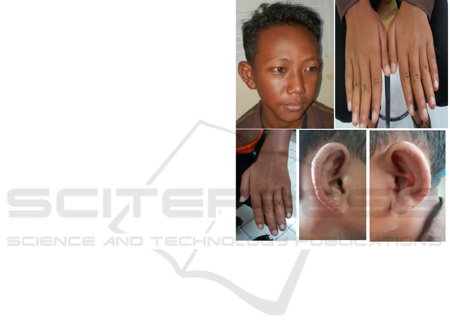
we found sparse eyebrows (madarosis) (see figure
1a). Dermatological examination revealed diffuse
hyperpigmentation macular with anesthesia on the
left hand (figure 1b and 1c). There were papules and
infiltrates on the ear lobes (figure 1d). The sensory
examination revealed anesthesia to touch and pain
stimulus in the skin lesions and area, which is
innervated by ulnar nerve and median nerve. On
palpation of the peripheral nerves, thickening and
tenderness found on the ulnar nerve. The motoric
examination revealed grade 3 weakness in abduction
movement, which is innervated by the ulnar nerve.
We also noted a decrease in sweat production in the
area with skin lesion during activities.
Figure 1. (a) Madarosis in eyebrows. (b) (c) a diffuse
hyperpigmentation macular, the size of a placard
accompanied by anesthesia on the left hand. (d) infiltrate
on the ear lobes.
The differential diagnosis in these patients is
multibacillary type leprosy, postinflammatory
hyperpigmentation, and tinea manus. Then in the
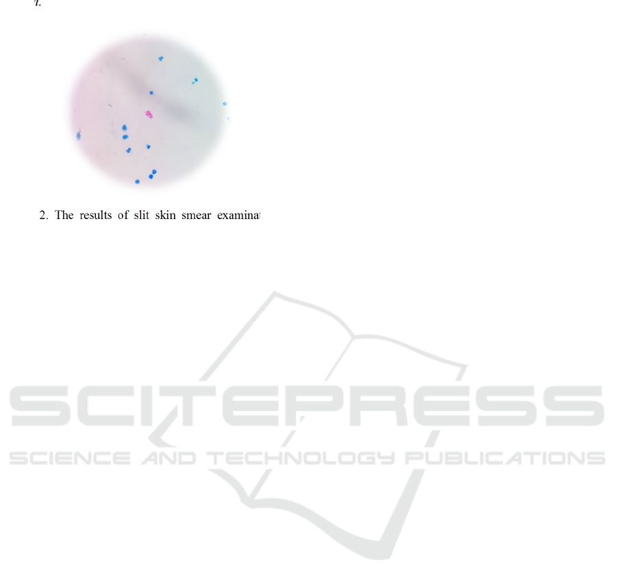
patient carried out a bacteriological examination of
acid-resistant bacteria with a skin smear slit on ear
lobes, bacteriological index results of the left ear
lobe (+) 4 and right ear lobe (+) 4 were obtained
(figure 2). A diagnosis of multibacillary leprosy as
defined by the World Health Organization (WHO)
was made. The patients, without a clear history of
exposure to infectious diseases, were subsequently
treated with multidrug therapy, consisting of
monthly doses of rifampicin 450 mg and clofazimine
150 mg, plus daily doses of dapsone 50 mg and
clofazimine 50 mg. The patient was educated to rest
enough and to control routinely every month,
besides doing daily care routinely. The prognosis of
the patient is quo ad vitam bonam, quo ad
ICTROMI 2019 - The 2nd International Conference on Tropical Medicine and Infectious Disease
408
functionam dubia ad malam, and quo ad sanationam
malam.
Figure 2. The results of slit skin smear examination in
patients showed (+) 4.
3 DISCUSSION
Leprosy in children under the age of 15 is a
significant epidemiological indicator. This is related
to the active transmission of disease in the
community, recalling that the leprosy control
program carried out is not
efficient.(WHO,2015;WHO,2016) In children, this
disease most often occurs at the age of 5-14 years
and only 5.8-6% of cases in children under five
years old. This is due to the relatively long
incubation period and delayed diagnosis of
indeterminate lesions. More common in boys,
possibly due to greater mobility and increasing
opportunities for contact.(Shetty et al,2013) Family
contacts are known to play an important role in the
development of leprosy in children. The risk of
developing leprosy in a person increases nine-fold if
the contact is between families. Furthermore, the
risk increases if contact with an MB patient, and the
incidence increases if the source of contact is his
mother (Singal et al, 2010; Jain et al, 2002).
In this patient, the source of contact was his father
who diagnosed with a multibacillary type of leprosy
since eight years ago. So that patients are at risk of
leprosy from birth because of the long incubation
period of leprosy with an average of 2-5 years but
can also be up to 40 years. (Kementrian Kesehatan
RI,2014) Risks are increasing because patients are in
contact with multibacillary leprosy patients, and
there is not BCG immunization history. In the study
of Richardus and Oskam, someone who had
received a BCG vaccine in his childhood received
protection by 57%.(Richardus et al,2019)
Leprosy in children is very difficult to detect,
usually due to errors in examining peripheral nerve
function. In a much younger child, it is more
difficult to check for changes in sensibility.(Barreto
et al,2017;Romero-Montoya et al,2014). The
diagnosis in this patient based on history, physical
and dermatological examination, and investigations.
Based on WHO Expert Committee on Leprosy, the
enforcement of the diagnosis of leprosy in children
is the same as that of adult patients, namely if there
is at least one of the followings cardinal signs:
hypopigmented or erythematous skin lesions with
loss or disturbance sensation, peripheral nerve
involvement characterized by thickened or enlarged
peripheral nerve with nerve disorders and presence
of acid-fast bacilli in a slit-skin smear.(Lee et
al,2012) The patient complaint blackish patch with
loss of sensations on the left hand since two years
ago. After one year, there was an appearance of
papules on the ears and sparse eyebrows followed by
swelling of left little finger one month ago. Physical
examination revealed sparse eyebrows (madarosis),
and dermatological examination revealed diffuse
hyperpigmentation macular with anesthesia on the
left hand. There were papules and infiltrates on the
ear lobes. The sensory examination revealed
anesthesia to touch and pain stimulus in the skin
lesions and area, which is innervated by ulnar nerve
and median nerve. Thickened and tenderness found
on the ulnar nerve and muscle weakness grade 3 was
found on the left hand, which is innervated by the
ulnar nerve. We also noted a decrease in sweat
production in the area with skin lesion during
activities. Ziehl-Neelsen staining of slit skin smear
revealed acid-fast bacilli with Bacteriological Index
4+. Positive skin smears have been reported in less
than 10% cases. The skin smear positivity has been
shown to increase with age. (Singal et al, 2010)
Ridley and Jopling classification based on
clinical, histopathological, and immunological
criteria can be used for classifying leprosy in adults
and children as well. In most children, the most
leprosy spectrum is borderline tuberculoid (BT)
types with a prevalence of 42-78%. However, a
large proportion of early cases of childhood leprosy
remain AFB negative because most of them are TT,
BT, or indeterminate
(Singal et al, 2010)
A
simplified classification based on the number of
lesions and total peripheral nerve involvement was
given by WHO in 1998; paucibacillary-PB) and
multibacillary-MB. (Wisnu et al,2016;WHO,2016;
Kementrian Kesehatan RI,2014). School children
lesions are very preliminary, and the present
challenge is to diagnose them with very mild
Multibacillary Leprosy in a Child
409

symptoms, with no reactions nor disabilities, still on
PB form. (Barreto et al,2017) Multibacillary leprosy
presents as more than five skin lesions with
hypoesthesia or anesthesia, symmetrical nerve
thickening, and nerve function deficits, madarosis,
leonine facies, and deformities in an advanced
stage.(Bryceson et al,1990). The patient was
diagnosed as multibacillary leprosy due to having
madarosis and slit skin smear revealed acid-fast
bacilli with Bacteriological Index 4+. The
differential diagnosis of tinea manus can be ruled
out because the lesions in tinea are usually well-
defined with elevated or more active edges even
though they are accompanied by dry skin and affect
only one part of the body. However, KOH
verification was negative. Postinflammatory
hyperpigmentation can be excluded because there is
no history of injury or infection that occurred in the
patient's left hand before.
The patient then prescribed with multi-drug
therapy for children with MB leprosy and advice on
daily care routinely. In Indonesia, MDT for Children
is divided into under five years, 5-9 years, 10-15
years, and more than 15 years. In patients treated
with MDT MB children, which consists of
rifampicin 450 mg every month, clofazimine 150 mg
at the beginning of the month and 50 mg per day,
and dapsone 50 mg per day. The prognosis of the
patient is good due to no disabilities and deformities
were seen.
4 CONCLUSION
Early diagnosis and treatment is a fundamental
strategy to prevent leprosy transmission. Leprosy in
children below 15 years old is a robust indicator of
the active source of infection in the community
where they live. Subclinical infection among
children is considered a sentinel for hidden
prevalence in the general population, as well. Early
diagnosis in children can be hard, even for those
with experience in dealing with this disease, because
of the full range of clinical aspects of the skin
lesions and mainly due to the difficulty of
performing the clinical peripheral nerve evaluation.
The younger the child, the more difficult the changes
in sensitivity are to evaluate. Ongoing research is
trying to develop better diagnostic tests and to
advance chemoprophylaxis and immunoprophylaxis
approaches. However, for now, we must maintain
leprosy expertise and improve the health
professionals training for leprosy diagnosis
REFERENCES
Abeje T, Negera E, Kebede E, Hailu T, Hassen I, Lema T,
et al. 2016. Performance of general health workers in
leprosy control activities at public health facilities in
Amhara and Oromia states. Ethiopia BMC Health Serv
Res. 16:122.
Barreto JG, Andrey M, Frade C, Filho FB. 2017. Leprosy
in Children. Pediatr Infect Dis. (June):23-28.
doi:10.1007/s11908-017-0577-6
Bryceson A, Pfaltzgraff RE. 1990. Leprosy, 3
rd
ed. New
York: Churchill Livingstone.
Jain S, Reddy RG, Osmani SN, et al. 2002. Childhood
leprosy in an urban clinic, Hyderabad, India: clinical
presentation and the role of household contacts. Lepr
Rev.73:248-53.
Kaur I, Kaur S, Sharma VK, Kumar B. 1991. Childhood
leprosy in northern India. Pediatr Dermatol. 8:21-4
Singal A, Chhabra N, 2010. Childhood Leprosy. In: Kar
HK, Kumar B, editor. IAL Textbook of Leprosy. New
Delhi: Jaypee Brothers Medical Publishers (P) Ltd.. P.
360-369.
.Kementerian Kesehatan RI, Direktorat Jenderal
Pengendalian Penyakit dan Penyehatan Lingkungan.
2014. Pedoman Program Pengendalian Penyakit
Kusta. Jakarta: Direktorat Jenderal Pengendalian
Penyakit dan Penyehatan Lingkungan.
Lee D.J., Rea T.H., Modlin R.L. Leprosy. 2012. In:
Goldsmith L.A., Katz S.I., Gilchrest B.A., Paller A.S.,
Leffell D.J., Wolff K. (Eds.): Fitzpatrick’s
Dermatology In General Medicine. 8
th
edition. New
York: McGraw-Hill Companies. p.2253-63.
Richardus JH, Oskam L. 2019. Protecting people against
leprosy : Chemoprophylaxis and immunoprophylaxis.
Clin Dermatol. 33(1):19-25.
doi:10.1016/j.clindermatol.2014.07.009
Romero-Montoya IM, Beltrán-Alzate JC, Ortiz-Marín DC,
DiazDiaz A, Cardona-Castro N. 2014. Leprosy in
Colombian children and adolescents. Pediatr Infect Dis
J. 33:321–2.
Shetty VP, Ghate SD, Wakade AV et al. 2013. Clinical,
bacteriological, and histopathological characteristics
of newly detected children with leprosy: a population-
based study in a defined rural and urban area of
Maharashtra, Western India. Indian J Dermatol
Venereol Leprol.79:512-7.
WHO. Global Leprosy Strategy 2016–2020: Accelerating
towards a leprosy-free world. New Delhi (India):
World Health Organization, Regional Office for South-
East Asia; 2016. This document gives a special focus
on early case detection on children before visible
disabilities occur. One of the main targets is zero
disabilities among new pediatric patients by 2020.
World Health Organization. 2016. Global Leprosy Update,
2015: Time for Action, Accountability, and Inclusion.
Geneva: Weekly Epidemiological Record. 19: 405-420.
Wisnu IM, Sjamsoe-Daili E, dan Menaldi SL. 2016.
Kusta.. In: Menaldi SLSW, Bramono K, dan Indriatmi
W. (eds.) Ilmu Penyakit Kulit dan Kelamin. Edisi 7.
Jakarta: Badan Penerbit FK UI. p. 87-102.
ICTROMI 2019 - The 2nd International Conference on Tropical Medicine and Infectious Disease
410
