
Perianal Giant Condyloma Acuminatum with HIV
Treated with Surgical Excision
Irvin Aldikha
1*
, Rr. Widya Kusumaningsih
1
, Asih Budiastuti
1
, Muslimin
1
,
Meira Dewi Kusuma Astuti
2
1
Department of Dermatovenereology, Medical Faculty of Diponegoro University / Dr. Kariadi Hospital, Semarang
2
Department of Pathological Anatomy, Medical Faculty of Diponegoro University / Dr. Kariadi Hospital, Semarang
*
Corresponding author
Keywords: Giant condyloma acuminatum (GCA), HPV
Abstract: Giant condyloma acuminatum (GCA) is a large condyloma caused by Human papillomavirus (HPV) infection
mostly type 6 and 11, that is locally invasive and does not metastasize. Homosexual is at risk for HIV infection
and acquiring condyloma acuminatum with 53% of prevalence rate. Currently, there is no gold standard in
managing the GCA case. A 20-year-old man presented with a single wart on the perianal area for four months
previously. He had a past unprotected sexual history and multiple male partners. Physical examination
revealed a cauliflower-like verrucous tumor on the perianal area, 8 x 6 x 3 cm in size. The anti-HIV screening
was reactive. The histopathological examination showed hyperplastic stratified keratinized squamous
epithelium with papillomatous growth, acanthosis and koilocytosis, supported the diagnosis of condyloma
acuminatum. Due to the size of the tumor, the location of the tumor, and the patient's immune status; the
patient was treated with surgical excision combined with ARV therapy (tenofovir, lamivudine, efavirenz).
The third-month post-surgery evaluation showed no sign of recurrence. Surgical excision and ARV therapy
in perianal GCA patient with HIV yielded a satisfactory result. Regular evaluation after surgery is required to
identify and prevent recurrence or metastasize potential.
1 INTRODUCTION
Giant condyloma acuminatum (GCA), also known as
Buschke-Lowenstein tumor (BLT) is a large
condyloma of the anogenital region caused by HPV
infection primarily type 6 and 11, that is locally
invasive and does not metastasize.
1
Immunosuppressive condition, especially in HIV
patients, are ten times susceptible to condyloma
acuminatum (Indriatmi et al, 2018).
HIV-positive
men who have sex with men (MSM) have a higher
risk to acquire condyloma acuminatum, with the
prevalence rate of 53%. (Heukelom,2016) GCA has
a risk for transformation into an aggressive squamous
cell carcinoma (Kose et al, 2016).
Therapeutic option for this condition is various,
such as topical therapy, surgery, and systemic therapy
(Murtiastutik et al, 2008). Currently, there is no gold
standard in managing the GCA case (Mistrangelo et
al, 2018). Recurrences after therapy often occur. A
study found that condyloma acuminatum relapsed as
much as 12,9% on HIV-infected patients, compared
to 9,3% on non-HIV-infected patients (Indriatmi et al,
2018).
This case report aims to report a case of perianal
giant condyloma acuminatum with HIV, which was
treated with excisional surgery and anti-retroviral
drugs (ARV).
2 CASE
The patient was an unmarried, 20-year-old male, of
Javanese race (Indonesian nationality), who came to
Dermatovenereology Clinic at Dr. Kariadi General
Hospital Semarang with a perianal wart since four
months previously. Initially, the size was as small as
a corn kernel; eventually, it grew bigger; it was not
painful or itchy and did not easily bleed. The patient
also felt uncomfortable when sitting. His wart has not
been treated, and it was his first time experiencing
this kind of condition. According to his sexual
history, he had anogenital sexual intercourse with
Aldikha, I., Kusumaningsih, R., Budiastuti, A., Muslimin, . and Astuti, M.
Perianal Giant Condyloma Acuminatum with HIV Treated with Surgical Excision.
DOI: 10.5220/0009989403750379
In Proceedings of the 2nd International Conference on Tropical Medicine and Infectious Disease (ICTROMI 2019), pages 375-379
ISBN: 978-989-758-469-5
Copyright
c
2020 by SCITEPRESS – Science and Technology Publications, Lda. All rights reser ved
375

multiple male partners about a year before these
symptoms appeared. He did not use condoms during
intercourse. There was no history of a genital wound,
receiving a blood transfusion, or using injected
narcotic drugs. There was no family history of a
similar condition. The patient was unemployed.
Health services cost was covered by BPJS (national
health coverage insurance). The socioeconomic status
appeared to be below average.
On physical examination, the patient was fully
conscious but experiencing mild pain. He had body
height of 164 centimeters and the bodyweight of 57
kilograms, blood pressure was 110/70 mmHg, heart
rate 84 times/minute, respiratory rate 20 times/minute
and the axillary temperature was 36,6°C. Upon
perianal examination, we found a cauliflower-like
tumor with a size of 8 x 6 x 3 cm, with verrucous
surface and flesh-like color.
Laboratory examination revealed reactive Anti-
HIV screening test with CD4 amount of 220
cells/mm
3
, 16,1 g/dL hemoglobin, and all other blood
tests were within average values. On rectoscope
examination, the intrarectal mucosa was reddish and
no mass found. Histopathology test showed
hyperplastic stratified keratinized squamous
epithelium with papillomatous growth, acanthosis,
koilocytosis; dermis consisted of hyperemic fibrous
connective tissue along with scattered lymphocytes,
histiocytes, and PMN leucocytes; there were no signs
of malignancy. The histopathology result was
suggestive of condyloma acuminatum.
The diagnosis of this patient was perianal giant
condyloma acuminatum with HIV co-infection. The
patient was treated with excisional surgery 1 cm
around the border of the lesion and given oral anti-
retroviral (ARV) therapy consisted of efavirenz 600
mg, lamivudine 300 mg, and tenofovir disoproxil
fumarate 300 mg. Post-operative therapy for this
patient was tranexamic acid 500 mg injection three
times daily and ketorolac 30 mg injection three times
daily. The post-surgery wound healed well. The
patient was discharged from the hospital with an
excellent general condition and no signs of
hemorrhage.
On the third month of evaluation, there were no
complaints and signs of recurrences. We suggested
him to do another hospital visit at six months after
surgery or if the lesion reappeared.
3 DISCUSSION
Classification and nomenclature of broad and
extensive condyloma acuminatum remain
controversial. Some authors argue that the
classification of GCA applies if the size is more than
2,5 cm.
6
GCA is a sexually transmitted disease that is
presumably caused by HPV infection, mostly HPV
type 6 and 11. HPV type 6 and 11 were found in 66%
and 33% of the cases of GCA. (Kose et al, 2016). The
characteristics of GCA are as follows: slow-growing
lesion, locally invasive, and a verrucous surface that
cannot spontaneously heal..(Kim et al, 2018)
The patient was a 20-year-old male, with
unprotected sexual history and had multiple male
partners. From the literature, we found that the
incidence of GCA was 0,1%, and a male was more
susceptible to GCA than women (2,3:1).(Kim et al,
2018) The mean age at presentation is 44 years old
(Kauffman et al,2018) Other possible risk factors are
smoking, multiple sex partners, anaerobic infections,
local chronic inflammation, and immune
deficiency.(Diani et al,2015) The patient was a
homosexual which increases his susceptibility to
acquiring HIV infection and condyloma acuminatum,
even at younger ages. The most common presenting
signs of GCA are perianal mass (47%), pain (32%),
perianal abscess or fistula (32%) and bleeding (18%).
Pruritus, difficulty in walking and defecation have
also been reported.
10
This patient also reported
discomfort when sitting, but without any pain.
The diagnosis of GCA can be made based on
clinical manifestations. Upon physical examination,
we found a cauliflower-like perianal tumor with a size
of 8 x 6 x 3 cm, with verrucous surface and flesh-like
color. From the literature, we found that giant
condyloma acuminatum could manifest as a large,
exophytic mass with cauliflower-like shape and
irregular surface.(Guttadauro et al,2015)
GCA is
commonly seen in the anogenital region.(Akdag et
al,2018)
GCA, unlike simple condyloma, it is locally
aggressive and destructive. (Kose et al, 2016).
In this
case, the lesion initially appeared four months ago,
and the size increased gradually until it reached the
current size. The quick progressivity of GCA might
be related to the immunosuppressed state of the
patient (CD4 levels of 220 cells/mm
3
), that
significantly affects the process of diminishing HPV
infections on the patient. HPV infections on GCA can
only be ascertained by the finding of HPV DNA using
polymerase chain reaction (PCR) method or Hybrid
Capture 2 (HC2) test. The examination using 3 to 5
percent of acetic acid was not performed as it was
prone to false-positive. (Indriatmi et al, 2018).
A biopsy could potentially be conducted if the
clinical findings were uncertain, such as cases in
immunocompromised patients, condyloma
acuminatum that has been unsatisfactorily treated in
ICTROMI 2019 - The 2nd International Conference on Tropical Medicine and Infectious Disease
376

the past, cases of pigmented warts, warts with
ulcerations and to exclude the possibility of
malignancies. (Indriatmi et al, 2018). Upon
histopathology examination, we found stratified
keratinized squamous epithelium with papillomatous
growth, acanthosis, and koilocytosis. The dermis
consisted of fibrous connective tissue that was
hyperemic, along with scattered lymphocytes,
histiocytes, and PMN leucocytes. There were no signs
of malignancy found within the lesions. This finding
is consistent with the literature, where
histopathological findings on condyloma
acuminatum are characterized by acanthosis and
papillomatosis on Malpighi layer, thickening, and
elongation of rete ridges, with parakeratosis on the
cornified layer. On stratum corneum, we can find
mitosis, nucleus koilocytosis, and mononuclear
inflammatory cells that infiltrated into the dermis.
4
The histopathology appearance of GCA is similar to
the normal condyloma acuminatum, but it has to be
distinguished from squamous cell carcinoma. We can
differentiate this condition from squamous cell
carcinoma with the aid of histopathological findings
because we did not find signs of malignancy (such as
infiltration of basal membranes, a vast amount of
mitosis, invasion of blood vessels and metastatic
lymphatic lesions) in this patient. (Murtiastutik et al,
2008).
Untreated GCA can be locally very destructive,
extending into the pelvic organs and bony structure,
causing secondary infections, bleeding and its
complication.(Kauffman et al,2018)
Giant condyloma acuminatum can be treated
using topical therapeutic modality (podophyllin,
fluorouracil, or radiotherapy), surgery (cryotherapy,
CO
2
laser surgery, electrosurgery or excisional
surgery), and systemic therapeutic modalities such as
chemotherapy, immunotherapy or aminolevulinate
acid. (Murtiastutik et al, 2008).
Because there is no
gold standard in managing GCA cases, the chosen
treatment for GCA is determined by size, amount,
location of the lesion, patient preference, cost, side
effects and the experience of the attending doctor.
(Mistrangelo et al, 2018)
The patient was treated with excisional surgery 1
cm from the margin of the lesion using a scalpel and
then continued with step-by-step cauterization from
perianal region to the inner mucosa of the rectum
(below the dentate line). According to the literature,
surgery was found to be the primary therapeutic
modality for GCA with a success rate of 63 to 91%,
even after recurrence.(Bessi et al, 2019) Although the
post-surgery recurrence rate is still at 50-60%, the
surgical method still has the lowest recurrence risk
compared to any other therapeutic modality of this
condition. On several cases of GCA treated with
massive excisional surgery, there were no recurrences
after a notable period of observation. Excisional
surgery with 1-cm disease-free margins seems to
guarantee the lowest rate of recurrence.(Guttadauro et
al,2015)
The application of topical therapy with podofilin
or fluorouracil is no indication for this patient.
Podofilin or fluorouracil has a poor outcome in GCA.
(Kose et al, 2016). Systemic therapies, although
possible, actually were rarely used for treating
patients with GCA. This was one of the reasons that
encouraged us to choose excisional surgery to treat
this patient.
The patient also got tranexamic acid 500 mg
injections three times a day and ketorolac 30 mg three
times a day by intravenous route as the post-operative
therapy. Post-surgery wound healed well during the
hospital stay. We discharged the patient on the fifth-
day post-surgery, with the good general condition and
without any signs of hemorrhage. We suggested the
patient visit the hospital one week after the surgery
for routine evaluation, or if the lesion reappeared.
Recurrence is a big problem with giant
condyloma, particularly in the immunocompromised
patient. The estimate of recurrence rate is 66%.
(Atkinson et al,2014) The third-month post-surgery
evaluation in this patient showed no complaint and
recurrence sign. We then suggest the patient do re-
evaluation in the sixth months after surgery.
According to the literature, some authors recommend
to see patient with a history giant condyloma every
six months in the first two years after surgery and then
annually. The average time of recurrence is
approximately ten months. (Atkinson et al,2014)
Another literature stated that recurrence of the lesion
usually happens within the first three months after
therapy. (Murtiastutik et al, 2008).
The prognosis for this patient are as follow: quo
ad vitam and quo ad sanam are dubia ad malam. This
is caused by the inability of the immune system to
prevent the entry of pathogens due to the ongoing
HIV infection. Not only this will aggravate the
current STD condition or make the STD resistant to
the therapy, but the patient might potentially get
infected with another pathogen that might risk the
patient’s life. The patient had quo ad cosmeticam
prognosis of dubia ad bonam because the excisional
surgery can remove the existing lesions, and the risk
of scarring is quite low.
Perianal Giant Condyloma Acuminatum with HIV Treated with Surgical Excision
377
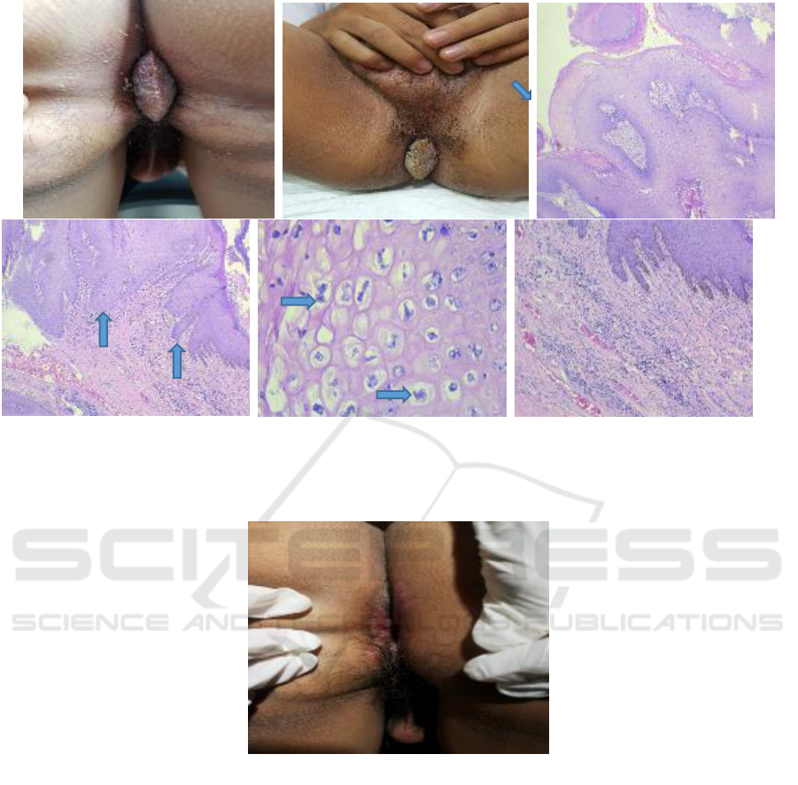
Figure 1. Results of physical and histopathology examination. A & B. Physical examination on August 23
rd
, 2018 at the
Dermato-venereology Clinic, Dr. Kariadi General Hospital Semarang; C. hyperplastic stratified keratinized squamous
epithelium with papillomatous growth [Hematoxylin & eosin (H&E), 40x]; D. acanthosis (H&E, 40x); E. koilocytosis (H&E,
400x); F. dermis consisted of inflammatory cells (H&E, 100x).
Figure 2. The patients' condition three months after surgery on January 29
th
, 2019, at the Dermatovenereology Clinic, Dr.
Kariadi General Hospital Semarang.
4 CONCLUSION
Surgical excision and ARV therapy in perianal GCA
patient with HIV yielded a satisfactory result. The
prognosis of the patient, including quo ad vitam and
quo ad sanam are dubia ad malam, but quo ad
cosmeticam is dubia ad bonam. Regular evaluation
after surgery is required to identify and prevent
recurrence or metastasize potential.
REFERENCES
Akdag O, Yildiran G. 2018. Malign differentiation of a
large Buschke Loewenstein tumor in penis. Surg J.
4:53-4.
Atkinson AL, Pursell N, Sisay A. 2014. The giant
condyloma (Buschke Lowenstein Tumor) in the
immunocompromised patient. Case rep obsgyn.1-4.
Bessi ME, Dougaz W, Jonez M, et al. 2019. A giant
anorectal condyloma is not synonym of malignancy. J
Gastrointest cancer. 1-3.
Diani M, Boneschi V, Ramoni S, et al. 2015. Rapidly
invasive Buschke Lowenstein tumor associated with
A
B
C
D
E
F
ICTROMI 2019 - The 2nd International Conference on Tropical Medicine and Infectious Disease
378

Human Papillomavirus types 6 and 52. Am sex transm
dis. 42(10):547-8.
Guttadauro A, Chiarelli M, Macchini D, et al. 2015.
Circumferential anal giant condyloma acuminatum : a
new surgical approach. Dis of colon and rectum.
58(4):49-52.
Heukelom SV. 2016. Condylomata acuminata of HIV-
positive men may harbour focal areas of dysplasia:
relevant implications for the management of human
papillomavirus-induced disease in high-risk patients.
Br J Dermatol. 175:735-43.
Indriatmi W, Daili SF. 2018. Kutil anogenital pada infeksi
HIV/AIDS. In: Hidayati AN, Daili SF, Niode NJ, et al,
editors. Manifestasi dan tatalaksana kelainan kulit dan
kelamin pada pasien HIV/AIDS. 1
st
ed. Jakarta: FKUI.
p. 66-72.
Kauffman LC, Alexandrescu DT. 2018. Giant condyloma
acuminata of Buschke and Loewenstein. eMedicine
Spec Derm Viral Inf. Available from:
https://emedicine.medscape.com/article/1132178-
overview#showall [accessed 25 February 2018].
Kim HG, Kesey JE, Griswold JA. 2018. Giant anorectal
condyloma acuminatum of Buschke–Löwenstein
presents difficult management decisions. Surg J. 4:1-4.
Kose R, Tas S. 2016. Treatment of a giant condyloma
acuminatum by surgical excision. Firat Med J.
21(1):54-6.
Koukoura O, Klados G, Strataki M, et al. 2015. A rapidly
growing vulvar condyloma acuminatum in a young
patient. BMJ case rep. 1-2.
Mistrangelo M, Dal CI, Volpatto S, et al. 2018. Current
treatments for anal condylomata acuminata. Minerva
chir. 73(1):100-6.
Murtiastutik D. 2008. Kondiloma akuminata. In: Barakbah
J, Lumintang H, Martodihardjo S, editors. Infeksi
Menular Seksual. 1
st
ed. Surabaya: Airlangga
University Press. p. 170-9.
Perianal Giant Condyloma Acuminatum with HIV Treated with Surgical Excision
379
