
Vogt Koyanagi Harada Syndrome
Ernawati Hidayat
1*
, Milka Wulansari
1
, Yosep Ferdinand Rahmat Sugianto
1
, Galih Sari Damayanti
1
Meira Dewi Kusuma Astutik
2
1
Department of Derrmatovenereology, Faculty of Medicine,
Diponegoro University/Dr.Kariadi Hospital
2
Department of Anatomic Pathology, Faculty of Medicine, Diponegoro University/Dr.Kariadi Hospital
*Corresponding author
Keywords: VKH, vitiligo, uveitis
Abstract: Vogt-Koyanagi-Harada syndrome (VKHS) is a rare autoimmune disorder involving pigmented multiorgan.
VKHS reported 6-8% in Asia,1-4 % in North America, and 2-4 % in Brazil of all uveitis. Diagnosis is made
based on American Uveitis Society (AUS) criteria; no history of eye trauma, and minimum 3 of 4 signs:
bilateral chronic iridocyclitis, uveitis, neurologic signs (tinnitus, neck stiffness, CNS symptoms), and
dermatologic signs (alopecia, poliosis, vitiligo). A 51-year-old female presented with generalized
hypopigmented-non pruritic patches since 8-year-old. Visual impairment was reported one month before.
Patches of white hair were found on the forehead and eyelashes. Tinnitus and frequent headache were
reported. There was no history of eye trauma. Ophthalmologic examination revealed bilateral panuveitis and
retinal detachment on the right eye. Histopathologic examination showed no melanin pigment in the basal
layer. The patient was treated with systemic methyl-prednisolone and topical steroid creams. Prognosis is ad
malam for ad sanam and ad cosmeticam. The diagnosis was based on AUS criteria and histopathologic
examination. The precise etiology for VKHS is challenging to establish. Treatment required long term
steroid administration and routine follow up to assess the progression of this disease.
1 INTRODUCTION
Vogt Koyanagi Harada Syndrome (VKHS), initially
described as an uveomeningoencephalitic syndrome,
is a rare granulomatous inflammatory disease that
affects pigmented structures, such as eye, inner ear,
meninges, skin, and hair (Geel et al, 2016; Lavezzo
et al, 2016). The disease is defined as a non-
infectious, bilateral, and granulomatous panuveitis
that occurs with or without extraocular
manifestations, and it typically affects young adults
(Ando et al, 2018).
In 1906, Vogt reported a patient
with atraumatic, idiopathic uveitis, poliosis, and
alopecia, a syndrome that in time would be
associated with his name. In 1926, Harada reported
five cases of bilateral posterior uveitis and retinal
detachment. In 1929, Koyanagi reported 16 patients
with headache, fever, dysgeusia, vitiligo, poliosis,
alopecia, bilateral anterior uveitis with occasional
exudative retinal detachment. Koyanagi published a
review article associating the posterior eye
involvement unequivocally with auditory and
integumentary manifestations. In 1932, Babel
suggested that these cases represented a single
entity, which was then named Vogt-Koyanagi-
Harada Disease (Geel et al, 2016; Lavezzo et al,
2016; Ando et al, 2018).
VKHS more frequently affects individuals of
pigmented skin, such as Asians, Middle Easterners,
Hispanics, and Native Americans (Ando et al, 2018;
O’Keefe et al, 2018; Silpa-Archa et al, 2016; Emily
et al, 2018).
The incidence of VKHS varies. Among
all cases of uveitis, it was reported 6-8% in Asia,1-4
% in North America, and 2-4 % in Brazil of all
uveitis. In China, VKHS is one of the most common
uveitis entities. In the United States, the incidence of
VKHS is approximately 1.5 to 6 per 1 million
patients, while in Japan, it is seen in approximately
800 new patients each year. Most studies have found
that women were affected more frequently than men
and that most patients were in the second to fifth
decades of life at the onset of the disease. However,
children and the elderly may also be affected.
Women account for 55 to 78 % of VKHS patients in
348
Hidayat, E., Wulansari, M., Sugianto, Y., Damayanti, G. and Astutik, D.
Vogt Koyanagi Harada Syndrome.
DOI: 10.5220/0009988503480351
In Proceedings of the 2nd International Conference on Tropical Medicine and Infectious Disease (ICTROMI 2019), pages 348-351
ISBN: 978-989-758-469-5
Copyright
c
2020 by SCITEPRESS – Science and Technology Publications, Lda. All rights reserved
the United States and approximately 38 % in Japan,
showing a global variation in gender predilection
(Lavezzo et al, 2016; Giannakourasa et al, 2017).
The exact etiology of VKHS is still a matter of
inquiry. The most accepted mechanism involves
autoimmune aggression against antigens associated
with melanocytes in a genetically susceptible
individual after a virus infection trigger.
Immunological and histopathological studies suggest
that VKH is an autoimmune inflammatory condition
mediated by CD4þ T cells that target melanocytes.
These activated T cells likely initiate the
inflammatory process through generation of
cytokines, IL 17 and IL 23,35 in individuals with
altered tolerance to melanocytes from deficient T
regulatory cells (Lavezzo et al, 2016; Emily et al,
2018; Baltmr et al, 2017).
Diagnosis is made based on American Uveitis
Society (AUS) criteria; no history of eye trauma or
surgery, and minimum 3 of 4 signs: Bilateral chronic
iridocyclitis, posterior uveitis (multifocal exudative
retinal or RPE detachments; disc hyperemia or
edema; or “sunset glow fundus”, which is a yellow-
orange appearance of the fundus due to
depigmentation of the RPE and choroid), Neurologic
signs (tinnitus, neck stiffness, cranial nerve or
central nervous system symptoms or cerebral spinal
fluid pleocytosis) and Cutaneous findings (alopecia,
poliosis or vitiligo) (Geel et al, 2016; Lavezzo et al,
2016; O’Keefe at al, 2017; Coutinho et al, 2017).
Early diagnosis and prompt immunosuppressive
treatment with corticosteroids and other
immunosuppressives can halt the progression and
prevent recurrence and vision loss. The mainstay of
treatment of VKHS is prompt, high-dose systemic
corticosteroids, followed by slow tapering of oral
corticosteroids throughout a minimum 6-month
period (Lavezzo et al, 2016; Joanne et al, 2014).
Localized lesions of vitiligo can be treated with a
high-potency fluorinated corticosteroid for 1–2
months. Treatment can be gradually tapered to a
lower potency corticosteroid (Birlea et al, 2012).
2 CASE
A 12-year-old girl came to Dermato-venereology
Departement Dr.Kariadi Hospital with generalized
hypopigmented-non pruritic patches since 8-year-
old. Visual impairment was reported one month
before. Patches of white hair were found on the
forehead and eyelashes. Tinnitus and frequent
headache were reported. There was no history of eye
trauma or surgery. Ophthalmologic examination
revealed bilateral panuveitis and retinal detachment
on the right eye. No similar history or symptoms in
his family. Physical examination showed body
height 165 cm, and bodyweight 65kg, blood pressure
is 120/70 mmHg, respiratory rate 18x/m, heart rate
88x/m, and axilla temperature 36°C. Skin lesions are
generalized hypopigmented macules, poliosis on
forehead and eyelashes.
The routine laboratory examination result was
typical. Histopathologic examination showed no
melanin pigment on the basal layer. The working
diagnosis is Vogt Koyanagi Harada Syndrome. The
patient was given oral methylprednisolone 16mg 2-
0-1 long term and followed by tapering off and
fluticasone propionate 0,05%/ twice daily.
Prognosis, quo ad sanam dubia ad malam, quo ad
kosmetikam dubia ad malam.
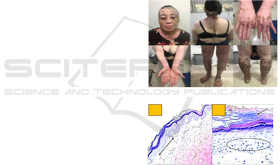
Figure 1. Generalized hypopigmented patches
Figure 2. Histopathologic examination A) no melanin
pigment on the basal layer. B) Hyperkeratosis and no
melanin pigment
3 DISCUSSION
The diagnosis of Vogt Koyanagi Harada Syndrome
in the patient was established by history, physical
examination, and histopathological examination.
Anamnesis a 51-year-old female presented with
generalized hypopigmented-non pruritic patches
since 8-year-old. Visual impairment was reported
A
B
Vogt Koyanagi Harada Syndrome
349

one month before. Patches of white hair were found
on the forehead and eyelashes. Tinnitus and frequent
headache were reported. There was no history of eye
trauma or surgery. Ophthalmologic examination
revealed bilateral panuveitis and retinal detachment
on the right eye. No similar history or symptoms in
his family. VKHS presents clinically in 4 different
phases: prodromal, acute uveitis, convalescent, and
chronic recurrent.
The prodromal phase may present a viral
infection and last anywhere between a few days to a
few weeks. In this phase, before ocular involvement,
clinical manifestations are predominantly
extraocular and include headache (82%),
meningismus (55%), fever (18%), nausea (9%),
vertigo (9%), orbital pain, auditory disturbances,
photophobia, and tinnitus. However, some patients
present with typical clinical features of VKH
without the prodromal symptoms. The acute uveitic
phase is following the prodromal phase, blurring of
vision develops in patients in both eyes, although the
involvement of 1 eye may be delayed. The
convalescent phase is Several weeks to months after
the acute uveitic phase, depigmentation of the
choroid, vitiligo, and poliosis occurs. The
convalescent phase usually lasts for months. Chronic
recurrent intraocular inflammation develops in some
of the patients and is characterized by exacerbations
of granulomatous anterior uveitis that is usually
resistant to systemic steroid therapy. This chronic
recurrent phase usually develops 6 to 9 months after
initial presentation and is also marked by
complications such as retinal pigment epithelium
(RPE) proliferation, subretinal fibrosis subretinal
neovascular membranes, posterior subcapsular
cataract, posterior synechiae, open-angle glaucoma
and, occasionally, angle-closure glaucoma, as well
as band keratopathy
(Lavezzo et al, 2016; Silpa-
Archa et al, 2016)
Physical examination revealed skin lesions are
generalized hypopigmented macules, poliosis on
forehead and eyelashes. Based on literature VKHS
have founded skin and hair changes: alopecia,
vitiligo, and poliosis.(Geel et al, 2016;Lavezzo et al,
2016; Silpa-Archa et al, 2016)
Histopathologic examination, in this case,
showed no melanin pigment in the basal layer. A
skin biopsy is rarely necessary to confirm the
diagnosis of vitiligo. Generally, histology shows an
epidermis devoid of melanocytes in lesional areas
and sometimes sparse dermal, perivascular, and
perifollicular lymphocytic infiltrates at the margins
of early vitiligo lesions and active lesions, consistent
with cell-mediated immune processes destroying
melanocytes in situ.(Birlea et al,2012)
The patient was given oral methylprednisolone
16mg 2-0-1 long term and followed by tapering off
and fluticasone propionate 0,05% cream/ twice
daily. Based on literature treatment of VKHS is
prompt, high-dose systemic corticosteroids,
administered either orally (prednisone 1–1.5 mg/kg
per day) or through a short course of intravenous
delivery (methylprednisolone 1000 mg per day,
intravenously, during 3 days), followed by slow
tapering of oral corticosteroids throughout a
minimum 6-month period. Timing to initiate
therapy, corticosteroid dosing, and duration of
therapy are the key factors in reducing the chance of
recurrences. (Lavezzo et al, 2016.Joanne et al,2014)
Localized lesions of vitiligo can be treated with a
high-potency fluorinated corticosteroid (e.g.,
clobetasol propionate ointment, 0.05%) for 1–2
months. Treatment can be gradually tapered to a
lower potency corticosteroid (e.g., hydrocortisone
butyrate cream, 0.1%). (Birlea et al,2012) Prognosis,
quo ad sanam dubia ad malam, quo ad kosmetikam
dubia ad malam.
4 CONCLUSION
Has been reported a case of Vogt Koyanagi Harada
Syndrome in a 51-year-old female presented with
generalized hypopigmented-non pruritic patches
since 8-year-old. Visual impairment was reported
one month before. Patches of white hair were found
on the forehead and eyelashes. Tinnitus and frequent
headache were reported. Physical examination
revealed Skin lesions are generalized
hypopigmented macules, poliosis on forehead and
eyelashes. Histopathologic examination, in this case,
showed no melanin pigment in the basal layer. The
mainstay of treatment of VKHS is prompt, high-
dose systemic corticosteroids, followed by slow
tapering of oral corticosteroids throughout a
minimum 6-month period.
Localized lesions of
vitiligo can be treated with a high-potency
fluorinated corticosteroid for 1–2 months. Treatment
can be gradually tapered to a lower potency
corticosteroid. Prognosis quo ad sanam dubia ad
malam, quo ad kosmetikam dubia ad malam.
ICTROMI 2019 - The 2nd International Conference on Tropical Medicine and Infectious Disease
350

REFERENCES
Ando T, Kato H, Mochizuki K, Ozawa K, Goshima S,
Matsuo M. 2018. MR findings of the orbit in patients
with Vogt–Koyanagi–Harada disease. Feb 23;1-6.
Baltmr A, , Lightman S, Netzer OT. 2016. Vogt Koyanagi
Harada syndrome- current perspectives. Clinical
Ophthalmology. 10 2345–2361
Birlea AS, Spritz RA, Norris DA Vitiligo. 2012. In: Wolff
K, Goldsmith LA, Katz SI, Gilchrest BA, Paller AS,
Leffell DJ, editors. Fitzpatrick's dermatology in
general medicine. 8
th
Ed. New York : Mc Graw Hill.
1680-90p
Coutinho I, Pedrosa C, Santos C, et al. 2017. A challenged
case of Vogt_Koyanagi_Harada syndrome: when
dermatological manifestations came first. Springer
Science Business Media Dordrecht. 1- 6
Emily Su, Vikash S. Oza, Paul Latkany. 2018. A case of
recalcitrant pediatric Vogt-Koyanagi-Harada disease
successfully controlled with adalimumab. Journal of
the Formosan Medical Association. 1-6
Geel NV, Speeckaert R. 2016. Acquired Pigmentary
Disorders. In: Burns T, Breathnach S, Cox N,
Griffiths C. Rook’s Textbook of Dermatology. 8
th
Ed.
Volume 1. Oxford: Blackwell scientific Publication.
88.43p.
Giannakourasa P, Andreanosa K, Giavia B, Diagourtas A.
2017. Optical Coherence Tomography Angiography:
Employing a Novel Technique for Investigation in
Vogt-Koyanagi-Harada Disease. Case Rep
Ophthalmol. 8:362–369
Joanne YW Ng, Fiona OJ Luk, Timothy YY Lai, Chi-Pui
Pan. 2014. Influence of molecular genetics in Vogt-
Koyanagi-Harada disease. Journal of Ophthalmic
Inflammation and Infection. 4:20
Lavezzo MM, Sakata VM, Morita C, Rodriguez
EE, Abdallah SF, da Silva FT, Hirata CE, Yamamoto
JH. 2016. Vogt-Koyanagi-Harada disease: review of a
rare autoimmune disease targeting antigens of
melanocytes. Orphanet J Rare Dis.1-21
O’Keefe GA, Rao NA. 2017. Vogt-Koyanagi-Harada
disease. Surv Ophthalmol. 62(1):1–25
Silpa-Archa S, Silpa-Archa N, Preble JM, Foster CS.
2016. Vogt- Koyanagi-Harada syndrome: perspectives
for immunogenetics, multimodal imaging, and
therapeutic options. Autoimmun Rev. 15(8):809–819
Vogt Koyanagi Harada Syndrome
351
