
Multiple Genital Ulcer on a Male Patient Due to Fungal
Balanoposthitis Suspect of Candida Albicans Infection Mimicking
Genital Herpes: A Case Report
Wresti Indriatmi
1*,
, Giorgio Barnes Komala
1
1
Department of Dermatovenereology, Medical Faculty of Universitas Indonesia, Jakarta, Indonesia
*Corresponding author
Keywords : Genital ulcer, balanoposthitis, case report, candida albicans
Abstract: Genital ulcer can be caused by an infectious or non-infectious diseases. The appearance of genital ulcer can
mimick one to another causative etiologies, so it becomes difficult to differentiate. We report one case of
34-year-old male, uncircumcised, with a painful multiple genital ulcer, he only have a sexual intercourse
with his wife, but conduct an oral sex. So our working diagnosis was genital herpes and we treated the
patient with valacyclovir 500 mg, twice daily, for 7 days. But turns out, the result of the culture for
microorganism was shown an unspecified fungal colonies growth, instead of bacteria. And also, after one-
week treatment, the ulcer became more profound and felt itchy rather than pain. We changed our working
diagnosis to balanoposthitis due to fungal infection with a suspicion of Candida albicansand treated the
patient with fluconazole 150 mg, single dose, bifonazole 1% cream and hydrocortisone 1 % cream twice
daily. After 5 days with a new regimen of treatment, almost all the ulcer was healed, no itchy or pain
sensations. With this case report, we hope that as a clinicians, we can be more careful and thorough in
examining a patient with a genital ulcer. A KOH examinations can be a consideration for an additional
diagnostic tools.
1 INTRODUCTION
Sometimes, we find a genital ulcer case in our daily
practice. This condition can be differentiate by the
etiologies, infectious or non-infectious. The most
common cause for the infectious etiology are from
sexually transmitted infections (STI), such as herpes
simplex virus (HSV), syphilis (Treponema pallidum),
chancroid (Haemophilus ducreyi), lympho
granuloma venereum (Chlamydia trachomatis),
fungal infection (e.g., Candida species) and others.
In America, the most common cause of genital ulcer
are HSV type 1 and 2, followed by syphilis and
chancroid. (Roet MA et al., 2013; Ballard RC, 2008)
Usually, ulcer is accompanied with pain or
uncomfortable sensation. That’s why finding the
right etiology in the most efficient time, is so
important.
We report one case of genital ulcer that was
caused by a fungal etiology. At first, we thought
about an atipical genital herpes symptom but later on
the manifestations was changed. Balanoposthitis due
to candida infection only occurs 20% of all
balanoposthitis cases. (Edwards EK et al.,
2013).That’s why it is quite important for us, as
clinicians, to be more careful and examine patient
more thoroughly in the future. Thus, we can
consider a KOH examination for an additional
simple diagnostic tools to make a decision.
2 CASE
A 34-year-old man, Japenese origin, presented with
a multiple genital ulcer since 2 days before (Figure
1). He felt moderate pain upon pressure and also
uncomfortable sensation while urinating. No history
of discharge from urethra. He said that his last
sexual intercourse is only with his wife. He also did
an oral sex. He hasn’t give any topical medicine or
taking an oral medication for his conditions. No
fever or other prodromal symptoms. On the physical
examinations, the penis was uncircumcised and there
were multiple ulcers, around 10 – 15 ulcers,
Indriatmi, W. and Komala, G.
Multiple Genital Ulcer on a Male Patient Due to Fungal Balanoposthitis Suspect of Candida Albicans Infection Mimicking Genital Herpes: A Case Report.
DOI: 10.5220/0009987903250328
In Proceedings of the 2nd International Conference on Tropical Medicine and Infectious Disease (ICTROMI 2019), pages 325-328
ISBN: 978-989-758-469-5
Copyright
c
2020 by SCITEPRESS – Science and Technology Publications, Lda. All rights reserved
325
approximately 1 mm – 5 mm diameters, mostly on
the preputium and a few on a coronary sulcus. The
ulcer itself was shallow, clean, moist, no induration,
confluence, tender, no active discharge, and edema
surrounding the ulcer. No lymph nodes enlargement
on the inguinal area.
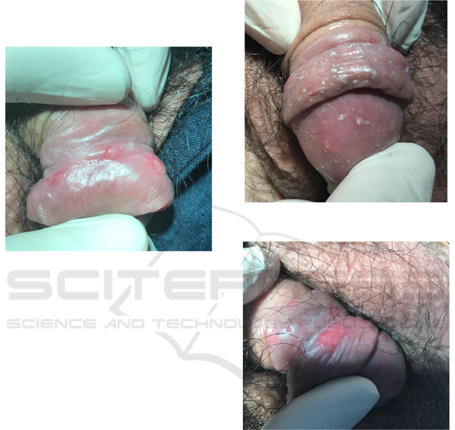
Figure 1.Clinical image of multiple ulcers on preputium
and a few on coronary sulcus.
We did some laboratory examinations for this
patient, consist of HSV-1 IgM, HSV-2 IgM, VDRL,
TPHA, Anti-HIV, resistance and microorganism
culture examination from the base of the ulcer, and
complete urinalysis. The complete urinalysis just
showed a slightly increase in white blood cells count
(5-7/Hpf). So, our working diagnosis was initial
lesion of genital herpes. We gave him valacyclovir
500 mg, twice daily, for 7 days.
One week later, the patient came for a second
visit. The rest of the laboratory result was finished,
all were within normal limit except for the resistance
and microorganism culture examination, showed an
unspecified fungal colonies. He also complains that
the ulcer was getting worse, itchy instead of pain
and there’s an active discharge from his ulcer. He
also stated that his wife also complain of itchy in her
genital. From the physical examination, the numbers
of the ulcers was increased, become a deep red
colour, some confluenced with each other, and a
thick curdy white-yellowish exudate (Figure 2 and
3). So, we changed our working diagnosis from
HSV infection to fungal balanoposthitis, with
suspicion of Candida albicansinfection. We treated
him with a single dose of fluconazole 150 mg orally,
topical bifonazole 1% cream, twice daily and topical
hydrocortisone 1% cream, twice daily. The
hydrocortisone was applied right after the bifonazole
cream.
Figure 2.A thick curdy white-yellowish exudates covering
most of the glans penis.
Figure 3. After being cleaned with normal saline, the ulcer
was showed more deep red color and there was a few new
small ulcers, on the glans of the penis and coronary sulcus.
Five days later, the patient came in again for a
follow up. The pain was minimal, no itchy, no
discharge and the number of ulcers were less than
before. Physical examination showed no ulcer,
erythema and discharge (Figure 4). So we
considered the treatment was completed.
ICTROMI 2019 - The 2nd International Conference on Tropical Medicine and Infectious Disease
326
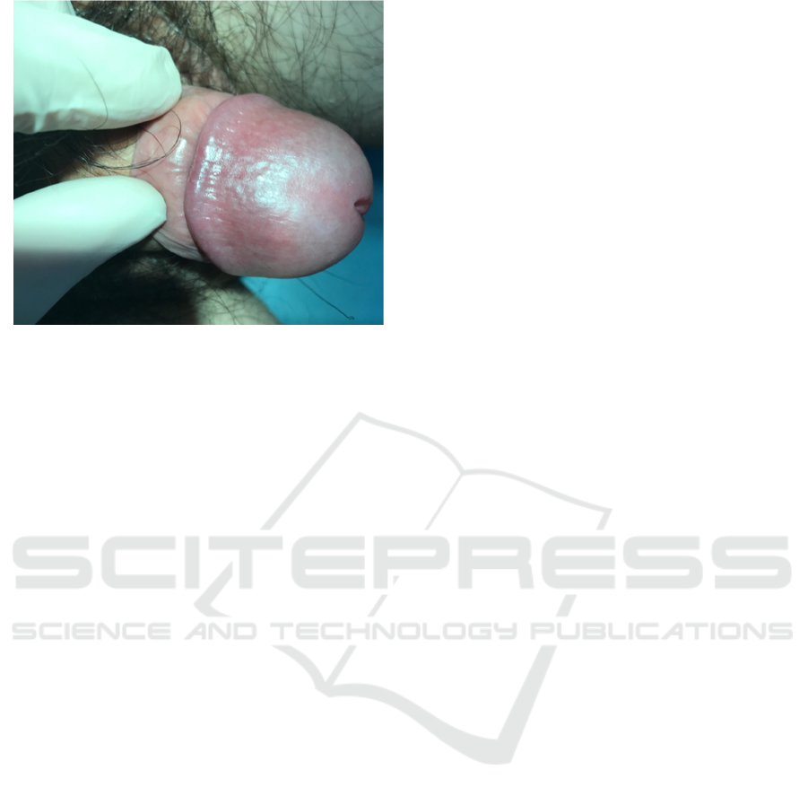
Figure 5. Healed of multiple ulcer after anti fungal
treatment.
3 DISCUSSION
A multiple genital ulcer usually caused by sexual
transmitted infection (STI), such as HSV, syphilis
and chancroid. Risk factors for genital ulcers are
lack of male circumcision, multiple sex partners (life
time or current), nonrecognition of ulcers in
prodormal stage, serodiscordant sex partners,
unprotected sexual contact and uprotected skin to
skin contact with ulcers. In genital ulcer case, we
should ask for more complaints in addition to the
symptoms of STI. Moreover, the past medical and
sexual history is important to assess the risk
behavior of the patient with STI. .(Roet MA et al.,
2013; Ballard,2008)
Balanoposthitis is defined as inflamation of the
glans or the prepuce.(Edwards EK et al., 2013;
Griffiths et al., 2016). A lot of condition can affect
the glans condition, from infectious to non-
infectious. Infectious etiologies such as Candida
species, Streptococci, anaerobes, Staphylococci,
Trichomonasvaginalis, herpes simplex virus etc. For
non-infectious such as, lichen sclerosus, lichen
planus, psoriasis, zoon balanitis, eczema to
premalignant condition, such as bowen’s disease,
bowenoid papulosis. (Ballard RC, 2008) But, all
cases of balanoposthitis was associated with poor
hygine and uncircumcised.(Edwards EK et al., 2013;
Habif TP, 2016; Griffiths et al., 2016). In this case,
the patient had a history of unprotected sex, he
confessed he only have sexual intercourse with his
wife. Patient’s history matches the risk factors for a
genital ulcer and also balanoposthitis.
Genital ulcer has several characteritics according
to the causative agent.(Roet MA et al., 2013; Ballard
RC,2008; Kundu RV, 2012; Habif TP, 2016; Farida
Z 2015). Genital HSV infection usually begins as
multiple vesicular lesions, located inside the foreskin,
labia, vagina, or rectum. Vesicles may rupture
spontaneously, becoming painful, shallow ulcers.
Sometimes there’s a prodormal symptoms, around
20% of the case. Primary syphilis usually begins
with a single, painless, well-demarcated ulcer
(chancre) with a clean base and indurated border.
Chancroid ulcers are usually deep, nonindurated,
bleeds easily, painful and usually cover with
yellowish grey exudate. The ulcers occur on the
prepuce and frenulum of the penis in men or on the
vulva or cervix in women.(Kundu RV et al., 2013;
Habif TP, 2016; Griffiths et al., 2016)
Candida balanoposthitis less than 20% of cases
of balanoposthitis3, and the most common pathogen
is Candida albicans.
9
It can give a manifestation as
maculopapular lesions with diffuse erythema, edema,
ulcerations, and fissuring of prepuce, also itchy
sensation.(Edwards EK et al.,2013; Habif TP, 2016
In our patient, with a multiple shallow genital ulcer,
painful in the beginning, it’s really similar with the
herpes simplex ulcer. That’s why we diagnosed this
patient as genital HSV infection, at first. But later,
when the symptoms become itchy and especially the
culture showed a fungal growth, it become more
convincing to suspect of Candida albicansinfection.
It’s important to determine a causative etiology
for genital ulcer. Laboratory evaluation of an initial
genital ulcer should include culture or polymerase
chain reaction, testing for HSV infection, HSV type-
specific serology, serologic testing for syphilis, and
culture for H. Ducreyiin settings with a high
prevalence of chancroid. For candidalbalanoposthitis,
the work ups are sub-preputial culture and KOH
examinations.
1-3
This patient was done a serology test
for syphilis, HSV, HIV, culture examination, gram
examinations and complete urinalysis. We didn’t do
a KOH examination, because of the manifestations,
we haven’t thought about fungal infection in the first
place. But it can be a learning experience for
clincians when facing this kind of cases in the future.
The treatment for genital ulcers is depend on the
causative agent but for candida balanoposthitis, the
recommended topical regimens are clotrimazole
cream 1% and miconazole 2% cream. For the
alternative regimen, are fluconazole 150 mg
3,10
,
orally (if the symptom is severe) or nystatin 100.000
units/gram
3
(if resistance or allergy to imidazoles).
Although there’s one case report in China, 2016,
about a Candida albicansresistance towards
Multiple Genital Ulcer on a Male Patient Due to Fungal Balanoposthitis Suspect of Candida Albicans Infection Mimicking Genital Herpes:
A Case Report
327

fluconazole, so they’re implied to treat C.albicans
infection according to the drug sensitivity test.(Hu Y
et al., 2017).Topical imizadole can also be applywith
hydrocortisone 1% if there’s a sign of marked
inflammation.(Edwards EK et al., 2013).For this
patient we gave himfluconazole 150 mg, single dose,
orally, because we thought the discharge from the
candida is alot and there’s also a marked inflamation
around the ulcer. We also gave bifonazole 1% for
thetopical antifungal and hydrocrotisone 1% for the
inflamation. Balanoposthitis is often reccurenton a
poor hygene person, so the main definitive therapy
for this is circumcision.( Habif TP, 2016)
4 CONCLUSION
Genital ulcer is just a clinical manifestation which
sometimes can be difficult to diagnose properly.
Balanoposthitis is an inflamation in glans or prepuce,
but turns out, it can also manifest as a genital ulcer.
So, when it’s confusing to make a diagnosis for
genital ulcer, KOH examinations can be considered
as additional work up to find the causative etiology
of balanoposthitis in the future, especially when the
patient is uncircumcised.
REFERENCES
Ballard RC. 2008. Genital ulcer adenopathy syndrome.In:
Holmes KK, Sparling PF, Stamm WE, Piot P,
Wasserheit JN, Corey L et al (eds.). 4
th
Sexual
Transmitted Diseases. New York: McGraw-hill; 1999 -
1208
Edwards EK, Bunker CB, Ziller F, Meijden WIVD. 2013
European guideline for the management of
balanoposthitis.Int j STD & AIDS; 2014; 0(0):1-12
Farida Z, Nilasari H. 2015. Ulkus genital. In: DailiSE
Makes WIB Infeksi Menular Seksual, Pedoman Praktis
Diagnosis dan Tata laksana. Kementrian Kesehatan
Republik Indonesia.27-31. .
Griffiths CEM, Barker J, Bleiker T, Robert C, Creamer D.
2016. 9
th
Rook’s Textbook of Dermatology. Oxford:
Blackwell;111.23.
Habif TP. 6
th
Clinical Dermatology: A Color Guide to
Diagnosis and Therapy. Amsterdam: Elsevier; 2016:
516-29.
Hu Y, Hu Y, Lu Y, Huang S, Liu K, Han X, Mao Z et
al.2017. A case report of Penile Infection Caused by
Fluconazole- and Terbinafine- Resistant Candida
albicans.Mycopathologia; 182 (3-4): 397-402.
Kundu RV, Garg, A. 2012. Yeast Infections: Candidiasis,
Tinea (pityriasis) versicolor, and Malassezia
(pityrosporum) folliculitis. In: Goldsmith LA, Katz SI,
Gilchrest BA, Paller AS, Leffell DJ, Wolff K et all
(eds.). 8
th
Fitzpatrick Dermatology in General
Medicine. New York: McGraw-Hill; 2298-2301.
Pudjiati SR, Rusetiyanti N. Kandidosisgenitalis. Daili SF,
Nilasari H, Makes WIB, Zubier F, Rowawi R, Pudjiati
SR.2017. Infeksi Menular Seksual edisi ke-lima.
Depok: Badan penerbit FKUI;249-6
Roett MA, Mayor MT, Uduhiri KA. 2013.Diagnosis and
management of genital ulcer.Indian j of clinprac. 2013;
24(6): 507-15.
Wolff K, Johnson RA, Saavedra AP, Roh EK.2017. 8
th
Fitzpatrick’s Color Atlas and Synopsis of Clinical
Dermatology. New York: McGraw-Hill;602-3.
ICTROMI 2019 - The 2nd International Conference on Tropical Medicine and Infectious Disease
328
