
Cutaneous Sarcoidosis with Nail Manifestations:
A Rare Finding
Firman Parrol
1*
, Karin Rachmani
1
, Sri Linuwih Menaldi
1
, Sondang P. Sirait
1
, Eliza Miranda
1
1
Department of Dermatology and Venereology, Faculty of Medicine, University of Indonesia/Dr. Cipto Mangunkusumo
National Central General Hospital, Indonesia
Keywords: Sarcoidosis, nail sarcoidosis, cutaneous sarcoidosis
Abstract: Sarcoidosis is a multisystem granulomatous disease, characterized by naked granuloma lesions with
multiorgan involvement such as the lung, skin, eye, liver, peripheral lymph nodes and, nail. Cutaneous
sarcoidosis is one of the most common findings, while nail sarcoidosis presents with a very low
incidence. We report a 46-years-old male patient, referred to the Department of Dermatology and
Venereology CiptoMangunkusumoHospital with enlarged erythematous plaque lesions on the ear and
nose since three months ago. Our patient also showed dystrophy and onycholysis of
toenails.Erythematous plaque lesions are a hallmark of chronic sarcoidosis, whilenail involvement is
closely linked with chronic and systemic sarcoidosis. Furthermore, skin and nail biopsies also showed
naked granuloma lesions. These findings strengthen our evidence of sarcoidosis, specifically chronic
sarcoidosis. Althoughnail sarcoidosis is found in our patients, there is internal organ involvement.
Therefore, a stepwise approach needs to be done to diagnose nail sarcoidosis.
1 INTRODUCTION
Sarcoidosis is a systemic granulomatous disease
with multiple organ involvement. Sarcoidosis may
manifest in organs such as the lung, skin, eye, liver,
cardiovascular, gastrointestinal, urogenital, kidney,
peripheral lymph nodes, and nail.(
Haimovic A
,
2012).
On the skin, sarcoidosis can be classified into
specific and non-specific lesions. If skin biopsy
revealed naked granulomas, it is classified as
specific lesion. Whereas, non-specific lesions
usually seen in erythema nodosum. (Mana J et al.,
2012).
Cutaneoussarcoidosis is one of the most
common form of sarcoidosis. Lesions most often are
found on the head and neck, both symmetrically or
asymmetrically on any part of the skin and mucosa.
Although incidences are very low, sarcoidosis can
also appear in other locations, one of them is nail.
Mana J et al., 2012).Despite its rarity, dermatologists
must consider nail sarcoidosis as a serious problem
because its chronic nature and related to systemic
disease.(Momen SE et al., 2013).
In this paper we
report cutaneous sarcoidosis with nail manifestation
which is a rare finding. However, systemic
involvement is not yet discovered.
2 CASE
A 46-years-old man was referred with enlarged
erythematous plaque lesions in the ear and nose
since three months ago. The lesion first appeared
two years agoon the right ear with nummular size.
Since then, the lesion spread to the whole auricle of
left ear. Three months later, erythematous plaque
lesion also appeared on the anterior nares. The
patient observed that the lesion size was progressing
over time. There was no history of pain, itching, loss
of sensation, or bleeding. The patient then came to
dermatologist and he was given mupirocin cream
and wet dressing of potassium permanganate for one
year. However, the condition did not resolve after
the treatment.
After one year of the initial lesion on the ear,
patient complained a disfiguration on the nail. Initial
changes were observed in both toes. Both of the
nails were damaged accompanied by swollen toes.
This was also observed on the fourth right toes and
the fifth left toes. There were no history of pain, itch,
bleeding or trauma before. The patient was referred
to dermatologists who diagnosed him with fungal
infection of the nail. He was given loprox® for six
months but there was no improvement. The patient
Parrol, F., Rachmani, K., Menaldi, S., Sirait, S. and Miranda, E.
Cutaneous Sarcoidosis with Nail Manifestations: A Rare Finding.
DOI: 10.5220/0009987403030306
In Proceedings of the 2nd International Conference on Tropical Medicine and Infectious Disease (ICTROMI 2019), pages 303-306
ISBN: 978-989-758-469-5
Copyright
c
2020 by SCITEPRESS – Science and Technology Publications, Lda. All rights reserved
303
then referred to another dermatologist who
suspected him with Leprosy. The patient then
transferred to CiptoMangunkusumoCentral General
Hospital for slit skin smearexamination. There was
no history of chronic cough, dyspnea, heart disease,
fever, arthralgia, or ocular disorder. There were no
night sweats or significant weight reduction. There
was no history of any similar disease in patient’s
family. In 2013, patient was diagnosed with
astrocytoma at the spinal cord. According to patient,
the diagnosis was made based on MRI. The
astrocytoma paralyzed patient from the waist to the
feet. The sensory, motoric and autonom nerves were
damaged.
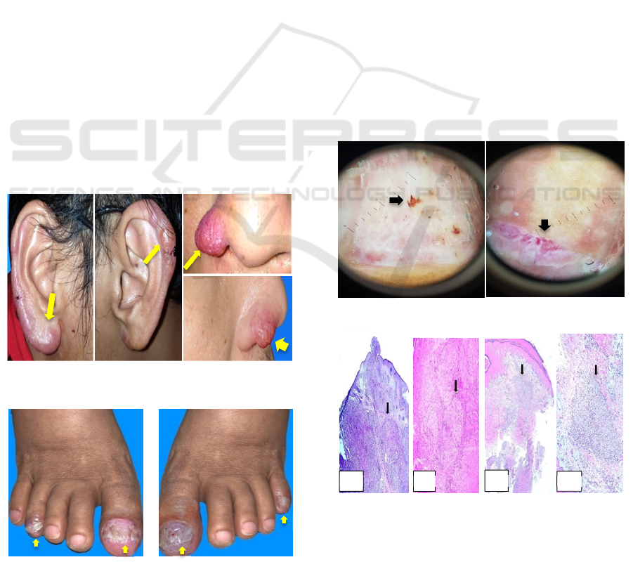
Physical examinations showed multiple
erythematous plaques with rough scales were seenin
both ears. The lesions werevary in size from
lenticular to nummular, arranged discreteand some
of them was confluent with well demarcated
borders. On the nosethere was a solitary nummular
circumscribed erythematous plaque with soft
consistency. (Figure 1) Dermoscopic examination
revealedorange color on skin lesion.
Onycodistrophy, onycholysis and digital clubbing
were observed on the fourth and first finger of the
right foot and the first and fifth finger of the left
foot. (Figure 2) On dermoscopic evaluation, there
were splinter hemorrhageon the nail bed. Multiple
atypical vesselswere found on the fourth nail of the
right foot. (Figure 3)
Figure 1. Multiple erythematous plaques were seen on
both ear and nose. (Arrows)
Figure 2. Nail dystrophy and onycholysis. (Arro.ws)
Based on history taking and physical examination, the
differential diagnosis were lupus vulgaris, cutaneous
sarcoidosis and cutaneous lymphoma. In order to
confirm our diagnosis, the Mycobacterium
tuberculosis (MTB),Mycobacterium Other Than
Tuberculosis (MOTT) culture and polymerase chain
reaction (PCR) obtained from skin tissue was done.
The results of MTB and MOTT culture were
negative, but PCR was positive for Mycobacterium
tuberculosis. Histopathological examination of the
nose revealed the epidermis of had flat rete ridges.
Epithelioid granuloma was observed in the dermis
with multiple giant cells surrounded by sparse
lymphocytes infiltrates. Nearby tissue was dominated
by inflammatory cells and areas of fibrosis. (Figure
4) On the other hand, histopathological examination
of the nail bed revealed acanthotic and irregular
elongation of the rete ridges. Epithelioid granuloma
was also observed in the dermis of the nailbed and the
proximal nail fold under the matrix. (Figure 4)
ZiehlNiehlsen staining showed no sign of acidfast
bacilli on the nose and nail specimen. Chest x-ray
examination revealed no sign of sarcoidosisor TB
infection in the lung, while x-ray examination on
patient's foot showed erosion on distal phalanx of
both thumb with osteolytic process on metatarsal and
phalanx of first finger, distal tibia and tarsal. No sign
of destruction was found.
Figure 3. Splinter hemorrhage and multiple atypical
vessels on the toes. (Arrows)
Figure 4. A, B. Epithelioid granuloma was observed in the
dermison plaque of the nose with multiple giant cells
surrounded by sparse lymphocytes infiltrates. (Arrows).C,
D. Epithelioid granuloma was also observed in the dermis of
the nailbed and the proximal nail fold under the matrix.
(Arrows)
A
B
C
D
ICTROMI 2019 - The 2nd International Conference on Tropical Medicine and Infectious Disease
304

3 DISCUSSION
Cutaneous sarcoidosis are classified into specific
lesions with histopathological findings of naked
granulomas, and nonspecific lesion that appear from
a process that does not form granulomas.(
Haimovic
A,2012) Skin manifestation are present in one fifth
of sarcoidosis. Differentiating these lesions is
important because cutaneous lesions have a
prognostic significance. (Mana J et al., 2012).
Non-
specific lesion such as erythema nodosum typically
associated with good prognosis and spontaneous
resolution while maculopapular lesions and
subcutaneous lesions are linked with remission of
systemic disease in two years. (Mana J et al., 2012).
Whole plaques and lupus pernio are closely
associated with chronic disease. (Mana J et al.,
2012).
The last type of lesions are disfiguring and
can have cosmetical, social, and psychological
impact. (Mana J et al., 2012).
Cutaneous lesions are found on our patient’s
nose and both ears. These lesions are identified to be
erythematous plaque lesions. It is known that plaque
lesions are typically persistent and associated with
chronic forms of sarcoidosis. (Mana J et al., 2012).
It
is consistent with the findings in our patients, whose
lesions appeared since two years ago, therefore
indicating a chronic form of sarcoidosis. Relatively
symmetric plaques and nodules that occur on the
nose, earlobes, cheeks, and digits, are consistent
with lupus pernio, which has a tendency for systemic
involvement. Nail changes are hardly found in
sarcoidosis.(Momen SE et al., 2013).However, when
present, it is closely associated with systemic
sarcoidosis. The prevalence of nail sarcoidosis was
reported (1.6%) of 188 patients with cutaneous
sarcoidosis.(Velen NK et al., 1987) Despite its
rarity, dermatologists must consider nail
sarcoidosisas a sign of chronic systemic disease.
.(Momen SE et al., 2013).One paper reviewed 33
patients with nail sarcoidosis to describe the changes
of nail.
3
The most common findings included nail
dystrophy, longitudinal ridges, subungual
hyperkeratosis, onycholysis, nail hyperkeratosis and
nail loss consecutively. Confirmation of sarcoidosis
was made from histopathological findings from of
the nail biopsies, which were the presence of naked
granulomatous. Among 33 patients, seven patients
are noted to have granulomatous infiltrates of the
dermis.(Losada-Campa et al., 1995; Kalb RE et al
1985).
In our patient, we also findonycodistrophy,
onycholysis and digital clubbing. Histopathological
examination revealed typical naked
granulomas.(Fernandez-Faith E et al., 2007).These
findings point us towards nail sarcoidosis.
Wakelin and James stated that the findings of
surrounding skin changes of erythematousplaques
over the proximal nail folds might direct towards a
diagnosis of sarcoidosis (Haimovic A et al 2012)
These skin changes are caused by the formation of
granulomas which have microcompressive effects in
the dermal compartment between the nail plate and
the phalanx.(Wakelin SH et al., 1995).Despite the
nail changes and granuloma lesion in the biopsy of
nail, one should have more evidence to confirm the
diagnosis of sarcoidosis in this patient. Nail
involvement in sarcoidosis is rare and is usually a
marker of chronic disease.Although most of the
cases reported are associated with digital bone cysts,
sarcoiddactylitis, and lupus pernio,cases without
these associations have also been described
proposed that most nail changes are secondary to
granulomatous compression of nail structures(Velen
NK et al., 1987).
Sarcoidosis is known to be “the great imitator”
because of its diverse manifestations and the ability
to resemble other cutaneous disease. .(
Haimovic
A,2012).Therefore, a stepwise approach of diagnosis
needs to be taken in every patient with
granulomatous inflammation.(Chopra S et al., 1999).
A stepwise approach to diagnose sarcoidosis in this
patient is divided into two parts. First, excluding
alternative cause of granuloma. Second, find at least
one additional organ involvement. Alternative
known cause of granuloma is very wide, including
tuberculosis, atypical mycobacteriosis, fungal
infections, reaction to foreign bodies, leishmaniasis,
rheumatoid nodules, and Melkersson-Rosenthal
syndrome. (Aranegui et al., 2010).
However, in this patient, another known cause
of granuloma are unlikely. Granulomatous lesion is
highly caused by sarcoidosis according to patient’s
history, negative culture of MTB, and MOTT, and
normal chestx-ray examination. Although PCR
showed positive for MTB,it will not affect the
diagnosis because positive result can still be found
in 80% of cutaneous sarcoidosis patient. Several
studies have shown the presence of Mycobacterium
DNA in sarcoid granuloma, suggesting previous
exposure to Mycobaterium, which might have
induced a granulomatous reaction. The common
problem raised during PCR assays is the high risk of
false positive results due to common laboratory
contamination or presence of killed or dormant
bacilli in the patient specimens. However, the key to
the diagnosis of tuberculosis is a positive result on
tissue culture which is the gold standard of
Cutaneous Sarcoidosis with Nail Manifestations: A Rare Finding
305

diagnosis. In our patient, tissue culture of MTB and
MOTT showed negative result. Therefore,
confirming cutaneous and nail sarcoidosis as our
diagnosis.(Makeshkumar V et al,2014;Brownell et al
2011).
Classic sarcoidal lesions in the small bones of the
hands and feet are well characterized and diagnosed
with conventional radiographs, on which they
demonstrate the familiar “lacy” lytic
appearance.(Moore SL et al., 2003). X-ray
examination on patient's foot showed erosion on
distal phalanx of both thumb with osteolytic process
on metatarsal and phalanx of first finger. This x-ray
finding is suggested as extracutaneous manifestation
of sarcoidosis. Another suspected systemic
manifestation in this patient was astrocytoma. This
finding raised question whether it is an astrocytoma
or granulomatous inflammation mimicking
astrocytoma. Neurosarcoidosis itself has a
predilection at the base of the brain or spinal cord
with cranial neuropathies are the most common
manifestation. Therefore, we plan to refer the patient
to Neurology Department to establish the diagnosis.
We also refer the patient to the Internal Medicine
Department to perform more diagnostic evaluation
to find involvement of other organs. We treat the
patients using clobetasol propionate cream 0.05%
twice a day for the lesion in the ear and nose. Lesion
in the nail is treated using clobetasol propionate
ointment 0.25%. Several literatures have reported
the efficacy of topical steroid to improve the
appearance of nail changes in 5 (15%) of the 33
patients. (Haimovic A et al., 2012; Aranegui et al.,
2010).
4 CONCLUSION
Cutaneoussarcoidosis is one of the most common
organs involved and appear in one third of
sarcoidosis cases. Cutaneoussarcoidosis lesion has
its prognostic significance. Different lesion could
have a different prognostic implication. In the other
hand, nail sarcoidosis is a rare manifestation of
sarcoidosis. Although it is rare, one must consider
the systemic involvement of sarcoidosis if the nail is
affected because its chronic and systemic nature. A
stepwise approach must be used to diagnose every
patient with nail involvement in sarcoidosis.
Documentation of additional organ involvement,
such as the lung, lymph nodes, eye, heart, and
nervous system is essential to diagnose nail
sarcoidosis which commonly observed with multi
organ involvement.
REFERENCES
Haimovic A, Sanchez M, Judson MA, Prystowsky S.
Sarcoidosis: a comprehensive review and update for
dermatologist. Part 1.Cutaneous disease.J Am
AcadDermatol. 2012;66:699e1-18.
Mana J, Marcoval J. Skin manifestations of
sarcoidosis.Presse Med. 2012;41:e355-74.
Momen SE, Al-Niaimi F. Sarcoid and the nail: review of
the literature. Clin and ExpDermatol.2013; 38: 119-
25.
Veien NK, Stahl D, Brodthagen H. Cutaneous sarcoidosis
in Caucasians. J Am AcadDermatol. 1987; 16: 534–40.
Losada-Campa A, De la Torre-Fraga C, de Gomez
Lian˜oA,Cruces-Prado MJ. Histopathology of nail
sarcoidosis.ActaDermVenereol.1995; 75: 404–5.
Kalb RE, Grossman ME. Pterygium formation due to
sarcoidosis.Arch Dermatol1985; 121: 276–7.
Wakelin SH, James MP.Sarcoidosis: nail dystrophy
without underlying bone changes. Cutis 1995; 55:
344–6.
Haimovic A, Sanchez M, Judson MA, Prystowsky S.
Sarcoidosis: a comprehensive review and update for
dermatologist. Part 2.Extracutaneous disease.J Am
AcadDermatol. 2012; 66: 719e1-10.
Chopra S, Vega-López F. Skin granulomas in clinical
practice. In: James DG, Zumla A, editors. The
granulomatous disorders. Cambridge, UK: Cambridge
University Press; 1999. p. 485-527.
Aranegui B, Garcia-Cruz A, de la Torre C, Gonza´lez-
Valladares MG. Trachyonychia and sarcoidosis. J Am
AcadDermatol 2010; 63: 59–60
Makeshkumar V, Madhavan R, Narayanan S. 2014.
Polymerase chain reaction targeting insertion sequence
for the diagnosis of extrapulmonary tuberculosis.
Indian J Med Res
Brownell I, Ramírez-Valle F, Sanchez M, Prystowsky S.
Evidence for Mycobacteria in Sarcoidosis. Am J
Respir Cell Mol Biol. 2011 Nov;45(5):899–905.
Moore SL, Terstein AE. Musculoskeletal sarcoidosis:
spectrum of appearance at MRImaging. Radiographics
2003; 23; 1389-99.
Fernandez-Faith E, McDonnell J. Cutaneous sarcoidosis:
differential diagnosis. ClinDermatol. 2007;25(3):276–
87.
ICTROMI 2019 - The 2nd International Conference on Tropical Medicine and Infectious Disease
306
