
A Case Report: Classical Clinical Presentation of Scrofuloderma
Confirmed with Novel Laboratory Workups
Riris Asti Respati
1*
, Rahmiati
2
, Teffy Nuari
1
, Rahadi Rihatmadja
1
, Eliza Miranda
1
,
Sri Linuwih Menaldi
1
1
Department of Dermatology and Venereology Faculty of Medicine Universitas Indonesia/
Dr. Cipto Mangunkusumo National Central General Hospital, Indonesia
2
Department of Anatomical Pathology Faculty of Medicine Universitas Indonesia/
Dr. Cipto Mangunkusumo National Central General Hospital, Indonesia
Keywords: Histopathology, IGRA, scrofuloderma, lymphadenitis, Xpert MTB/RIF
Abstract: Scrofuloderma, can develop on the skin through direct extension from an endogenous source of tuberculosis
such as lymph node. This report presents a case of scrofuloderma in its classical presentation in a 41-year-
old man, who suffered from a three-week history of a painless swelling that ulcerated. Physical examination
revealed two tender nodules on the left anterior neck and armpit. The diagnosis was confirmed bythe
presence of an underlying focus of infection tuberculous lymphadenitis, positive results ofMycobacterium
tuberculosis interferon-gamma release assay (IGRA), Xpert MTB/RIF, and histopathological examination.
Treatment with anti-tuberculosis regiment resulted in an excellent outcome.
1 INTRODUCTION
Cutaneous tuberculosis is a chronic infection of the
skin by Mycobacterium tuberculosis, and less
commonly by M. bovis and M. atypical. (Santos JB et
al., 2014). It is relatively rare and accounts for 1% of
extrapulmonary tuberculosis. In the Asian
community, most cases are between the ages of 10
and 50 years. (Ho SCK, 2003). Scrofuloderma, also
known as tuberculosis cutis colliquative, is a
common form of cutaneous tuberculosis followed by
other formse.g., tuberculosis verrucosa cutis and
lupus vulgaris(Sethi et al., 2012; Haase O et al.,
2014).
The development of clinical manifestations in
scrofuloderma should be understood as the result of
interactions among the environment, agent, and host.
(Santos JB et al., 2014).It most commonly occurs in
the neck from underlying tuberculosis in a deeper
structure, usually a lymph node, bone, and joint.
Starting with lymphadenitis, lymph node attachment
with surrounding tissue causes peri adenitis, which
becomes doughy and progresses to liquefaction.A
cold abscess is formed, and the skin erodes to form a
discharging sinus and fistula formation. The fistula
estuary extends and forms an ulcer. It is
characterized by elongated, irregularly shaped,
granulated tissue at the base, and covered with
seropurulent discharge.The ulcer may be healed with
scarring (Santos JB et al., 2014; Sethi et al.,
2012;Lai-Cheong JE et al.,2007).
Diagnostic steps usually begin with history
taking, physical examination, and confirmation tests,
e.g., polymerase chain reaction, tissue culture,
tuberculin skin test, and histopathology.
2,6
Treatment
of scrofuloderma is similar to pulmonary
tuberculosis. World Health Organization (WHO)
recommends regimens consisted of four drugs,
isoniazid (H), rifampicin (R), pyrazinamide (Z) and
ethambutol (E) given to intensive and continuation
phases.(Ho SCK, 2003;Kar S et al., 2011).
The following report showcases several
techniques, some with their shortcomings, that may
be utilized to improve the accuracy of diagnosis in a
clinically characteristic presentation of scrofuloderma.
2 CASE
A 41-year-old man presented with a three-week
history of multiple ulcerson the upper chest and neck
(Figure 1).It started from a painless neck masses six
Respati, R., Rahmiati, ., Nuari, T., Rihatmadja, R., Miranda, E. and Menaldi, S.
A Case Report: Classical Clinical Presentation of Scrofuloderma Confirmed with Novel Laboratory Workups.
DOI: 10.5220/0009987302990302
In Proceedings of the 2nd International Conference on Tropical Medicine and Infectious Disease (ICTROMI 2019), pages 299-302
ISBN: 978-989-758-469-5
Copyright
c
2020 by SCITEPRESS – Science and Technology Publications, Lda. All rights reserved
299
months before a consultation, that gradually
increased in number and size. They suppurated and
broke down, forming ulcers with granulating tissue.
There was no other trauma.
Systemic examination did not reveal drenching
night sweats and unexplained pyrexia. Nonetheless,
approximately2 kilograms of weight loss in the last
two months was admitted. He had neither significant
childhood lung disease or tuberculosis background
and had received BCG vaccination.
Physical examination from the upper chest and
bilateral supraclavicular skin revealed multiple
ulcers measured approximately 4x3 cm, with
suppuration and granulation tissue at the base and
deeply undermined edges.
Routine laboratory work-ups were unremarkable
except for an elevated erythrocyte sedimentation rate
(ESR) 25 mm/hr. Mycobacterium tuberculosis
interferon-gamma release assay (IGRA)was positive
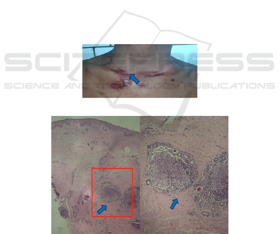
(1.61 IU/mL). Histopathological examination
showed epithelioid granulomas with a variable
number of Langhans giant cell and lymphocytes
(Figure 2)
On a subsequent visit, there were newtwo fixed
tender swelling in the left anterior neck and left
axillary (Figure 3). Physical examination showed
multiple nodular swelling measuring approximately
4x3 cm and 3x3 cm, fixated, doughy, without signs
of acute inflammation. The patient was consulted to
pulmonology division.
Further investigations had been carried out, such
as fine-needle aspiration biopsy (FNAB). The result
showed abscess material with lymphocytes,
histiocyte, neutrophil, with marked caseation
necrosis area. Mycobacterium tuberculosis was
detected using the Xpert MTB/RIF. Chest x-ray was
normal. Another workup, including polymerase
chain reaction (PCR) for Mycobacterium
tuberculosis, mycobacterial culture, Mycobacterium
other than tuberculosis culture, sputum culture, and
tuberculin skin test showed negative findings.
Furthermore, the patient started standard anti-
tuberculosis treatment consisting of four drugs,
rifampicin, isoniazid, pyrazinamide, and ethambutol.
After two months of therapy with anti-tuberculosis
regiments, he reported improved symptoms. No new
nodules had evolved (Figure 4).
Figure1. An ulcerated lesion with purulent fistulain the clavicular region.
Figure 2. Epithelioid granulomas, 100x (A), 400x (B) (hematoxyllin-eosin)
A
B
ICTROMI 2019 - The 2nd International Conference on Tropical Medicine and Infectious Disease
300
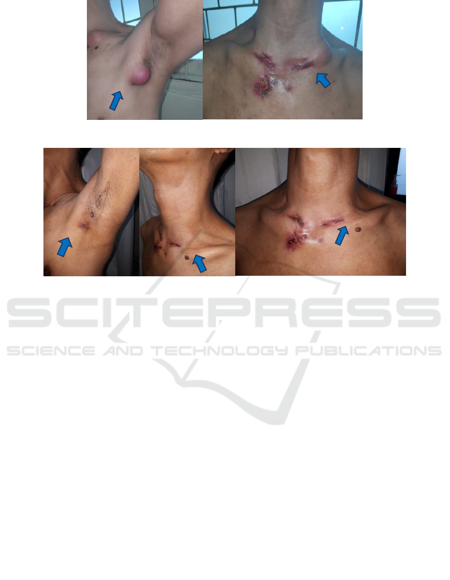
Figure 3. Cervical and axillary tender fixed masses
Figure 4. Lymphadenopathy has improved after two months of treatment.
3 DISCUSSION
Scrofuloderma is increasingly recognized as the
most common form of cutaneous tuberculosis in
adults (Pasmayathy L et al., 2008). This patient was
41 years old, included inthe profoundly affected
population who often have scrofuloderma. The
presentation of cutaneous tuberculosis depends on
the pathogenicity of the mycobacteria, route of
infection, and the level of host cell-mediated
immunity (CMI). Scrofuloderma lesions begin as
subcutaneous nodules which become doughy and
progressive liquefaction.(Santos JB et al., 2014;Ho
SCK,2003;Frankel A et al., 2009).The fistula estuary
extends and forms an ulcer. It is characterized by
suppuration and granulation tissue at the base and
deeply undermined edges.
2
In our case, they started
as painless swelling overlying his upper chest and
neck six months ago. The masses gradually enlarged
into cold abscess formation. It suppurates and breaks
down, forming an ulcer with granulation tissue at the
base covered with purulent discharge.
Scrofuloderma is caused by dormant tuberculosis
reactivation. There is contiguous involvement of
overlying skin from the underlying focus infection
such as tuberculous lymphadenitis. Scrofuloderma
from tuberculous lymphadenitis often affects the
neck, axillae, chest wall, and groin. (Ho SCK, 2003;
Aliaagaoglu et al., 2015). As can be seen in the
present case, the area of predilection was the neck
and axillae, which were the familiar site of
scrofuloderma. Moreover, there were two tender
swellings occurred in the left supraclavicular and
axillary region, which were tuberculosis
lymphadenitis. The left supraclavicular and axillary
nodes receive lymphatic drainage from the thoracic
duct causing of a lesion at multiple sites. In
scrofuloderma, the host has moderate cell-mediated
immunity. Following infection, macrophages that
circulate to lymph nodes and then haematogenic
spread to other parts of the body phagocyte the
mycobacteria. (Santos JB et al., 2014). Macrophages
act as antigen-presenting cells and interact with T
lymphocyte. (Ho SCK, 2003). When the
mycobacteria survive, in which they divide within
the macrophages. It is inducing the production of
cytokines such as IL-6, IL-12, IL-1α, and IL-1β,
resulting in the recruitment of monocytes,
lymphocytes, neutrophils, and dendritic cells.
1
The
persistent presence of these interleukins stimulating
macrophages will ultimately lead to their
differentiation into epithelioid and giant cells, which
will be more or less organized into granulomas
A Case Report: Classical Clinical Presentation of Scrofuloderma Confirmed with Novel Laboratory Workups
301

according to individual host factors. (Santos JB et
al., 2014) This explains the histopathology finding
that may show marked caseation necrosis and
abscess material with mixed inflammatory
infiltrations dominate the center of the lesion. (Santo
s JB et al., 2014.Besides, a presence of characteristic
tubercular granulomas with epithelioid cells in the
dermis is observed in 57%–96% of the samples.
(Rahman et al., 2018) showed only 16.7% cases
whereas granuloma with caseous necrosis found in
the dermis. On the other hand, 55.6% the majority of
the cases showed granuloma without caseous
necrosis.
12
Although not typical, a histopathological
examination from skin showed the presence of
epithelioid cell granulomas with a variable number
of Langhans giant cell and lymphocytes infiltrates in
the dermis. Central caseation necrosis was found
from FNAB, therefore support the diagnosis of
tuberculosis lymphadenitis. Some acid-fast bacilli
can be found (Ho SCK, 2003). This patient’s
bacteriological examination showed no acid-fast
bacilli. This is in line with the literature which stated
that bacteriological examination did not always find
acid-fast bacilli although a higher bacterial load. (Ho
SCK, 2003). Mycobacterial culture is the gold
standard for determining the presence of active TB
infection. (Ho SCK, 2003). However it is not always
possible to obtain a positive result. Positivity is
lower in an exclusively cutaneous presentation, that
is around 23% (Santos JB et al., 2014; Frankel A et
al., 2009).Polymerase chain reaction is used
primarily as a complement to clinicopathological
evaluation. It was reported in another study that one
out of three scrofuloderma patients had a positive
PCR finding(Santos JB et al., 2014;Tan WP et al.,
2007). Positive PCR result is not always obtained. In
the diagnosis of cutaneous TB, the sensitivity and
specificity of PCR vary greatly from literature.
Detection of Mycobacterium tuberculosisby a PCR
in this patient turned out to be negative.
A diagnosis of tuberculous lymphadenitis with a
cutaneous extension (scrofuloderma) has been
made.It was confirmed by history taking, clinical
features, a positive result on IGRA, Xpert MTB/RIF,
and histopathological findings. The patient was
quickly started on an anti-tubercular treatment
regimen that included isoniazid, rifampicin,
ethambutol, and pyrazinamide. The cutaneous lesion
regressed, and the ulcer starts healing.
4 CONCLUSION
In this patient, scrofuloderma occurs as a result of
extension from underlying tuberculous
lymphadenitis. By proper history taking and
morphologic features examination, a preliminary
diagnosis can be made, that must be followed by the
best methods available. Here our case had shown
that although mycobacterial culture and PCR test
failed to yield positive findings, IGRA, Xpert
MTB/RIF, and histopathological evaluations
provided the conclusive results. That the patient
responded well to treatment was another proof of the
infection.
REFERENCES
Santos JB, Figueiredo AR, Ferraz CE, Oliveira MH, Silva
PG, Medeiros VL. Cutaneous tuberculosis:
epidemiologic etiopathogenic and clinical aspects –
Part I. An Bras Dermatol. 2014;89(2): 219–29.
Ho SCK, Cutaneous tuberculosis: clinical features,
diagnosis, and management. H K Dermatol Venereol
Bull. 2003;11:130–8.
Sethi A. Tuberculosis and infections with atypical
mycobacteria. In: Goldsmith LA, Katz Si, Gilchrest
BA, Paller AS, Leffel DJ, Wolff K. Fitzpatrick’s
Dermatology in general medicine. 8th ed. New York:
McGraw-Hill. 2012: p.2225–40.
Haase O, Von Thomsen AJ, Zillikens D, Solbach W,
Kahle B. Recurrent abscesses of the neck:
scrofuloderma. JAMA Dermatol. 2014;150(8):909–10.
Lai-Cheong JE, Perez A, Tang V, Martinez A, Hill V,
Menage HP. Cutaneous manifestations of tuberculosis.
Clin Exp Dermatol. 2007;32(4):461–6.
Santos JB, Figueiredo AR, Ferraz CE, Oliveira MH, Silva
PG, Medeiros VL. Cutaneous tuberculosis: diagnosis,
histopathology and treatment - Part II. An Bras
Dermatol. 2014;89(4): 545–55.
Kar S, Krishnan A, Gangane N, Preetha K.
Scrofuloderma-a case series from rural India. Indian J
Tuberc. 2011;58(4):189–95.
Pandmavathy L, Lakshmana R, Ethirajan N, Manohar U,
Krishnaswamy BK. Scrofuloderma: a
clinicopathological and epidemiological study. Indian
J Dermatol Venereol Leprol. 2008;74(6):700.
Frankel A, Penrose C, Emer J. Cutaneous tuberculosis: a
practical case report and review for the dermatologist.
J Clin Aesthet Dermatol. 2009;2(10):19–27.
Aliaagaoglu C, Atasoy M, Albayrak H, Ozdemir S, Yanik
ME, Aktas A. Scrofuloderma: 30 years of experience
from eastern Turkey. Int J Dermatol. 2015;54(5):612–
3.
Tan WP, Tang MB, Tan HH. Scrofuloderma from the
acromioclavicular joint presenting as a chronic ulcer in
an immunocompetent host. Singapore Med J.
2007;48(9):243–5.
Rahman MA, Asaduzzaman ATM, Mahmud MM,
Chowdhury AM, Rahman MM, Haque MA, et al. Role
of polymerase chain reaction in the diagnosis of
cutaneous tuberculosis. J Pac Assoc Dermatol.
2018;28(3):277–83.
ICTROMI 2019 - The 2nd International Conference on Tropical Medicine and Infectious Disease
302
