
Diagnostic Utility of Epithelial Membrane Antigen (EMA) and
Calretinin (Cal) in Malignant Pleural Mesothelioma: A Case Report
Noni Novisari Soeroso
1*
, Ade Indriya
1
, Andika Pradana
1
, Rahmat Hidayat
1
1
Department of Pulmonology and Respiratory Medicine, Faculty of Medicine, Universitas Sumatera Utara,
Jl. Dr. Mansyur No. 5 Medan 20155, Sumatera Utara, Indonesia
Keywords: Calretinin, epithelial membrane antigen (EMA), malignant pleural mesothelioma, pleuroscopy
Abstract: Malignant pleural mesothelioma (MPM) is a rare disease, but the incidence of this disease has increased
over time. The diagnosis of MPM can be established based on radiological and pleuroscopic findings. The
immunohistochemistry of EMA and Calretinin may confirm the diagnosis of MPM. We reported a case of
70-years-old man, worked as a construction worker for 20 years came with severe left chest pain and
shortness of breath for 1 month. Chest X-ray shows left moderate pleural effusion, and thoracic CT-scan
result revealed mass in left pleura. Pleuroscopy had been performed and mass in the parietal pleura was
found with the histopathology reports suggestive for mesothelioma. Immunohistochemistry staining with
EMA and Calretinin were both positive, confirming the diagnosis of malignant pleural mesothelioma. He
was then scheduled for chemotherapy. However, his condition worsened over time and finally passed away.
1 INTRODUCTION
Mesothelioma is cancer that originates from
mesothelial cells lining the pleural cavity,
peritoneum, pericardium and tunica vaginalis.
Pleural mesothelioma is most commonly found in all
cases of mesothelioma (80% of cases) and asbestos
is believed to be one of the causes of pleural
mesotheliomas.
Australia is one of the countries with the highest
level of mesothelioma cases in the world. The
predicted mortality rate in the UK is estimated at
around 90.000 by 2050. In Japan, around 100.000
deaths over the next 40 years has been predicted.
Men have a risk three times greater than women in
cases of mesothelioma. However, no definite data of
mesotheliome prevalence in Indonesia (American
Cancer Society, 2018).
Malignant pleural mesothelioma is mostly
caused by exposure to asbestos in the workplace.
This is more common in older people, but
ssometimes may occur in younger people (Husain et
al., 2018).
When mesothelioma is diagnosed at an ealry
stage before invading lymph nodes and othe parts of
the body, treatment is more effective and prognosis
is more promising. However, the diagnosis of
mesothelioma appears very challenging. In fact, it is
often misdiagnosed as less serious conditions like
fibrous pleuritis. Thus, more diagnostic modalities
will provide help to estabilish the diagnosis of
mesothelioma accurately. We reported a patient
diagnosed with malignant pleural mesothelioma
based on positive immunohistochemistry findings of
epithelial membrane antigen and calretinin.
2 CASE REPORT
A 70-year-old man, heavy smoker, came with
shortness of breath for 1 month before being
hospitalized, getting worse when coughing or deep-
breathing. Non-productive cough has been
experienced for 3 months. He also felt severe left
chest pain in the last 1 month with VAS 5-6. He
used to work as construction worker. He also
experienced decreased appetite and loss of body
weight about 5 kg in 1 month. He has never taken
anti-tuberculosis drugs. While admission, his vital
sign were within normal limit except for increased
respiratory rate and SpO2 92% in room air.
Complete blood count showed anemia (9.3 mg/dl).
Arterial blood gas shows mild hypoxemia and
respiratory alkalosis (pH: 7.48; pCO2: 38.9 mmHg;
pO2: 70 mmHg; HCO3: 26.9 mmol / L; BE: 2.7
mmol / L; SaO2: 93%). Chest X-ray showed
Soeroso, N., Indriya, A., Pradana, A. and Hidayat, R.
Diagnostic Utility of Epithelial Membrane Antigen (EMA) and Calretinin (Cal) in Malignant Pleural Mesothelioma: A Case Report.
DOI: 10.5220/0009863402050207
In Proceedings of the 2nd International Conference on Tropical Medicine and Infectious Disease (ICTROMI 2019), pages 205-207
ISBN: 978-989-758-469-5
Copyright
c
2020 by SCITEPRESS – Science and Technology Publications, Lda. All rights reserved
205
moderate left pleural effusion. Thoracentesis was
performed and about 1000ml fluid was drained, with
pleural fluid cytology result was chronic
inflammatory smear. Thoracic CT scan revealed a
suspicion for left pleural tumors in the apex and
medial to lower field along with massive left pleural
effusion and left inferior lobe atelectasis (Figure
1A).
Pleuroscopy was then performed and multiple
masses in the parietal pleura and visceral pleura
were found, continued with biopsy (Figure 1B).
Histopathologic finding supported the diagnosis of
mesothelioma (Figure 2A). Immunohistochemistry
examination with Pancytokeratin, EMA (Figure 2B)
and calretinin (Figure 2C) were conducted with
positive result, confirming the diagnosis of stage
IIIB malignant pleural mesothelioma. He was then
scheduled for chemotherapy using Cisplatin 60mg
BSA and Etoposid 100 mg BSA. However, his
condition worsened over time and he eventually
passed away.
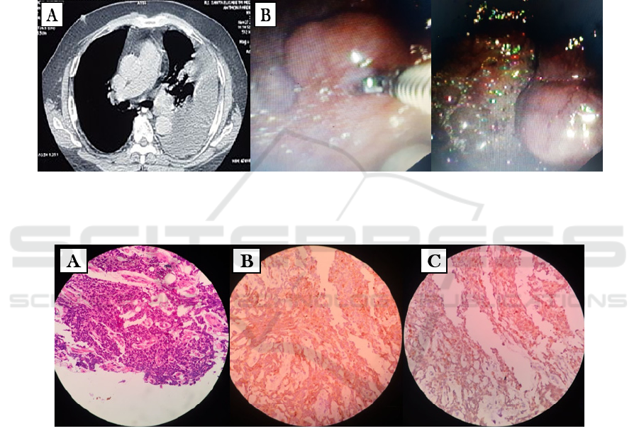
Figure 1: (A) Thoracic CT scan revealed a suspicion for left pleural tumors in the apex and medial to lower field along with
massive left pleural effusion and left inferior lobe atelectasis. (B) Multiple masses in the parietal pleura and visceral pleura
were found.
Figure 2: (A) Histopathologic finding supported the diagnosis of mesothelioma, Immunohistochemistry examination with
Pancytokeratin, EMA. (B) and calretinin. (C) were conducted with positive result, confirming the diagnosis of malignant
pleural mesothelioma.
3 DISCUSSION
Asbestos exposure is highly related to the incidence
of malignant pleural mesothelioma (MPM). It may
affect mesothelial, a thin membrane lining the
organs in the body including the thoracic cavity
(pleura), heart (pericardium) and abdominal cavity
(peritoneum). (American Cancer Society, 2018).
MPM is mostly caused by exposure to asbestos
in the workplace. Pleural mesothelioma is more
common in people over 70 years old, but sometimes
it may affect younger people. Our patient reported in
this case was 70 years old and had a prolonged
exposure to asbestos in the workplace as a
construction worker, thus raised a high suspicion for
mesothelioma.
Asbestos can cause mesothelioma through four
mechanisms. The first mechanism is pleural
irritation. Thin and long fibers (width <0.25 μm and
length> 0.8 μm) penetrating the alveolar epithelium
towards the pleural cavity will cause repeated
ICTROMI 2019 - The 2nd International Conference on Tropical Medicine and Infectious Disease
206

irritation of the mesothelial surface and local
inflammation. The second mechanism is related to
the disorder of the mitotic process. The third
mechanism is the formation of oxygen radicals
which are associated with high iron content in
asbestos fibers. And the last mechanism is
stimulation of macrophages by asbestos fibres to
secrete various cytokines and growth factors that
will induce inflammation and promotion of
malignancy, including tumor necrosis factor-α
(TNFα), interleukin-1β (IL-1β), transforming growth
factor-β (TGF β) and platelets derived from growth
factors (PDGF) (Mossman et al., 2013).
Patients with MPM often feel shortness of breath
and severe chest pain, just like what our patient
reported. The pain is most oftenly localized
accompanied with pleural effusion. Additional
symptoms such as cough, malaise, and decreased
appetite along with weight loss and fever without
any sign of infection (American Cancer Society,
2018).
Diagnostic approaches include chest X-ray and
thoracic CT scan to determine the location of tumor
and metastasis. Cytology of pleural fluid, peritoneal
or pericardial fluid along with tissue biopsy may
help to confirm the diagnosis (American Cancer
Society, 2018; Cancer Council, 2015).
Tumor markers for MPM can be detected by
immunohostochemistry examination with Epithelial
Membrane Antigen (EMA) and Calretinin. Both
give high positive results for mesothelioma. Several
studies stated that the sensitivity and spesificity of
EMA for MPM are 91.8% and 100% respectively
(Nautiyal et al., 2017). Calretinin is currently used
as a marker for mesothelial cells both benign and
malignant and more than 95% are positive for
epitheloid-type mesothelioma. Calretinin is used
primarily to differentiate mesothelioma from
carcinoma or other malignant metastases, especially
those with a similar histopathologic findings with
mesothelioma from tissue biopsy or cytology.
However, other studies have shown that calretinine
is not only positive for mesothelial cells, but may
also be positive in other malignancies such as
metastatic adenocarcinoma or squamous cell
carcinoma (Husain et al., 2018). Barberis et al stated
that anticalretinin staining of pleural fluid yielded
100% positive in malignant mesothelioma and 23%
positive in a metastasis adenocarcinoma (Nautiyal et
al., 2017; Husain et al., 2018).
As a conclusion, we reported a case of malignant
pleural mesothelioma diganosed with the positive
immunohistochemistry findings of EMA and
Calretinin. The use of both modalities may yield a
better sensitivity and spescificity level to confirm the
diagnosis accurately.
FUNDING
No grant support or funding from public institutions
or private enterprises was received for this case
report.
ACKNOWLEDGEMENTS
The researcher would like to thank Universitas
Sumatera Utara Hospital which have allowed the
retrieval of medical history data.
REFERENCES
American Cancer Society. 2018. Malignant Mesothelioma.
[online]. [cited 2016 Mei 14]. Available from:
www.cancer.org/cancer/malignantmesothelioma/detail
edguide/malignant-mesothelioma
Cancer Council. 2015. Understanding Pleural
Mesothelioma. A guide for people with cancer, their
families and friends. [online]. [2016 Mei 14].
Available from:
https://www.cancer.org.au/content/about_cancer/eboo
ks/CAN4798%20Understanding%20Pleural%20Mesot
helioma%20web.pdf
Husain, A. N. et al., 2018. Guidelines for pathologic
diagnosis of Malignant Mesothelioma: 2017 Update of
the consensus statement from the International
Mesothelioma Interest Group. Archives of Pathology
and Laboratory Medicine. 142(1): 89-108. doi:
10.5858/arpa.2017-0124-RA.
Mossman, B. T. et al., 2013. New insights into
understanding the mechanisms, pathogenesis, and
management of malignant mesotheliomas. American
Journal of Pathology. 182(4): 1065-77 doi:
10.1016/j.ajpath.2012.12.028.
Nautiyal, N. et al., 2017. Diagnostic utility of Epithelial
Membrane Antigen (EMA) and Calretinin (CAL) in
effusion cytology. Journal of Clinical and Diagnostic
Research. 11(5): EC36-EC39. doi:
10.7860/JCDR/2017/24339.9888.
Diagnostic Utility of Epithelial Membrane Antigen (EMA) and Calretinin (Cal) in Malignant Pleural Mesothelioma: A Case Report
207
