
Hemiparesis in Pediatric Tuberculous Meningitis: A Case Report
Sylvia Cahyadi
1
, Jeanette Marchi
1
, Rina Amalia Caromina Saragih
2*
, Lesmana Syahrir
3
1
General Practitioner, Siloam Dhirga Surya Hospital, Jalan Imam Bonjol No 6 Medan. North Sumatera, Indonesia
2
Pediatric Emergency & Intensive Care Division, Child Health Department, Faculty of Medicine, Universitas Sumatera
Utara, Jl. Abdul Hakim No.1, Medan, North Sumatera, Indonesia
3
Department of Pediatrics. Siloam Dhirga Surya Hospital, Jalan Imam Bonjol No 6 Medan, North Sumatera, Indonesia
Keyword: Tuberculous meningitis, pediatric tuberculous meningitis, hemiparesis
Abstract: Tuberculous meningitis (TBM) can occur as the sole manifestation of tuberculosis (TB) or concurrent with
pulmonary or other extrapulmonary sites of infection. It causes high mortality and morbidity. Patients with
TBM develop typical symptoms and signs of meningitis. Cranial nerve palsies, hemiparesis, paraparesis,
and seizures are common and should raise the possibility of MTB as the etiology of meningitis. We present
a case of TBM in an 11-year-old who was admitted to High Care Unit Siloam Dhirga Surya Hospital,
Medan, Indonesia with a decreased level of consciousness, sudden weakness of a right extremity, headache,
nuchal rigidity, and history of fever and cough. Chest X-ray showed miliary TB and head CT scan showed
hydrocephalus. Laboratory results were leukocytosis, hyponatremia, and positive TB IGRA. The patient
was then treated with normal saline infusion, anti TB regimen, an antibiotic, and oral corticosteroid. With
regular admission of the anti-tuberculosis drug, oral corticosteroid, and physiotherapist, the patient showed
improvement in his motoric function. The clinical symptom of TBM may appear as hemiparesis without a
seizure. Proper treatment of tuberculous meningitis may lead to a better outcome.
1 INTRODUCTION
Indonesia has one of the highest burden of
tuberculosis (TB) globally (WHO, 2018). It is
estimated that childhood TB constitutes 10 to 20%
of all TB cases in high burden countries, accounting
for 8 to 20% of TB-related deaths. Approximately,
25% of pediatric TB cases are extrapulmonary, with
tuberculous meningitis (TBM) being the most severe
form. Worldwide, tuberculous meningitis (TBM)
accounts for the majority of the deaths due to TB
(Israni, et al., 2016).
Tuberculous meningitis may present at any age
but is less common at the extremes of life. The peak
incidence is in children between 2 and 4 years of
age. Most early symptoms relate to underlying
pulmonary TB present in most infants who develop
TBM as a complication of primary infection (Chin,
2014).
The most commonly recorded signs and
symptoms of TBM were an altered level of
consciousness (90.1%), meningism (77.2%), fever
(68.2%), and loss of appetite (61.4%). Focal
neurological signs included unilaterally non-reactive
pupils (13.6%), other cranial nerve palsies (22.7%),
limb paresis (27.3%), and aphasia (18.2%)
(Rohlwink, et al., 2016).
2 CASE PRESENTATION
An 11-year-old boy was admitted to the High
Care Unit (HCU) Siloam Dhirga Surya Hospital
with a decreased level of consciousness, headache,
and sudden weakness on the right extremities. The
patient had been hospitalized before with fever and
cough. There was a history of fever and cough for
two weeks and weight loss in one month. No history
of close contact with active TB patient, but his father
was suffering from chronic cough without receiving
any medication.
Physical examination revealed somnolence
(GCS 14), slurred communication, and normal
temperature. On neurological examination, there was
nuchal rigidity, clonus on the right extremities,
positive Babinski sign, and no muscle contraction in
the right extremities. No presence of seizure. The
chest was clear bilaterally.
Cahyadi, S., Marchi, J., Saragih, R. and Syahrir, L.
Hemiparesis in Pediatric Tuberculous Meningitis: A Case Report.
DOI: 10.5220/0009862801770179
In Proceedings of the 2nd International Conference on Tropical Medicine and Infectious Disease (ICTROMI 2019), pages 177-179
ISBN: 978-989-758-469-5
Copyright
c
2020 by SCITEPRESS – Science and Technology Publications, Lda. All rights reserved
177
Laboratory test showed leukocytosis (11.820
/mm
3
) with elevated of neutrophil (74.9%) and
Erythrocyte Sedimentation Rate (ESR). The patient
was hyponatremic (127 meq/L) and had positive TB
IGRA.
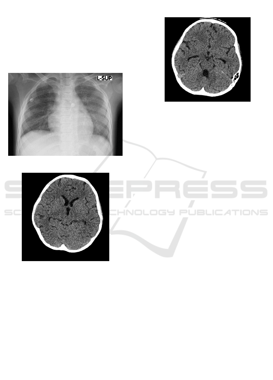
Chest X-ray showed miliary TB (Figure 1), and
the CT of the brain showed mild hydrocephalus
which suggests meningitis (figure 2 and figure 3).
Tuberculin test was negative (0 mm). The lumbar
puncture could not be performed technically.
Figure 1: Chest X-Ray.
Figure 2: Head CT scan shows mild hydrocephalus in the
third ventricle.
The patient was then treated with four anti-
tuberculosis drugs (isoniazid, rifampicin,
pyrazinamide, and ethambutol), oral corticosteroid,
mannitol infusion, and normal saline infusion.
Antibiotic meropenem was added to the treatment.
Previously before admitted to HCU, the patient had
been given ceftazidime as an antibiotic. The patient
showed improvement after 3 days in the HCU. His
consciousness began to improve, no headache and
nuchal rigidity and maintained a stable of
hemodynamics. The patient still had right
hemiparesis. He was moved to the ward and was
discharged with the anti-tuberculosis regimen and
oral corticosteroid.
Figure 3: Head CT scan shows mild hydrocephalus in the
fourth and temporal ventricle.
The patient was then scheduled for having
physiotherapist regularly twice a week. With regular
administration of the anti-tuberculosis drug, oral
corticosteroid, and physiotherapist, the patient
showed an improvement in his motoric function.
Two weeks after being discharged from the hospital,
he was able to walk by himself.
3 DISCUSSION
Neuro-tuberculosis is the most serious complication
of TB in children. Among the various forms of
neuro-tuberculosis, TBM remains the most severe
and the most common in developing countries
(Torok, 2015). Tuberculous meningitis continues to
be an important cause of morbidity (especially
neurologic impairment) in children from resource-
poor countries (Israni, et al., 2016).
Owing to the suboptimal performance of
diagnostic tests of TBM, the diagnosis in children
relies on a thorough assessment of all the evidence
derived from a careful evaluation of medical history,
clinical examination, and relevant investigations.
Approximately 60% of children with TBM have
radiological evidence of pulmonary TB (Toorn &
Solomons, 2014).
Tuberculous meningitis is a subacute meningitis
illness, which presents in various stages. According
to the British Medical Council Staging System,
tuberculous meningitis can be divided into 3 stages.
The first stage consists of nonspecific symptoms of
low-grade fever, headache, irritability, drowsiness,
malaise, vomiting, photophobia, listlessness, and
poor weight gain/weight loss. The second stage
shows a sign of meningeal irritation with or without
ICTROMI 2019 - The 2nd International Conference on Tropical Medicine and Infectious Disease
178

slight clouding of consciousness with focal
neurological signs such as cranial nerve palsies or
hemiparesis. In advanced clinical stages, TBM
presents severe clouding of consciousness or
delirium, convulsions, and serious neurological signs
such as hemiplegia, paraplegia, involuntary
movement (Israni, et al., 2016).
Tuberculin skin test (Mantoux test) may be
nonreactive in 50% cases of CNS TB. Hence, it is
helpful in supporting the diagnosis of TBM when
positive, but an isolated positive Mantoux cannot be
used to label a case of TBM, as false positive/false
negative reactions are commonly known (Aulakh &
Chopra, 2018).
Tuberculous meningitis usually presents with a
Cerebrospinal fluid (CSF) of 10–500 cells/μL that are
polymorphs initially and lymphocytes later. A low
glucose <40 mg/dL (rarely<20 mg/dL) or a
CSF/plasma glucose ratio <50% or a high-protein
content (400–5000 mg/dL) is suggestive of the
diagnosis of TBM. The CSF lactate levels are usually
raised to 5–10 mmol/L (normal range, 1.2–2.1
mmol/L). Ziehl–Neelsen (ZN) staining for the smear
examination has a sensitivity of approximately 50%,
whereas a bacterial culture has a sensitivity of 60% to
70% (Aulakh & Chopra, 2018). CT scanning and
MRI of the brain may reveal hydrocephalus, basilar
meningeal enhancement, infarcts, edema, and
tuberculomas (Toorn & Solomons, 2014).
This patient presented with a decreased level of
consciousness, headache, nuchal rigidity, positive
Babinski sign, right hemiparesis. TB IGRA was
positive in this patient. He previously complained of
cough and fever for two weeks and weight loss for 1
month.
Chest X-ray also showed miliary tuberculosis,
and the CT of the Brain showed mild hydrocephalus
which suggests meningitis. From the clinical
examination, diagnostic tests, this patient can be
categorized into stage 2 of TBM.
WHO recommends a 12-month treatment plan
(2RHZE/10RH) for children with suspected or
confirmed TBM (Toorn & Solomons, 2014). We
gave anti-tuberculosis drugs to this patient according
to the WHO guideline. Meropenem was given due to
the possibility of bacterial meningitis that is evidently
suggested by leukocytosis and increased neutrophil
count. Hyponatremia occurs in up to 85% of children
with TBM and is thought to be secondary to either
syndrome of inappropriate antidiuretic hormone or
cerebral salt wasting. We also found hyponatremia in
this patient which was treated with normal saline
fluid. Corticosteroid oral was given to reduce the risk
of death and neurological deficit (Toorn &
Solomons, 2014).
The patient showed clinical improvement in his
motoric function after being given an anti-
tuberculosis drug regimen, oral corticosteroid, and
physiotherapist.
4 CONCLUSION
Patients with TBM develop typical symptoms and
signs of meningitis including headache, fever, and
stiff neck, although meningeal signs may be absent
in the early stage. The duration of symptoms before
presentation ranges from several days to several
months. In particular, in resource-limited settings,
TBM cases may present in advanced clinical stages,
with GCS scores of 10 or less. Cranial nerve palsies,
hemiparesis, paraparesis, and seizures are common
and should raise the possibility of tuberculous
meningitis as the etiology of meningitis (Chin,
2014).
This case shows that clinical symptoms of
tuberculous meningitis can appear as hemiparesis
without a seizure. Proper treatment of tuberculous
meningitis may lead to a better outcome.
REFERENCES
Aulakh, R., & Chopra, S. (2018). Pediatric tubercular
meningitis : A review. J Pediatr Neurosci, 13:373-
82.
Chin, J. H. (2014, June). Tuberculous Meningitis.
Neurol Clin Pract, 4(3):199-205.
doi:10.1212/CPJ.0000000000000023.
Israni, A. V., Dave, D. A., Mandal, A., Ranjan, R. D.,
Singh, A., & Sahi, P. K. (2016). Tubercular
meningitis in children: Clinical, pathological, and
radiological profile and factors associated with
mortality. J Neurosci Rural Pract, 7(3):400-404.
doi:10.4103/0976-3147.181475.
Rohlwink, U. K., Donald, K., Gavine, B., Padayachy, L.,
Wilmshurst, J. M., Fieggen, G. A., & Figaji, A. A.
(2016). Clinical characteristics and
neurodevelopmental outcomes of children with
tuberculous meningitis and hydrocephalus.
Developmental Medicine & Child Neurology,
58:461-468.
Toorn, R. v., & Solomons, R. (2014). Update on the
Diagnosis and Management of Tuberculous
Meningitis in Children. Semin Pediatr Neurol,
21:12-18.
Torok, M. (2015, March). Tuberculous meningitis :
advance in diagnosis and treatment. Br Med Bull,
113(1):117-31. doi:10.1093/bmb/ldv003.
WHO. (2018). Global Tuberculosis Report 2018.
Geneva: World Health Organization.
Hemiparesis in Pediatric Tuberculous Meningitis: A Case Report
179
