
Correlation of Gestational Sac Diameter, Fetal Heart Diameter, and
Fetal Head Diameter to Gestational Age of Local Cats (Felis
Domesticus) Pregnancy in Indonesia
Analis Wisnu Wardhana
1
, Aulia Firmawati
1
, Albiruni Haryo
2
, Kevin Ersananda
3
, Nabila Safira
3
,
Tiara Balqhis
3
1
Department of Anatomy and Histology, Faculty of Veterinary Madicine, Brawijaya University
1
Department of Reproduction, Faculty of Veterinary Medicine, Brawijaya University
2
Department of Pathology, Faculty of Veterinary Medicine, Brawijaya University
3
Student of Program Study of Veterinary Medicine, Faculty of Veterinary Medicine, Brawijaya University
Keywords: Domestic Cat, Gestational Sac Diameter, Fetal Head Diameter, Fetal Heart Diameter, Ultrasonography.
Abstract: Fetal development in domestic cat can be monitored using ultrasonography to control viability, maintain the
nutrition care, and drug use. Due to control the viability of foetuses, this study observed the correlation of
gestational sac diameter, fetal head diameter, and fetal heart diameter compared to gestational age. This study
was performed by using 9 queens that divided into three groups. The first group measured the gestational sac
diameter, the second group measured the fetal head diameter, and the third group measured the fetal heart
diameter. Gestational sac diameter and fetal head diameter are measured by specific formula, while the fetal
heart diameter calculated by measuring point to point of heart edge and compare to head diameter to verify
the gestational age. As the result, the gestational sac diameter, the feal head diameter, and the fetal heart rate
of domestic cat in Indonesia have correlation to gestational age and also can be used to estimate the gestational
edge.
1 INTRODUCTION
The Felis domesticus is local breed of cat in Indonesia
(Madyantari, 2016). It has short hair with different
color which depend on the alel frecuency. The
average of body lenght of this cat is 76 cm with 2-3
kg of female body weight and 3-4 kg of male body
weight (Mariandayani, 2012).
Pregnancy in cats and fetal development can be
detected using several methods such as abdominal
palpation, radiography, and ultrasonogrphy. The
abdominal palpation is the cheapest way to detect
pregnancy but only can detected in second trimester
of cat pregnancy and has low accuracy so that can
make a false pregnancy. The radiography has more
accuracy but only can determine the number of
fetuses and can not determine the viability of fetuses.
The ultrasonography is a high accuracy method to
detect pregnancy because of ability to evaluate the
viability, organ development, and number of fetuses
(Purohit, 2010).
Investigation of fetal development has been
studied in recent years, but it is rarely investigated in
domestic cat especially in Indonesia. Early detection
and investigation of fetal development are important
to maintain the nutritional care, prevent of dangerous
drug use for the fetal, and the viability of fetal its self
(Machun eat al, 2011). This aim of this study was to
evaluate the correlation between gestational sac
diameter, fetal head diameter, and fetal heart diameter
to gestational age using ultrasonography.
2 MATERIALS AND METHOD
The study used nine samples of queens and it is
divided into 3 groups, based on gestational age (GA)
(0-20 days, 21-40 days, and 41-60 days). Every group
have different method to find accurated gestation age.
First group will use gestation sac diameter (GSD) to
find the accuracy gestation age with specific formula
(figure 1). The second group was measured the fetal
heart diameter. The fetal heart diameter can be
Wardhana, A., Firmawati, A., Haryo, A., Ersananda, K., Safira, N. and Balqhis, T.
Correlation of Gestational Sac Diameter, Fetal Heart Diameter, and Fetal Head Diameter to Gestational Age of Local Cats (Felis domesticus) Pregnancy in Indonesia.
DOI: 10.5220/0009586300370040
In Proceedings of the 6th International Conference on Advanced Molecular Bioscience and Biomedical Engineering (ICAMBBE 2019) - Bio-Prospecting Natural Biological Compounds for
Seeds Vaccine and Drug Discovery, pages 37-40
ISBN: 978-989-758-483-1
Copyright
c
2020 by SCITEPRESS – Science and Technology Publications, Lda. All rights reserved
37
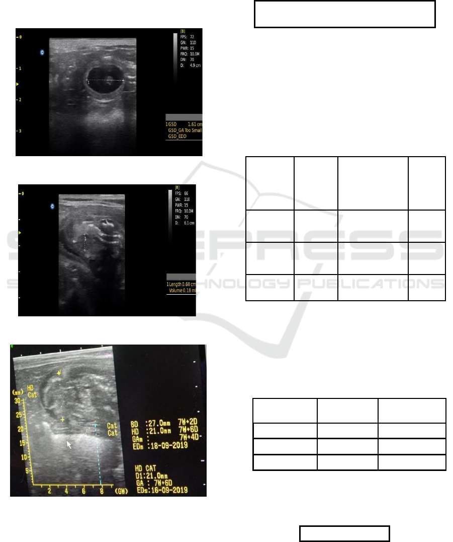
detected by freeze the image (figure 2) that is
characterized as hypoechoic with anechoic lumen and
then measuring point to point of the heart image.The
last group was measured head diameter. It can be
performed by detect the line between the outer edges
of head (the fartest edge) that has highest (white)
opacity (figure 3) and the gestational age can be
measure using the specific formula.
Figure 1. Measurement of Gestational Sac Diameter
Figure 2. Measurement of Fetal Heart Diameter
Figure 3. Measurement of Fetal Head Diameter
3 RESULT
Formulation for calculating gestation age in the
quuens using extrafetal structures in early pregnancy
(0-20 days) is:
GA = 1,368x – 11,566
x = inner chorionic circumference (mm)
The gestation sac diameter of 3 queens were
evaluated. The earliest sign of pregnancy in queen
is the presence of gestational sac, which is appeared
like a small circular anechoic structure at
ultrasound. There were no significant differences
between the three cats for GSD for early gestation
age (Table 1).
Table 1. Data of Gestation Sac Diameter (GSD) (cm) of
Domestic Cat during Pregnancy
Gestation
Results
Cats
Name Sac Formula
GA
(days)
Diameter
Gempi
1.61
GA = 1.368(16.1)
10
–
11,566
Titty 2.03
GA = 1.368(20.3)
16
–
11,56
Cippy
1.49
GA = 1.368(14.1)
8
–
11.566
Fetal heart diameter is measured by detecting point
to point of heart edge and compare to the head
diameter to verify the gestational age. The data were
collected in Table 2.
Table 2. Data of Fetal Heart Diameter (cm) of Domestic Cat
during Pregnancy
Cats Name
Days
Fetal Heart
Diameter
Gempi 21
0.6
Mimi
31
0.73
Meme
32
0.78
Correlation between fetus head diameter with
gestational age can be measured by the formula
below and the result is shown in Table 3.
GA = 25 x HD + 3
ICAMBBE 2019 - 6th ICAMBBE (International Conference on Advance Molecular Bioscience Biomedical Engineering) 2019
38

Table 3. Data of Fetal Head Diameter of Domestic Cat
during Pregnancy
Cat
Head
Diameter
Results
(cm)
Formula
GA
N
ame
(days)
Putty 2.0
GA = (25 x 2.0)
53
+ 3
N
ana 1.76
GA = (25 x
47
1.76) + 3
Betty 2.1
GA = (25 x 2.1)
55
+ 3
4 DISCUSSION
Gestational sac was first detected between days 8 and
12 of gestation in cat (Lopate, 2018). As this study
performed, the gestational sac diameter can be
measured in days 8 of gestational age. The formula to
measured the gestational sac diameter in early
gestational age between 0 - 20 days indicates that the
alteration of gestation sac diameter will affect
gestational age of the fetus. But, the gestational sac
not too elongate, the result of gestation age of
measured gestational sac 1.41 cm is 8 days and in 1.61
cm the gestation age at 10 days, and the result of
gestational age when gestational sac diameter in 2.03
cm is 16 days. So, if the gestational sac diameter
elongate 0,2 cm then the gestational age will be
increased 2-3 days. This is also explained in recent
study of Zambelli et al. (2002) that gestational sac
diameter has correlation to gestational age.
The first detection of fetal heart rate by
ultrasonography can be measured between 16-18
days of pregnancy (Zambelli et al.,2002). The
measurement of heart diameter is helpful for
determining the fetal viability (Oral H et al., 2007).
Fetal heart diameter is important to be measured for
ealier detection of heart abnormalities and screening
of fetal congenital heart disease (Sylwestrzak et al.,
2018). In this study, we collected data of heart
diameter in last trimester of pregnancy cats and
shown that in days 21 the fetal heart diameter is 0.6
cm, in days 31 showed 0.73 cm in fetal heart
diameter, and in days 32 showed 0.78 cm in fetal heart
diameter. From that data, we can see that fetal heart
diameter was correlated to gestational age, in which
the greater fetal heart diameter can be measured as the
older gestational age. This study revealed positive
correlation to study from Oral H et al. (2007) who
reported that heart rate were correlated with fetal age
and can be used to estimate the gestational age.
Beside determine the gestational age, the fetal heart
diameter were correlated to fetal size also as the study
of Giannico, et al. (2015). The increasing of fetal
heart diameter indicates the incresing of fetal size due
the development of organs, in which the bigger size
of fetal needed more blood supply to the whole body
so that the fetal heart diameter is increasing.
Fetal head diameter can be detected at 24 days of
gestational age in cat (Macun et al., 2011). The
diference of fetal head size can caused the deviation
measurement (Nyland, 2002). The data of this study
show that in 47 days, the diameter of the head is 1.76
cm, in 53 days, the diameter of head is 2 cm, and in
55 days of pregnancy the diameter of head is 2.1 cm.
This collected data explained that increasing diameter
of the head is indicate that the fetuses are getting
older. In recent study explained that the fetal head
diameter had correlation with the age of gestation
(Macun et al.,2011).
5 CONCLUSION
Monitoring of fetus development is important to
evaluate the fetus viabilty. In this study can be
conclude that gestational sac diameter, fetal heart
diameter, and fetal head diameter have correlation to
gestational age. These parameters can be used to
estimate of gestational age.
ACKNOWLEDGEMENTS
We many thanks to LPPM and Faculty of Veterinary
Medicine, Universitas Brawijaya for providing
research assistance funds and supporting the
completion of this research.
REFERENCES
Giannico, A. T., Mayumi, E., Aparecida, D., Sousa, M. G.,
& Froes T.R. (2016). Canine and Fetal
Echocardiography: Correlation for the Analysis of
Cardiac Dimension. Ve Res Commun 40: 11-19
Lopate, C. (2018). Gestation Aging and Determination of
Parturition Date in the Bitch and Queen Using
Ultrasonography and Radiography. Veterinary Clinics
of North America: Small Anim Practice 48(4): 617-638
Correlation of Gestational Sac Diameter, Fetal Heart Diameter, and Fetal Head Diameter to Gestational Age of Local Cats (Felis
domesticus) Pregnancy in Indonesia
39

Macun, H. C., Erat, S., & Arikan, S. (2011). Comparison of
Prenatal Development of Turkish Angora and Van
Cats. Veterinaarski Arhiv 81: 671-682
Madyantari, N., Hidayat, S. & Wahab T. (2016).
Perancangan Buku Ilustrasi Kucing Di Bandung. Jurnal
e-Proceeding of Art and Design. 3 (3): 746-753
Mariandayani, H. N. (2012). Keragaman Kucing Domestik
(Felis domesticus) berdasarkan Morfogenetik. Jurnal
Peternaan Sriwijaya 1(1):10-19
Nyland, T.G. & Mattoon J.S. (2002). Small Animal
Diagnostic Ultrasound. USA: Elsevier.
Oral, H., Pancarci, S. M., Gungor, O., & Kacar, C. (2007).
Determination of Gestational Ag by Measuring Feal
Heart Diameter with Transrectal Ultrasonograph in
Sheep. Medycyna Wet 63(12): 1558-1560
Purohit, G. (2010). Methods of Pregnancy Diagnosis in
Domestic Animal: The Current Status. WebMed
Central Reproduction 1(12): 1-26
Sylwestrzak, O. & Respondek-Liberska, M. (2018).
Echocardiographic Methods of Fetal Heart Size
Assesment-Heart to Chest Area Ratio and Transversal
Heart Diameter. Prenat Cardio 8(1): 20-23
Zambelli, D., Castagnetti, C., Belluzzi, S., & Bassi, S.
(2002). Correlation between the age of the conceptus
and various ultrasonographic measurements
during the first 30 day of pregnancy in domestic cats
(Felis catus). Theriogenology 57: 1981–1987.
ICAMBBE 2019 - 6th ICAMBBE (International Conference on Advance Molecular Bioscience Biomedical Engineering) 2019
40
