
Helminthiasis and Aspergillosis Suspect Examination in Pigeon
Albiruni Haryo and Rifqi Rahman
Laboratory Pathology Anatomy, Faculty of Veterinary Medicine, Brawijaya University
Keywords: Pigeon, Examination, Helminthiasis, Aspergillosis
Abstract: Pigeons are commonly maintained using a simple cage system, this system also easier to transmit disease
such as helminthiasis and aspergillosis to infect pigeons. The air of this study was to know and diagnose
changes in macroscopic and microscopic with histopathology method in pigeons. The organs examined are
proventriculus, intestine, liver, and skin. Macroscopic changes were seen in liver change color into
brownish-yellow on right lobes of the liver, hiperemi proventriculus, hemorrhage, and swelling intestine,
baldness, and crust in the upper neck skin. Microscopic changes seen in hepar are white blood cell
infiltration in triad portal, congesti and ulcer mucosa gland proventriculus, intestine shown epithelial
erosion, rupture villi, hemorrhage and hyperplasia of cell goblet and skin shown black colored infiltration in
dermis allegedly infected by Aspergillosis sp. From examination and observation, it can be concluded that
macroscopic and microscopic changes lead to the helminthiasis and suspected Aspergillosis sp.
1 INTRODUCTION
Pigeons are animals that are maintained extensively
with pure cage carrying management in purposes to
simplify preservation and inexpensive, but this
system aids the transmission of diseases such as
helminthiasis and aspergillosis more quickly. The
common helminthiasis hitting the pigeons are
Ascaridia Columbae, Capillaria sp, and Tetramers sp
(Alkharigy et al., 2018). A contaminated feed with
worm eggs or through ectoparasites such as fleas can
transmit the helminthiasis. In addition to
helminthiasis, some diseases such as Aspergillosis
are also easily transmitted to pigeon populations,
especially if the conditions are humid. Aspergillosis
is caused by Aspergillus sp, whose transmitted
through spores can be inhaled through breathing,
digestion, or sticking to the skin and infecting the
dove. Helminthiasis and Aspergillosis may cause
economic losses due to adult-stage worms living in
the digestive tract, causing blockages and interfering
with the absorption of nutrients from its pigeons.
This study aims to identify helminthiasis and
Aspergillosis based on visible macroscopic and
microscopic alteration. Macroscopic observation in
the neck skins that facing a loss of fur and blackened.
Microscopic results indicate aspergillosis infection in
the skin. Fungi Aspergillosis sp leads to
Aspergillosis. The acute Aspergillosis infiltrates the
dermis and tissue beneath, which could cause
necrosis in the infected area (Bernadeschi et al.,
2015).
2 MATERIALS AND METHODS
2.1 Instrument and Materials
The applied animal is a pigeon with an age of
around four months. The method used to observe
macroscopic alteration is the Necropsy method. For
histopathological observation, the utilized method is
the Histotechnique by doing network isolation and
followed by staining the preparations using
Hematoxylin-eosin staining, which is conduct at the
Anatomy Pathology Laboratory and Histology
Laboratory, Faculty of Veterinary Medicine,
Brawijaya University
2.2 Animal Preparation
The animal (pigeon), which is four months old
approximately, is euthanized by embolism through
magnum foramen. The 3 cc syringe loaded with air
and injected it to the animal's brain through the
foramen magnum.
Haryo, A. and Rahman, R.
Helminthiasis and Aspergillosis Suspect Examination in Pigeon.
DOI: 10.5220/0009586200330036
In Proceedings of the 6th International Conference on Advanced Molecular Bioscience and Biomedical Engineering (ICAMBBE 2019) - Bio-Prospecting Natural Biological Compounds for
Seeds Vaccine and Drug Discovery, pages 33-36
ISBN: 978-989-758-483-1
Copyright
c
2020 by SCITEPRESS – Science and Technology Publications, Lda. All rights reserved
33
2.3 Necropsy Implementation
Must ensure the total death from the animal (pigeon)
before doing a necropsy. Necropsies stages were
performed such cleansing into the incised area by
rinsing using the water flow afterward, the fixation
and incision were made to see organ abnormalities.
The standard necropsy procedure was carried out
according to Majo and Dolz (2011). The purpose of
a necropsy is carried out to assist overall the
examination of organ abnormalities, for
macroscopic abnormalities examination, and also
sampling for the histopathological preparations.
2.4 Histopathology Preparation
Hematoxylin Eosin staining prep will be using the
samples from selected organs that assumed having
abnormalities in the pigeons, which are liver,
intestine, skin, and proventriculus (Janqueira, 2007).
3 RESULTS AND DISCUSSION
The results of the liver macroscopic examination
showed a color change into yellow in the right lobe
(Figure 1.) Microscopic examination showed the
inflammatory cell infiltrate in the portal triad
(Figure 2.) According to Lope et al. (2017), the liver
has a complex hemodynamic groove. Blood from
the spleen, pancreas, and gastrointestinal flows into
the liver through the portal vein along with the
hepatic arteries. The inflammation of inflammatory
cells is an excessive accumulation of cells in the
tissues or blood vessels. Portal triads consist of
venules portal, arterioles portal, and bile ducts.
Blood flow from the portal vein is blood flow that
comes from the intestine, spleen, and rectum so that
it contains numerous antigens residual of the
intestinal bacteria such as lipopolysaccharide
endotoxins (LPS) and leukocytes when there is
inflammation due to the infection process in these
organs (Bogdanos. et al. 2013).
Figure 1. Pigeon's liver that appears turned into yellow in
the right lobe on the macroscopic examination (blue
arrow).
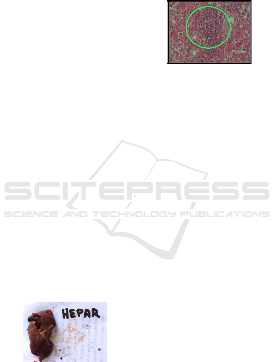
Figure 2. Histopathology of pigeon's liver tissue by
Hematoxylin Eosin staining (400x). Annotation : A.
Infiltration of inflammatory cells near the central vein
(black arrow). Inflammation of inflammatory cells in the
triad's portal (inside the green ring.
Macroscopic observations of proventricular
organs show hyperemia. The proventriculus is
seemed to have a dark red color (Figure 3). The
proventricular disorder is an anomaly occurring in
specific cases such as Newcastle disease.
Pathognomonic lesions of ND disease characterized
by the presence of petechiae and hemorrhage in the
mucosa proventriculus (Nakamura et al. 2010).
There was no hemorrhage, and petechiae
founded in the mucosa in the pigeon's case, but it has
hyperemia. Microscopic results showed congestion,
mucous gland ulceration, and ruptured
proventricular gland (Figure 4). Congestion is a
vascular size enlarged. Congestion generally occurs
when there was an expansion in tissue activity,
increased acid levels, CO2, also an infection or
tissue damage. It is due to vasodilation, and
increased blood flows into the area. Increased blood
flows to the gastrointestinal commonly occur during
the metabolic process. Erosion is a superficial
damaged on the surface of the tissue that could be
occurred by inflammation, trauma, or parasites that
break the extent of the mucosa and does not reach
the muscular mucosa. Ulcers are local lesions on the
skin or mucosal layer that show damaged superficial
epithelium and also in deeper tissues. The histology
shows superficial damages that indicate the
occurrence of mucosal gland erosions, and the
damage also exists unto the (mucosal gland). The
proventriculus histopathology shows that the
damage has spread into the proventricular glands
(Figure 4).
ICAMBBE 2019 - 6th ICAMBBE (International Conference on Advance Molecular Bioscience Biomedical Engineering) 2019
34
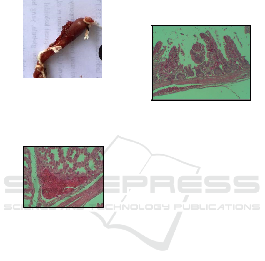
Figure 3. Proventricular hyperemia in pigeons becomes
dark red
The results of macroscopic observation of intestine
organs show obstruction with hyperemia. Nematode
worms which generate the obstruction are visible
after cross-section Microscopic observations
revealed epithelial erosion, villi rupture, goblet cell
hyperplasia, and hemorrhage (Figure 5).
Figure 4. Histopathology of proventriculus tissue with
Hematoxylin Eosin staining (400x) Annotation:
Congestion in proventricular tissue and erosion, ulcer
mucosal gland proventriculus
Helminthiasis damages can create Epithelial
erosion and villous rupture. The advances in
structure cells to goblet cell hyperplasia are the
body's defense attempts to react to the antigen or
epithelial damage. Healthy goblet cells are present in
intestine cells as cells that secreted mucin, which
serves to cover and protect the intestinal mucosa.
Damage to the intestinal epithelium causes more
goblet cells to produce mucin as a response to
epithelium damages (Djojodibroto, 2007).
Hemorrhage is an escape of blood from ruptured
blood vessels, and the bleeding can occur inside or
outside of the body. Increased blood pressure, blood
vessel infection, and inflammatory blood vessel
walls can provoke hemorrhage (Mohan, 2010).
Nematodes are one of the causes of bleeding in the
intestinal mucosa because it can damage the
epithelium and intestine blood vessels. Common
nematodes in pigeon intestines are Ascaridia
columbae, Capillaria sp, and Tetramers sp
(Alkharigy et al., 2018).
Figure 5. Histopathology of intestine tissue with
Hematoxylin Eosin staining (100x) Annotations: Ruptured
villi (black arrow). Goblet cell hyperplasia of villi
intestine and hemorrhage in the intestine villi.
4 CONCLUSIONS
Pigeon necropsy based on macroscopic and
microscopic changes leads to the assumption of
helminthiasis with types of nematodes and
Aspergillosis. Helminthiasis, especially nematode
intestinal, can cause changes both macroscopically
and microscopically in the liver and intestine, and
Aspergillosis can cause changes both macroscopic
and microscopic to the skin.
REFERENCES
Alkharigy, F. A., El Naas, A. S., Maghribi, A. A. 2018.
Survey Of Parasite in Domestic Pigeons (Columba
livia) in Tripoli, Libya. Open Veterinary Journal.
Vol.8(4) : 360 – 366
Bernardeschi, C., Foulet, F., Ortonne, N., Sitbon, K.,
Quereus G., Lortholary O., Chosidow, O., Bretagne, S.
2015. Cutaneus Invasive Aspergillosis: Retrospective
Multicenter Study of The French Invasive-
Aspergillosis Registry and Literature Review. Journal
of Medicine. Vol 94(26): e1018.
Bogadanos, D. P., Gao, B., Gershwin, M. E. 2013. Liver
Immunology. Journal of Compare Physiology 3 (2):
567 – 598
Butcher, G. D, and Miles, R. 2018. Avian Necropsy
Techniques.
Djojodibroto, D. 2007. Respirologi (Respiratory
Medicine).Penerbit Buku Kedokteran ECG: Jakarta.
Mohan, H. 2010. Textbook Of Pathology Sixth Edition.
Jaypee Brothers Medical Publisher: New Delhi
Helminthiasis and Aspergillosis Suspect Examination in Pigeon
35

Latimer, S. K., Pauline M. Rakich, P. M., Branson, W. R.,
Harrison, G. J., Harrison, L R. 1994. Avian Medicine:
Principles and Application. Wingers Publishing: USA.
Majo, N and Dolz, R. 2011. Atlas of Avian Necropsy.
Servet Publishing: USA
Nakamura, K., Ohta, Y., Abe, Y., Imai, K., Yamada, M.
2010. Pathogenesis of Conjunctivits caused by
Newcastle disease viruses in specific-pathogen-free
chickens, Avian pathology journal. 33(3):371 – 376.
Studdert, V. P., Gay, C. C. and Blood, D.C. 2012.
Saunders Comperehensive Veterinary Dictionary, 4th.
Sauders : Philadelphia.
ICAMBBE 2019 - 6th ICAMBBE (International Conference on Advance Molecular Bioscience Biomedical Engineering) 2019
36
