
Green Synthesis and Characterization of Silver Nanopaticles
Mahyuni Harahap
1,2
, Mia Oktavia
1,2
and Saharman Gea
1,2
1
Cellulosic and Functional Materials Research Centre, Universitas Sumatera Utara,
Jl. Bioteknologi, Medan, 20155, Indonesia
2
Chemistry Department, Faculty of Mathematics and Natural Science, Universitas Sumatera Utara,
Jl. Bioteknologi, Medan, 20155, Indonesia
Keywords: Green synthesis, silver nanoparticles, chemical reduction, TEM, UV-visible
Abstract: Green synthesis of silver nanoparticles (AgNPs) was carried out by chemical reduction method. The
reduction was carried out in a close and dark condition at 90
o
C for 4 h. Glucose was used as the reducing
agent, while starch was used as the stabilizing agent. The reduction of Ag
+
was indicated by color change in
the solution from colorless to brown. Synthesized AgNPs were characterized by UV-visible spectroscopy
and transmission electron microscopy, which showed that AgNPs had SPR value of 435 nm with spherical
shapes. Moreover, the dimension of AgNPs was approximately 9.15 ± 4.15 nm.
1 INTRODUCTION
Silver (Ag) is a promising chemical element to be
used in various applications such as medicine,
electronics, and household tools. It is classified as
a transition metal and has interesting properties.
However, the uses of silver are limited because it
oxidized spontaneously when exposed to free
oxygen molecules (Khorasani et al.,
2009)(Zheludkevich et al., 2004). Recently, the
production of silver in nanoscale has become
highly attractive due to nanoparticles physical,
chemical, and biological properties, which are
being studied through analytical techniques, i.e. x-
ray diffraction, x-ray photoelectron spectroscopy,
fourier-transform infrared spectroscopy, UV-vis
spectroscopy, transmission electron microscopy,
scanning electron microscopy, dynamic light
scattering, and localized surface plasmon
resonance (Salleh et al., 2020).
Silver nanoparticles (AgNPs) have unique
physical and chemical properties with their
surface-to-volume ratio, which enable modification
of their physical, chemical, and biological
properties. These properties have made AgNPs in
high demand for various applications such as in
health care, as well as for medical and industrial
purposes (Morones et al., 2005). The biological
activity of AgNPs is the most intriguing property
influenced by the particle size distribution, surface
chemistry and morphology, chemical composition,
agglomeration, capping agents, particle responses
in media, the release of ions, and the reducing
agents used in the synthesis of AgNPs (Prakash et
al., 2017).
Generally, the synthesis of AgNPs through
physical, chemical, and biological methods is
known as green synthesis. Additionally, the
synthesis is classified into top-down (consists of
mechanical grinding of silver bulks) and bottom-
up method (consists of chemical reduction, sono-
decomposition, and electrochemical methods) as
shown in Figure 1 (Slepička et al., 2020).
Figure 1. AgNPs preparation method.
Some studies reported green synthesis of
AgNPs by using eucalyptus hybrida (Safeda)
leaves was able to produce stable AgNPs in
solutions with uniform shapes. Moreover, by using
chemical reduction method, AgNPs produced had
good antimicrobial activity (Landage, Wasif and
Dhuppe, 2014). However, no AgNPs dimensions
were reported (Dubey, Bhadauria and Kushwah,
2009).
574
Harahap, M., Oktavia, M. and Gea, S.
Green Synthesis and Characterization of Silver Nanopaticles.
DOI: 10.5220/0010613700002775
In Proceedings of the 1st International MIPAnet Conference on Science and Mathematics (IMC-SciMath 2019), pages 574-576
ISBN: 978-989-758-556-2
Copyright
c
2022 by SCITEPRESS – Science and Technology Publications, Lda. All rights reserved
This study aimed to synthesize AgNPs by
chemical reduction process. Chemical synthesis
has been widely used as it can easily produce
AgNPs with higher yields and lower cost
compared to physical approach.
2 EXPERIMENTAL
Materials
AgNO
3
, starch and glucose were purchased from
Merck, Germany.
Chemical reduction of silver nanoparticles
AgNO
3
was used as a precursor for the synthesis of
AgNPs by chemical reduction method. Firstly,
AgNO
3
and glucose were mixed under constant
stirring with 1:1 ratio in a beaker glass. Then, 80
mL of the mixture were transferred to another
beaker glass and 20 mL of starch 1% was added.
The mixture was vigorously stirred in a close and
dark condition at 90 ℃ for 4 h. After that, the
sample was cooled at room temperature and
centrifuged at 5000 rpm for 10 minutes. The
product was collected for characterization (TEM
and UV-visible).
UV-visible
UV-visible spectra were recorded on an
ultraviolet/visible spectrophotometer (UV 1800
series, Shimadzu Scientific Instrument). The
samples were diluted and measured with
wavelengths between 200 and 800 nm.
Transmission
electron microscopy
The nanoparticles of AgNPs were investigated by
using a LoJeol 1200 EX transmission electron
microscope (TEM). The instrument was operated
using an accelerating voltage of 80 kV.
3 RESULTS AND DISCUSSION
UV-visible analysis
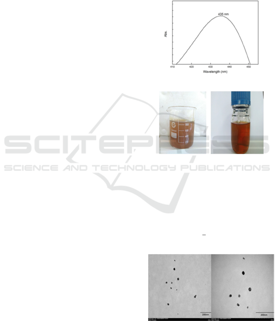
Surface plasmon resonance (SPR) of the
synthesized AgNPs was analyzed by using UV-
visible spectra between 200 nm and 700 nm. In
this study, the SPR value of AgNPs obtained was
435 nm (Figure 2). Previous study reported AgNPs
with SPR value from 410 nm to 425 nm (Handoko
et al., 2017). The success in AgNPs synthesis in
this study was indicated by the colour change from
Ag
+
reduction (Figure 3).The formation of AgNPs
is well-known to be indicated by the
transformation of solution from colourless to
brown. The change occurred in the solution
suggested the formation of AgNPs with the
excitation of SPR in silver metal nanoparticles
(Dubey, Bhadauria and Kushwah, 2009).
Figure 2 UV-visible spectra of silver nanoparticles.
Figure 3 Brown color from Ag
+
reduction.
Transmission electron microscopy
Images from TEM analysis of AgNPs are
presented in Figure 4. From the Figure, AgNPs
were shown to have formed spherical shapes,
which were also reported in other studies
(Shanmugam et al., 2018)(Handoko et al., 2017).
The dimension of AgNPs produced was calculated
by using image J analysis. The average size was
approximately 9.15 + 4.15 nm. Based on the
investigation, AgNPs solution was stable with up
to one-month storage.
Figure 4. TEM images of AgNPs.
Green Synthesis and Characterization of Silver Nanopaticles
575

4 CONCLUSION
Green synthesis of AgNPs using chemical
reduction was done by using AgNO
3
, glucose and
starch as the precursor and reducing agents. The
formation of AgNPs was indicated by the color
change in mixing solution from colorless to
become dark brown after 4 h of synthesis. UV-
visible analysis showed that SPR value of AgNPs
was at 435 nm. The spherical shapes of AgNPs
were confirmed by TEM analysis. In addition, the
dimensions of AgNPs were calculated by using
Image J analysis and obtained average diameter at
approximately 9.15 + 4.15 nm.
ACKNOWLEDGEMENT
The authors would like thank the Rector of
Universitas Sumatera Utara and all researchers at
Cellulosic and Functional Materials Research
Centre for the support given throughout the
research.
REFERENCES
Dubey, M., Bhadauria, S. and Kushwah, B. S. 2009.
Green synthesis of nanosilver particles from extract
of eucalyptus hybrida ( safeda ) leaf’, Digest Journal
of Nanomaterials and Biostructures, 4(3), pp. 537–
543.
Handoko, C. T., Huda A., Bustan H.D., Yudono B.,
Gulo F.2017. Green synthesis of silver nanoparticle
and its antibacterial activity’, Rasayan Journal
Chemistry, 10(4), pp. 1137–1144.
Khorasani, G., Hosseinmehr S.J., Azadbakhr M.
2009.Aloe versus silver sulfadiazine creams for
second-degree burns: A randomized controlled
study, Surgery Today. doi: 10.1007/s00595-008-
3944-y.
Landage, S. M., Wasif, A. I. and Dhuppe, P. 2014.
‘Synthesis of nanosilver using chemical reduction
methods’, International Journal of Advanced
Research in Engineering and Applied Sciences, 3(5),
pp. 14–22.
Morones, J. R.. ElechnigueraaJ.L., Carnacho A. 2005.
The bactericidal effect of silver nanoparticles,
Nanotechnology, 16, pp. 2346–2353. doi:
10.1088/0957-4484/16/10/059.
Prakash, V., Pawar J., Patel A.K., Henry R. 2017.
Analysis of nucleation and growth parameter of
silver nanoparticles for sensors, in 2017 1st
International Conference on Electronics, Materials
Engineering and Nano-Technology, IEMENTech
2017. doi: 10.1109/IEMEN TECH.2017.8076939.
Salleh, A., Naomi R., Utami N.D., Muhammed A.W.,
Mahmoudi E., Mustafa N., Fauzi M.B. 2020. The
potential of silver nanoparticles for antiviral and
antibacterial applications : a mechanism of action’,
nanomaterials, 10, p. 1566.
Saravanakumar, K., Chelliah R., Shanmugam, S. 2018.
Green synthesis and characterisation of biologically
active nanosilver from seed extract of Gardenia
jasminoides Ellis’, Photochemistry and
Photobiology. doi:
10.1016/j.jphotobiol.2018.05.032.
Slepička, P., Kasalkova N.S., SiegelJ., Kolska J., Svocik
V. 2020. Methods of gold and silver nanoparticles
preparation’, Materials, 13, p. 1. doi:
10.3390/ma13010001.
Zheludkevich, M. L. Gusakov A.G., Voropaef A.G.,
Vecher A.A.,KozyrskyE.N., Rasvovov S.A. 2004.
Oxidation of silver by atomic oxygen, Oxidation of
Metals. doi: 10.1023/b:oxid.00000 16275.96500.24.
IMC-SciMath 2019 - The International MIPAnet Conference on Science and Mathematics (IMC-SciMath)
576
