
Effect of Beluntas (Pluchea indica (L.) Less) Leaves Ethanol Extract
to Incision Wound and Healing in Mice (Mus musculus L.)
Emita Sabri and Rahma Handayani
Department of Biology, Faculty of Mathematics and Natural Sciences, Universitas Sumatera Utara, Medan, Indonesia
Keywords: Incision Wound, Mice, Pluchea indica (L.) Less, Wound Healing Process.
Abstract: A wound can be caused by punctures, collisions, bites or scratches of sharp object that can be avoid by
mecanism of wound healing. The use of herbal medicine is an alternative choice to wound healing because of
the relatively small side effects, one of which is Pluchea indica (L.) Less leaves ethanol extract because it
contains saponin, tannin, and terpenoid as anti-inflammatory and antibacterial to accelerate the process of
wound healing.
The aims of this research were to find out the effectiveness of ethanol extract of beluntas leaf
for duration and histological appearance process of wound healing on mice’s skin. This research used 25 male
mice that were divided into five different treatment groups. The treatment groups were treated with K-, K+
(povidone-iodine) and beluntas leaf ethanol extract with three concentration of 25%, 50% and 75% ointments.
Incision wound was made into 1 cm length, the ointments were applied onto the wound and observed for
twice a day in 14 days. Histological preparation was made to calculate epithelial thickness, lymphocytes and
fibroblasts. The data were analyzed statistically using SPSS. The result of this research showed beluntas
leaves ethanol extract 25% was a faster effect on the duration of wound healing which was 6.8 days.
Histological observations showed that beluntas leaf ethanol extract ointment concentration of 25% had the
most significant influence of the average epithelial thickness, while concentration of 75% had the most
significant influence of average number of lymphocytes and fibroblast. In conclusion, the beluntas leaves
extract ointment has a positive effect on the wound healing process.
1 INTRODUCTION
Wound is a condition that characterized by damage
some of normal body tissues such as epithelial tissue,
connective tissue, authority and skin which are often
followed by nerve tissue damage and rupture of blood
tissue. Wound can be caused by sctratches, collisions,
puncture, animal stings and other. To avoid futher
damage wound healing mechanism that begins with
inflammation (Abdurrahmat, 2014).
The mechanism of wound healing naturally have
several phases that are inflammation phase,
ploriferation phase and remodeling phase. Wound
healing process requires proper care. Right eksternal
condition and chemical compounds to protect wound
area from contamination of microorganism and build
the structure of the wound cover by itself (Handayani
et al., 2015).
Untritical wound healing treatment can make
inflammatory process becomes more longer, as a
result the wound area becomes infected and will
prolong the wound to heal (Sinaga dan Tarigan,
2012). Therefore some herbal preparations are needed
as alternative choices for the process of inflammation
and wound healing because have relatively smaller
side effects and herbal plants are abundant in nature
Indonesia has about 40.000 medicinal plants but
only about 25% have been explored by researchers
and used as traditional medicine. Traditiona
medicines use herbs derived from certain plants that
are harmless and can be taken in an urgent situation
(Marbun dan Restuasi, 2015). One of some plants that
people use as traditional medicinal plants is beluntas
(Pluchea indica (L.) Less).
Beluntas is plant of the Asteraceas family that life
in hard and rocky habitats. All parts of beluntas plant
are used for medicinal purposes, both roots, stems and
leaves (Dalimartha, 2008). In Thailand and Java, the
root is used as antipyretics, ulcers and sinusitis and
leaves are used as tuberculosis durgs, thrown body
odor and anti-inflammatory. In Indo-China leaves and
shoots which are crushed and mixed with alcohol are
used as rheumatism and scurvy (Purnobasuki, 2014;
Sudirman et al., 2017). Beluntas leaves as an anti-
Sabri, E. and Handayani, R.
Effect of Beluntas (Pluchea indica (L.) Less) Leaves Ethanol Extract of Incision Wound and Healing in Mice (Mus musculus L.).
DOI: 10.5220/0010138200002775
In Proceedings of the 1st International MIPAnet Conference on Science and Mathematics (IMC-SciMath 2019), pages 171-177
ISBN: 978-989-758-556-2
Copyright
c
2022 by SCITEPRESS – Science and Technology Publications, Lda. All rights reserved
171
inflmmatory and astringent because of saponin and
triterpenoid coumpound (Goyal and Agrrawal, 2013).
Based on the research of Widyawati et al. (2014),
the results of phytochemical screening of beluntas
leaves using ethanol as solvents produced chemical
compounds are saponin, tannin, terpenoid, flavonoid
and alkaloid with have high antioxidant activity.
Based on research by Puspitasari and Prayogo (2017)
antioxsidants can inhibit free radicals so can prevent
diseases caused by radical such as liver damage,
inflammation, diabetes, cancer, antiaging and wound
healing.
Some researchers have reported that beluntas
leaves have analgesic effects (Sibarani et al., 2013),
as larvicides (Muta’ali dan Purwani, 2015), as
antibacterial (Rahmi et al., 2015), as antidiarrheal
(Nurhalimah et al., 2015) and as anti-inflammatory
(Sudirman et al., 2017) but it still little research on
wound healing. Acording the phenomenon it is
necessary to examine whether the ethanol extract of
beluntas leaves (Pluchea indica (L.) Less) has an
effect on wound healing in mice (Mus musculus L.)
as information for the community so can be utilized
in the future.
2 MATERIALS AND METHODS
2.1 Preparation of Sample
This research was taken in Animal Structure
Laboratory, Natural Material Chemistry Laboratory,
Faculty of Mathematics and Natural Sciences,
University of North Sumatera. Sample was used 25
male mice, weight 25-30 g devide into five treatment
group were treated with K-, K+ (Povidone iodine), PI,
PII and PIII which were treated by beluntas leaf
extract ointment based on Febriana et al. (2015) have
been modified with each concentration respectively
25%, 50% dan 75%. Mice were placed in a cage made
from plastic material and covered by a gauze. Mice
were acclimatized for 1 week and fed by adlibitum.
2.2 Beluntas Leaves Extraction
The method of making extraction is maceration.
Frresh beluntas leaves were weighed and washed then
dried without direct sunlight. The dried leaves were
grinded to coaster powder then was macerated with
etanol 70% with ratio between powder and solvent is
1:10. Soaked for 6 hours with occasional shaking and
stirring, then soaked for 18 hours again. The maserate
was filtered and then repeated once again with a
solvent volume half of the first process. The maserate
was evaporated with rotary evaporator at 40
0
C
(KEMENKES RI, 2013). Beluntas leaf extraxt was
then screened to find out secondary metabolites.
2.3 Beluntas Leaves Ethanol Extract
Ointment Formulation
Acording to Kusumawardhani (2015), the
concentration of beluntas leaves ehanol extract
ointment were obtained by using the formula:
L
𝑥 100 % (1)
L : ointment concentration (%)
a : ethanol extract of beluntas leaves (g)
b : total weight (50 g)
Table 1: beluntas leaves ointment formulation.
Ingredients Wei
g
ht
(
m
g)
25% 50% 75%
Vaseline 37,5 25 12,5
Ethanol exctract of
b
eluntas leaves
12,5 25 37,5
2.4 Treatments of Sample
Mice were anesthetized then the dorsal of mice were
made shaved and a 1 cm long incision was made
through the skin. Mice were treated under grouping
dosing section and the ointment formulaton as
described. Treatments were started from day 1 to day
14 and ovserved in twice a day.
2.5 Histological Assessment
The mice skin was taken on the 14t
h
day after mice
were dislocated first. Histological assessment using
the paraffin method with Hematoxylin-eosin staining
(Suntoro, 1983).
2.6 Observation Parameters
The parameters used were time span of wound
healing for 14 days, number of lymphocyte cells,
number of fibroblasts and epithelial thickness on day
14
th
in skin mice. Microscopial axamination used
OptiLab Microscope Camera with magnifaction 100x
and 400x.
2.7 Data Analysis
The data ovatined were then analyzed statistically by
using SPSS software version 22.0 them using Anova
test and continued with Post hoc test.
IMC-SciMath 2019 - The International MIPAnet Conference on Science and Mathematics (IMC-SciMath)
172
3 RESULTS
3.1 The Time Span of Wound Healing
Effect of ehtanol extract of beluntas leaves for time
span of wound healing for 14 days can be seen in
Table 2.
Table 2: Average time span of wound healing.
Groups Average time span ± SD
K(-) 7.40
c
± 1.14
K(+) 8.40
ab
± 0.54
PI 6.80
a
± 0.83
PII 8.20
c
± 0.83
PIII 8.40
c
± 1.14
Based on Table 2, it can be seen that the fastest
wound healing was PI group with 6.8 days, while the
longest was K+ and PIII treatments with 8.4 days.
Data were tested with One Way ANOVA and
obatained significant results of 0.045 (p<0.05),
followed by Duncan test and the results showed that
the treatment group of 25% concentration of beluntas
leaves was the most significant effect treatment for
average time span of wound healing in mice, this
maybe caused by chemical subtances such as saponin
and tannin which made wound healing process faster
than usual.
Accordance with the research of Prasetyo et al.
(2010) that speed of wound healing was influenced
by drugs or compound that are given by stimulate the
growth new cells faster. Furthermore Sudiman et al.
(2017) said that tannin has a role in wound healing by
donating hydrogen atoms to bind and neutralize free
radicals so that reducing lipid autooxidation by
protecting cell membranes in reducing inflammatory
reaction. It is thought that this action keeps the cell
membranes from being damage of bacteria to repairs
during wound healing process. Parampasi dan
Soemarno (2012) also said that saponin act as
antibacterial that cause denaturation of proteins in
bacteria so that bacterial cell membranes will be
damage and finally lysis. The damage of bacterial
membrane can prevent conatmination of bacterial in
wound heling occurs.
Wound tretament of PII and PIII provide longer
healing effect than others because it maybe the
compound at that concentration did not working well
so the wound healing process taken longer. The
treaments also did not closed immediately due to
blockage to dried up and became a scab because the
concentration of extract that to high so there was a
blockage in the wound area and eventually made a
new wound and heal longer.
The researchers of Dalazen et al. (2005) using
Vernonia scorpoides said wound healing can be
hampered because the higher concentration of
exctract that given, then the higher cytotoxic effect
are caused. Putrianirma et al. (2019) said that the
concentration level of the solution can also inhibit
saponins to penetrate the cell membrane, also the
levels of saponins that are too high can cause cell
membrane permeability to increase so that the cell
dies. The picture of the wound healing process in
mice for 14 days can be seen in Figure 1
3.2 The Epithelial Thickness of Mice
Wound
Histological observations of ephitelial thickness
measured from stratum basale layer to the stratum
corneum obtained avarage epthitelial thickness in five
treatments can be seen in Table 3.
Table 3: Averages epthitelial thickness in mice wounds on
the 14
th
day.
Groups Averages of phitelial
thickness ± SD (µm)
K(-) 95.74
a
± 36.37
K(+) 101.83
a
± 37.78
PI 222.55
c
± 23.26
PII 180.40
bc
± 47.56
PIII 148.74
b
± 25.67
Based on Table 3, it can be seen that the highest
average number of ephitelial thickness on the day 14
th
was in the treatment PI group with 222.55 µm, while
the lowest of ephitelial thickness was in the K- group
with 95.74 µm.
Effect of Beluntas (Pluchea indica (L.) Less) Leaves Ethanol Extract of Incision Wound and Healing in Mice (Mus musculus L.)
173
Groups Day 1
th
Day 7
th
Day 14
th
K(-)
K(+)
PI
PII
PIII
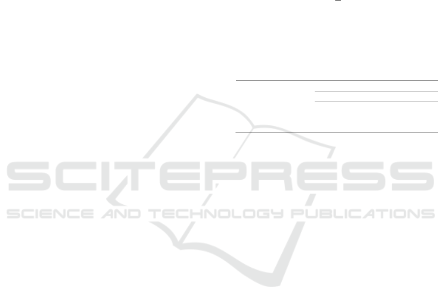
Figure 1: wound healing process of each group of treatment in mice (Mus Musculus L), day 1
th
( ) wounds red and swollen,
day 7
th
( ) wounds shrink and dry and day 14
th
( ) wounds healed.
Data were tested with One Way ANOVA and
obatained significant results of 0.00 (p<0.05),
followed by Duncan test and the results showed that
the treatment group of 25% (PI) and concentration
50% (PII) of beluntas ehanol extract had a significant
effect for epthitelial thickness. High epithelial
thickness in the treatment of beluntas ethanol extract
ointment showed a faster reepithelization in wound
healing process.
Reepithelization is an important step in wound
healing, the faster the process of reepithelization, the
faster the wound healed (Prasetyo et al., 2010).
Terpenoid compound enhance the wound healing
process caused was known to have high antimicrobial
and antioxidant effects thought be liable for wound
contraction and increase time span of epitheliazation
of skin tissue (Wijaya et al., 2014). Saponins’s act in
wound healing stimulating collagen type I which
action in increase process of epitheliazation tissue, as
an antimicrobial and accelerated epithelial cell
migration (Miladiyah and Prabowo 2012).
According to Parampasi and Soemarno (2013)
within 24 to 48 hours, epithelial cells move from edge
of the wound along the edge of incision in dermis and
precipitate compound of basale membrane along the
process. These cells coalesce in the middle line of
wound under the surface scap then producing an
epithelial layer of wound. Proliferation of epithelial
IMC-SciMath 2019 - The International MIPAnet Conference on Science and Mathematics (IMC-SciMath)
174
cell caused the epidermal layer thickened.
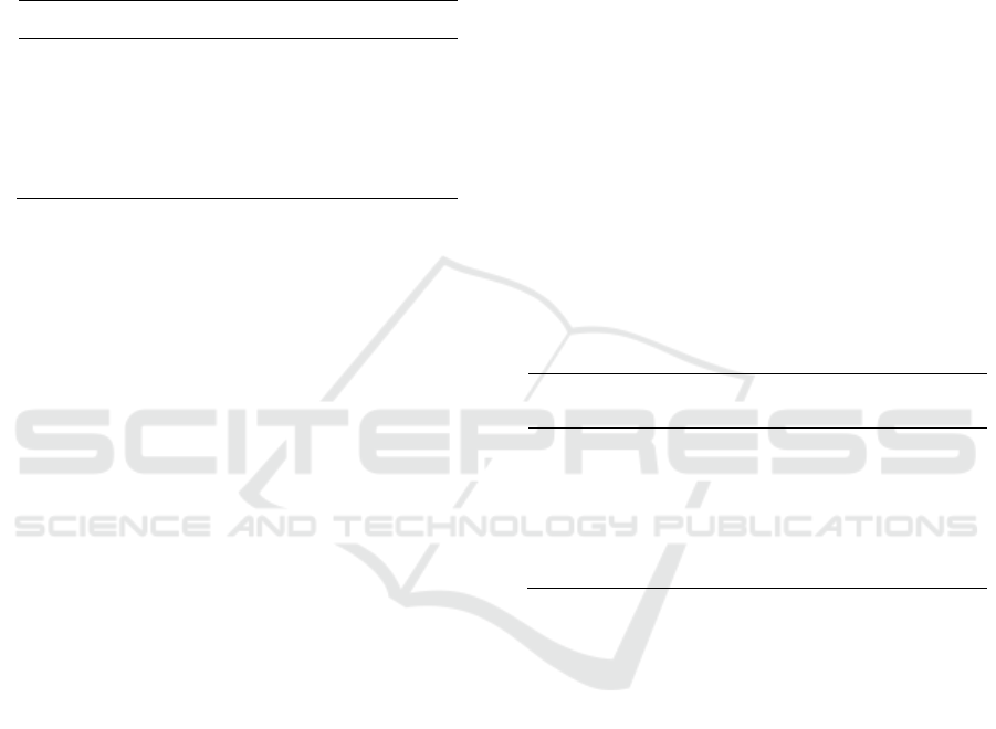
Histological preparation of epithelial thickness in
mice wound 14
th
day can be seen in Figure 2.
Figure 2: Histological examination of epithelial thickness
wound skin in mice (Mus musculus L.).
3.3 The Number of Lymphocytes in
Mice Wounds
Histological observations of the average number of
lymhpvytes on the 14th day of five treatments group
can be seen in Table 4.
Table 4: Average number of lymphocyte cells.
Groups Average number of lymphocytes ± SD
K(-) 5,64
a
± 3,02
K(+) 7,42
a
± 1,93
PI 12,24
b
± 2,82
PII 12,58
b
± 2,25
PIII 17,02
c
± 2,42
Based on Table 4, it can bee seen that the highest
average number of lymphocytes on the day 14th was
PIII treatment group with 17.02, while the lowest is
K- group with 5.64.
Data were tested with One Way ANOVA and
obtained significant results of 0,00 (p<0,05),
followed by Duncan test and the results showed that
the treatment group of ethanol extract beluntas leaves
of 75% concentration had a significant effect for
number of lymphocyte cells. The high number of
lymphocyte cells indicated a wound healing process.
The high number of lymphocyte cells in treatment of
beluntas leaves ethanol extract maybe due to
chemical compounds in the leaves of beluntas
affecting the number of lymphocytes in wound area.
Wibawani et al. (2015) that saponin and tannin
compound have antimicrobial property that can
prevent and control wound infections by destroying
pathogens and can reduce local inflammation and
tissue damage. Izzaty
et al. (2014) also said that role
of lymphocyte in wound healing process is release
lymphokines that very influential in inflammatory
process by affecting the aggregation and chemotaxix
of macrophages in wound area. Lymphokines is
important for stimulating and activating macrophages
in phagocytosis process, activated macrophages will
release cytokines which will activate lymphocytes.
Lymphocytes and macrophages stimulate each other
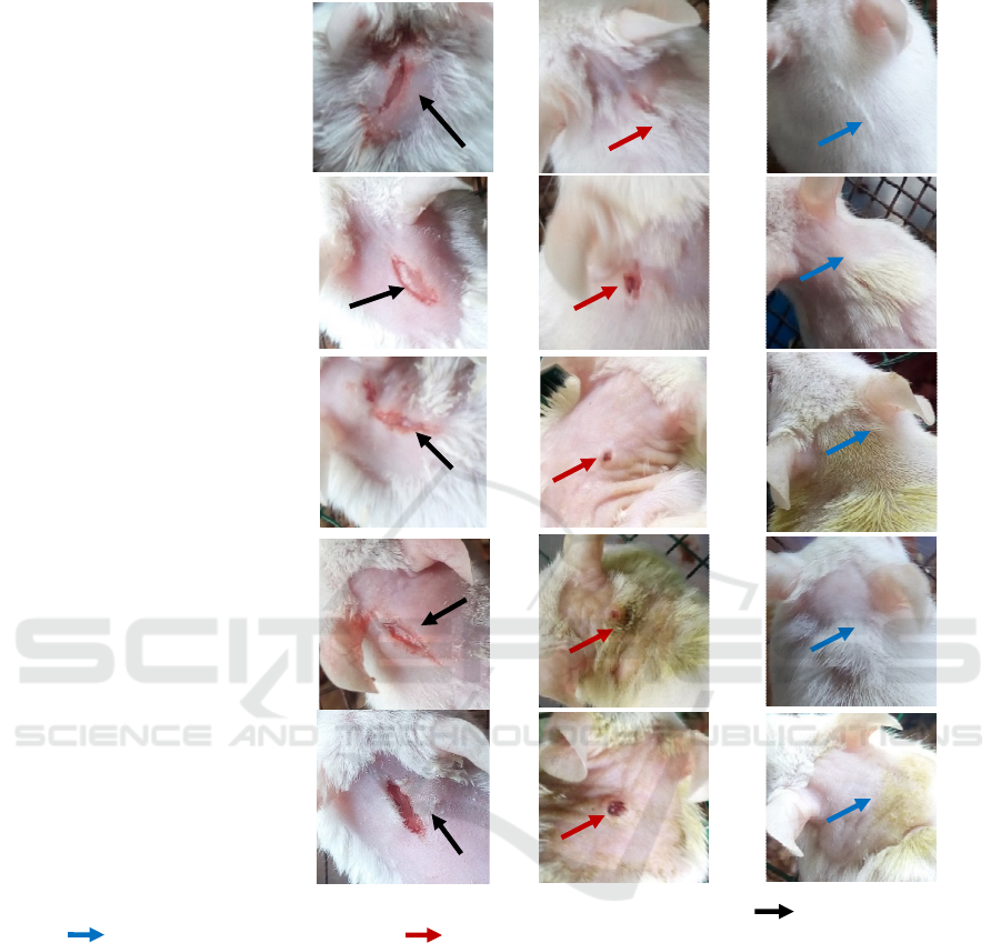
to eliminate bad subtances. Histological preparatons
of lymphocyte in the wound area 14th day can be seen
in Figure 3
Figure 3: Histological examination of lymphocyte cells
wound skin in mice (Mus musculus L.).
K(-)
K(+)
PI
PII
PIII
Effect of Beluntas (Pluchea indica (L.) Less) Leaves Ethanol Extract of Incision Wound and Healing in Mice (Mus musculus L.)
175
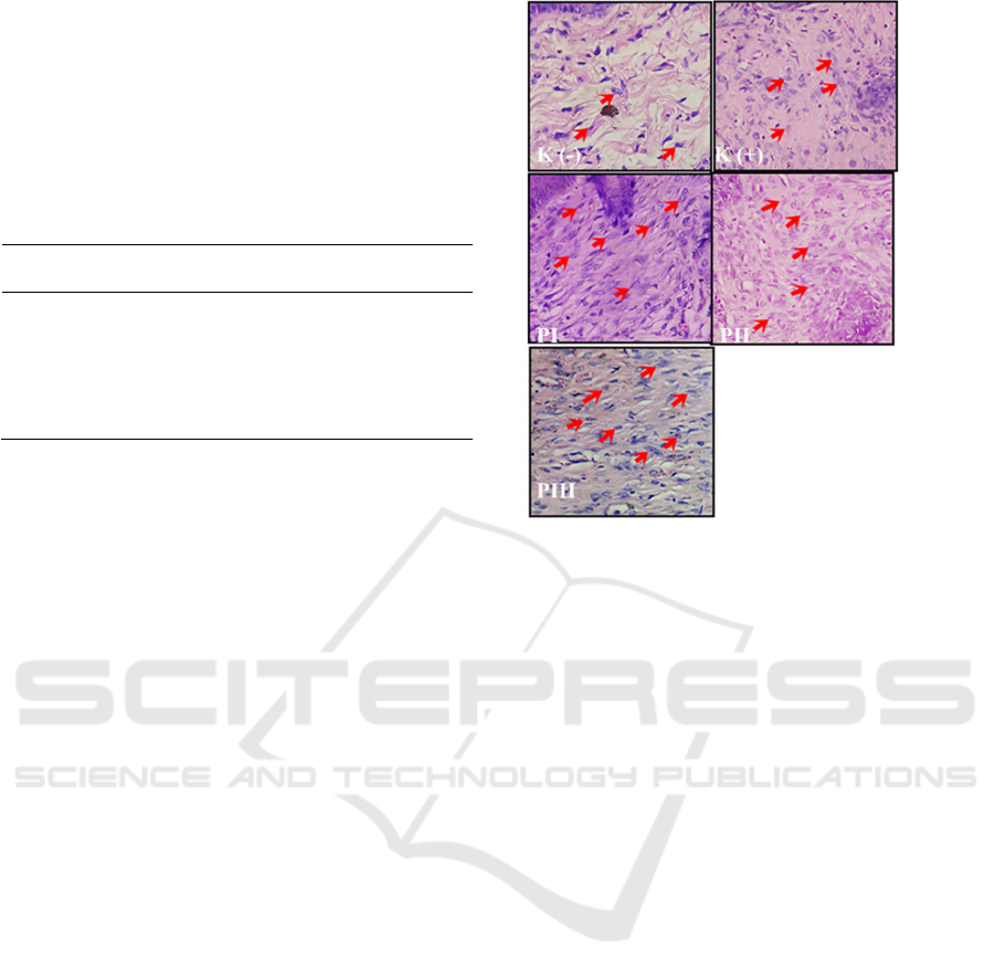
3.4 The Number of Fibroblasts in Mice
Wounds
Histological observations of the average number of
fibroblasts from five treatment groups can bee seen in
Table 5.
Table 5: Average number of fibroblasts in mice wounds 14
th
day.
Groups Averages number of
fibroblasts ± SD
K(-) 4,74
a
± 2,35
K(+) 12,32
b
± 3,02
PI 14,90
bc
± 5,20
PII 17,64
bc
± 2,46
PIII 20,16
c
± 5,34
Based on Table 5, it can be seen that the highest
average number of fibroblast on the 14th day was PIII
treatment group with 20.16 while the lowest was K(-)
treatment group with 4.74. Data were tested by One
Way ANOVA and obtained significant results of 0,00
(p<0,05), followed by Duncan test and the results
showed that the ethanol extract beluntas leaves (25%,
50% and 75%) had a significant effect for number of
fibroblast. The large number of fibroblast in treatment
of beluntas leaf extract may be caused chemical
compounds in beluntas leaves such as tannins.
This is consistent with the statement of Palumpun
et al. (2017) that tanin has a cellular mechanism
activity that is eliminate free radicals, increasing the
wounded connection by activating fibroblast. The
tannin-containing wound stimulates the proliferation
of fibroblasts and secretes collagen and proteoglycans
are the main components of the extracellular matrix
(ECM) and forms granulation tissue. Nurdiana et al.
(2016
said that fibroblast does important role in the
proliferation phase in wound healing process.
A good
wound healing process is characterized by an increase
in the number of fibroblasts induced by fibroblast
growth factor (FGF). The more number of fibroblasts,
the more collagen will be formed, thus accelerating
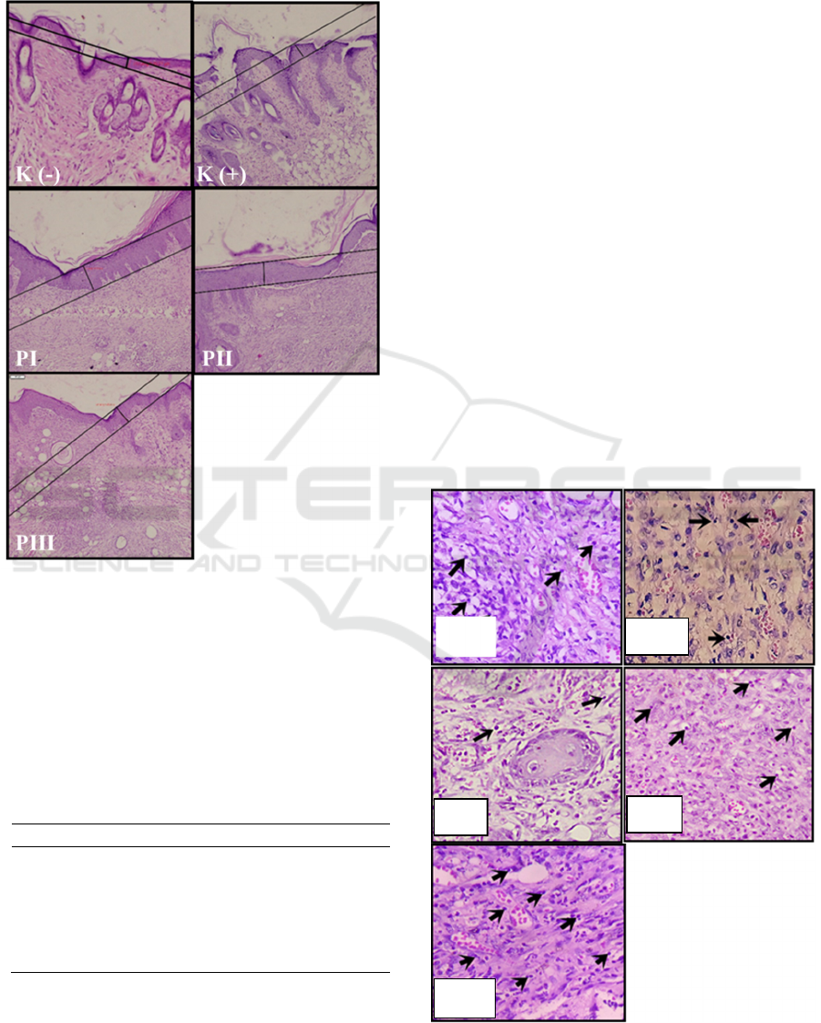
wound healing. Histological preparations of
fibroblast cells injured area day 14 can be seen in
Figure 4
Figure 4: Histological examination number of fibroblasts
wound skin in mice (Mus musculus L.).
4 CONCLUSION
Beluntas leaves extract ointment of 25%g gives a
faster effect for time span of wound healing and
average of epithelial thicknes,
while concentration of
75% had the most significant influence of average
number of lymphocyte and fibroblast of incision
wound healing in mice (Mus musculus L.)
REFERENCES
Abdurrahmat, AS., 2014. Luka, Peradangan dan
Pemulihan. Jurnal Entropi. 9(1): 721-840.
Dalazen, P., Uzoibian, BO., Ogbanya, KC., Nnaji, T., 2005.
Effect of Topical Application of Extract Root from
Vernonia scorpoides on Exsicional Wounds in Mice.
Farmacogn. 15: 82-87.
Dalimartha, S., 2008. Atlas Tumbuhan Obat Indonesia.
Jakarta. Pustaka Pembangunan Swadaya Nusantara.
Febriana, MH., Amintarti, S., Putra, A., 2015. Pengaruh
Ekstrak Daun Beluntas (Pluchea indica (L.) Less)
terhadap Pertumbuhan Bakteri Escherichia coli. Jurnal
Wahama-Bio. 13(1): 60-63.
Goyal, PK, and Anggrawal, RR., 2013. A Review on
Phytochemical and Biological Investigation of Plant
Pluchea. Indo America Journal of Pharmaceutical
Research. 3(4): 3379-3381
Handayani, E., Siswanto, E., Pangesti, AY., 2015. Uji
Aktivitas Ekstrak Etanol Gambir (Uncaria gambir
Roxb.) terhadap Penyembuhan Luka Bakar pada Kulit
IMC-SciMath 2019 - The International MIPAnet Conference on Science and Mathematics (IMC-SciMath)
176

Punggung Mencit Putih Jantan (Mus musculus). Jurnal
Ilmiah Manutung. 1(2): 134-139.
Izzaty, A., Dewi, N., Pratiwi, DIN., 2014. Ekstrak Haruan
(Channa striata) Secara Efektif Menurunkan Jumlah
Limfosit Luka. Dentofazial. 13(3): 176-179.
Kementerian Kesehatan Republik Indonesia., 2013.
Suplemen III Farmakope Herbal Indonesia Edisi I.
Kementerian Kesehatan RI. Jakarta.
Kusumawardhani, AD., Kalsum, U., Rini, IS., 2015.
Pengaruh Sediaan Salep Ekstrak Daun Sirih (Piper
betle Linn.) terhadap Jumlah Fibroblas Luka Bakar
Derajat IIA pada Tikus Putih (Rattus novergicus) Galur
Wistar. Majalah Kesehatan FKUB. 2(1): 16-28.
Marbun, EMA., dan Restuasi M., 2015. Pengaruh Ekstrak
Etanol Daun Buas-buas (Prema pubescens Bulme)
sebagai Antiiflamasi pada Edema Kaki Tikus Putih
(Rattus novergicus). J Biosains. 1(3): 107-112.
Miladiyah, I., and Prabowo, BZ., 2012. Ethanolic Extract
of Anredera cordifolia (Ten.) Steenis Leaves Improve
Wound Healing in Guinea pigs. Universa Medica.
31(3): 4-9.
Muta’ali, R., dan Purwani, KI., 2015. Pengaruh Ekstrak
Daun Beluntas (Pluchea indica (L.) Less) terhadap
Mortalitas dan Perkembangan Larva Spodoptera litura
F. Jurnal Sains dan Seni. 4(2): 55-56.
Nurdiana, Ulya I., Putra, PRA., 2016. Pengaruh Pemberian
Ekstrak Daun Melati (Jasminum sambac L. Ait)
Terhadap Jumlah Fibroblas Kulit dalam Penyembuhan
Luka Bakar Derajat II pada Tikus Putih (Rattus
novergicus) Galur Wistar. Jurnal Keperawatan. 4(1):
1-5.
Nurhalimah, H., Wijayanti, N., Widyaningsih, TD., 2015.
Efek Antidiare Ekstrak Daun Beluntas (Pluchea indica
L.) Terhadap Mencit Jantan yang Diinduksi Bakteri
Salmonella Thypimurium. Jurnal Pangan dan
Agroindustri. 3(3): 1083-1094.
Palumpun, EF., Wiraguna, AGP., Pangkahila, W., 2017.
Pemberian Ekstrak Daun Sirih (Piper betle) Secara
Topikal Meningkatkan Epidermis, Jumlah Fibroblas,
dan Jumlah Kolagen dalam Proses Penyembuhan Luka
pada Tikus Jantan Galur Wistar (Rattus novergicus).
Jurnal 1-Biomedik. 5(1): 1-7.
Parampasi, N., dan Soemarno T., 2013. Pengaruh
Pemberian Ekstrak Daun Pepaya Etanol 70% pada
Proses Penyembuhan Luka Insisi. Majalah Patologi.
22(1): 31-36.
Prasetyo, BF., Wientarsih, I., Priosoeryanto, BP., 2010.
Aktivitas Sediaan Gel Ekstrak Batang Pohon Pisang
Ambon dalam Proses Penyembuhan Luka Sayat pada
Mencit. Jurnal Veteriner. 11(2): 9.
Purnobasuki, H., 2004. Potensi Mangrove Sebagai
Tanaman Obat. Jurnal Biota. 9(2): 125-126.
Puspitasari, AD., dan Proyogo, LS., 2017. Perbandingan
Metode Ekstraksi Maserasi dan Sokletasi Terhadap
Kadar Fenolik Total Ekstrak Etanol Daun Kersen (Mu
tingia calabura). Jurnal Ilmiah Cendikia Eksakta. 2(1):
1-2.
Putrianirma, R., Triakoso, N., Yunita, MN., Yudaniayanti,
IS., Hamid, IS., Fikri, F., 2019. Efektifitas Ektrak Daun
Afrika (Vernonia amygdalina) secara Topikal untuk
Reepitelisasi Penyembuhan Luka Insisi pada Tikus
Putih (Rattus novergicus). Medik veteriner. 2(1): 30-35.
Rahmi, A., Cahyanto, T., Sujardo, T., Lestari, RI., 2015.
Uji Aktivitas Antibakteri Ektrak Daun Beluntas
(Pluchea indica (L.) Less) terhadap Propionibacterium
acnes Penyebab Jerawat. Edisi Juni. 9(1): 141-142.
Sinaga, M., dan Tarigan, R., 2012. Penggunaan Bahan Pada
Perawatan Luka. Jurnal Keparawatan Klinis. 3(2): 5.
Sibarani, VR., Wowor, PM., Awaloei, H., 2013. Uji
Analgesik Ekstrak Daun Beluntas (Pluchea indica (L.)
Less) pada Mencit (Mus musculus). Jurnal e-Biomedik.
1(1): 621-622.
Sudirman, RS., Usmar., Rahim, A., Bahar MA, 2017.
Aktivitas Antiiflamasi Ekstrak Etanol Daun Beluntas
(Pluchea indica L.) pada Model Inflamasi Terinduksi
CFA. Jurnal Farmasi Galenika. 3(2): 191-198.
Suntoro, SH., 1983. Metode Pewarnaan (Histologi dan
Histokimia). Penerbit Bhratara Karya Aksara. Jakarta.
Wibawani, L., Endang, SW., Utami, YW., 2015. Pengaruh
Pemberian Etanol Daun Melati (Jasminum sambac
L.Ait) secara Topikal terhadap Peningkatan Kontraksi
Luka Bakar Derajat II pada Tikus Putih (Rattus
novergicus) Galur Wistar. Majalah kesehatan FKUB.
2(4): 201-202.
Widyawati, PS., Budianta, TW., Kusuma, FA., Wijaya EL,
2014. Difference of Solvent Polarity to Phytichemical
Content and Antioxidant Activity of Pluche indica Less
leaves Extracts. International Journal of
Pharmacognosy and Phytochemical Research. 6(4):
850-855.
Wijaya, BA., Citraningtyas, G., Wehantous, F., 2014.
Potensi Ekstrak Etanol Tangkai Daun Talas (Colocasia
esculenta L.) Sebagai Alternatif Obat Luka pada Kulit
Kelinci (Oryctolagus cuniculus). Jurnal Ilmiah
Farmasi. 3(3): 2302-2493
Effect of Beluntas (Pluchea indica (L.) Less) Leaves Ethanol Extract of Incision Wound and Healing in Mice (Mus musculus L.)
177
