
Hybrid of the PMD Filter, the K-Means Clustering Method and the
Level Set Method for Exudates Segmentation
Syaiful Anam, Zuraidah Fitriah and Nur Shofianah
Mathematics Department, Brawijaya University, Veteran Street, Malang, Indonesia
Keywords: Diabetic Retinopathy, Exudates, Segmentation, Level Set Method, PMD Filter, K-Means Method.
Abstract: Diabetic retinopathy is an eye disease caused by diabetes mellitus. Early diagnosis of diabetic retinopathy is
necessary to avoid blindness. Exudate is one of the symptoms of the diabetic retinopathy. Ophthalmologists
use the fundus images of a patient to extract the exudates for evaluating the diabetic retinopathy. The
exudates segmentation of the fundus images is a difficult task for ophthalmologists because the fundus
images often have poor qualities, such as the boundaries between objects in a less clear, low contrast and
containing noise. There are many methods of segmentation, one of which is by using an active contour
model. One of the known active contour models is the level set method. It has been widely applied in many
applications in the image processing. However, it cannot work well on the noisy image. This paper proposes
the hybrid of the PMD filter, K-means clustering method and the level set method for segmenting exudates.
The PMD filter and K-means method are exploited to remove the noise. From the results of the
experimental results obtained that the hybrid of K-means clustering method and the level set method is able
to work better in segmenting fundus images than the standard level set method.
1 INTRODUCTION
The prevalence of diabetes mellitus has become
more rapidly in the middle income and low-income
countries. The number of people which have
diabetes mellitus significantly increased (Weng and
Hu, 2017). Diabetes is one of the metabolic diseases
that occur by increasing a blood sugar level in the
body. The increment of blood sugar level in the
body may occur when the body has a problem in
insulin secretion or make use of the formed insulin.
According to the World Health Organization,
diabetes mellitus is a disease which is characterized
by increasing a blood sugar level. It is accompanied
by metabolic disorders of carbohydrates, lipids, and
proteins. Diabetes mellitus may cause many
complication diseases, for example, vascular
complication. Classically the diabetes mellitus
vascular complication diseases are categorized into
two which are microvascular and macrovascular.
The most common diabetes mellitus microvascular
complication is diabetic retinopathy.
Diabetic retinopathy is a damage to the retinal
microvascular system due to prolonged
hyperglycemia. It may lead to blindness. Nowadays
in the western word, the diabetic retinopathy causes
the blindness in the working people (Semeraro et al.,
2015). Diabetic retinopathy is characterized by a
narrowing of retinal vessels. It is caused by the
accumulation of fluids and fatty material in the
retina. It causes bleeding in the retinal vessel so that
it leads to blurred vision. If this condition is left,
then it can cause severe vision damage as well as
blindness. The risk of diabetic retinopathy can be
prevented by detecting and controlling blood sugar,
blood pressure and lipids appropriately (Tarr et al.,
2013).
Ophthalmologists use the retinal images known
as the fundus image is to diagnose diabetic
retinopathy. From fundus, it can be seen the small
blood vessels, microaneurysm, and exudate,
however, it may be in low contrast. For diagnosis the
diabetic retinopathy, ophthalmologists usually use
the fundus image by evaluating the exudate which is
one of the symptoms of diabetic retinopathy.
Therefore, the extraction of the exudate on the
fundus image is needed (Madhukar et al., 2017). The
exudate extraction of the fundus image is a difficult
task for ophthalmologists because the fundus image
often has poor qualities. Therefore, a method that
automatically aided computers will help an
108
Anam, S., Fitriah, Z. and Shofianah, N.
Hybrid of the PMD Filter, the K-Means Clustering Method and the Level Set Method for Exudates Segmentation.
DOI: 10.5220/0008517901080116
In Proceedings of the International Conference on Mathematics and Islam (ICMIs 2018), pages 108-116
ISBN: 978-989-758-407-7
Copyright
c
2020 by SCITEPRESS – Science and Technology Publications, Lda. All rights reserved

ophthalmologist to recognize and extract the signs of
diabetic retinopathy disease.
Segmentation method is one of the methods
which can be used for extracting the exudate. The
image segmentation method will divide the image
into a separate set of regions with uniform texture
attributes, etc. (Dhivya and Anitha, 2014). It can be
applied for segmenting the fundus image area into
two parts i.e. the exudate and other areas. Many
image segmentation methods have been proposed.
The image segmentation method can be divided into
two categories, i.e. edge based method and the area-
based method (Airouche et al., 2014). The level set
method has been proposed for image segmentation.
It is developed by using variational models and
partial differential equations. This method has been
widely used successfully for segmenting the medical
images (Anam et al., 2013, Anam et al., 2014a,
2014b). The level set method has some advantages
over the other methods, i.e. the snake method
thresholding method and region growing method.
However, it cannot work well when the images
contain noise. The level set contour will stop
prematurely in the evolution process and it results in
unsatisfactory segmentation when it is applied for
the noisy image. Many representative conventional
noise reduction methods have been proposed, i.e. the
median filters (Russ, 2006), morphology analysis
(Soille, 1999), bilateral filters (Tomasi and
Manduchi, 1998). However, if they are applied for
fundus image, the exudate boundaries also become
dull unexpectedly. Above all those methods, Perona-
Malik Diffusion (PMD) filter is able to preserve an
edge effectively in the image smoothing. If the PMD
filter is applied to a fundus image, the exudate
boundaries can be preserved, but several noises
cannot be removed. K-means is a clustering method
which has been successfully applied for an
de image noise reduction or an image denoising
(Barca and Rumantir, 2008, Pandey and Bhadauria,
2016). Barca and Rumantir have proposed a
modified K-means method. It is able to eliminate the
noise in the multicolored motion capture image
sequences. While Pandey and Bhadauria have
proposed the method for removing the high-density
impulse noise in the image by using a modified k-
mean algorithm. For this reason, this research uses
the level set with the PMD filter and the K-means
method. The PMD filter and the K-means method
are used as preprocessing to avoid the stopping
premature of evolution curve in the level set. For
this reason, the level set, the PMD filter and K-
means methods for image segmentation will be
considered in this paper.
This paper proposes a hybrid of the PMD filter,
the K-means clustering method and the level set
method for segmenting exudate in fundus images.
The results of the exudate segmentation in the
fundus image will give the information of diabetic
retinopathy sign for ophthalmologists.
2 RELATED WORKS
This section will discuss several theories and the
previous research which is related to this research,
such as diabetic retinopathy and fundus image,
image segmentation, Perona-Malik diffusion filter,
K-Means, and level set method.
2.1 Diabetic Retinopathy and Fundus
Image
Diabetes mellitus is a chronic disease which is
caused by an obtained deficiency in the insulin
production, or by the ineffective insulin production.
Diabetic retinopathy is a microvascular complication
caused by diabetes mellitus. If it is not treated, it
causes blindness.
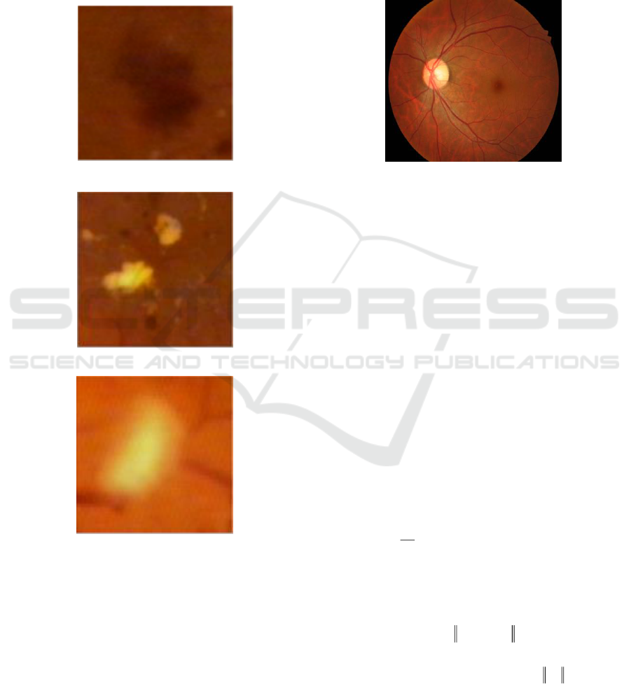
The first symptom of the diabetic retinopathy
appears microaneurysm. If microaneurysm is
broken, it can cause hemorrhage to be seen in Figure
1(a). After that, it seems hard exudate as shown in
Figure 1(b). Hard exudate is a leaky lipid formation
of weakened blood vessels. Along with the severity
of retinopathy disease, the blood vessels may
become inhibited causing microinfarct in the retina
called soft exudate as shown in Figure 1(c). The
diagnosis of diabetic retinopathy using a fundus
image is necessary because the disease is
progressive, the example of the fundus image can be
seen in Figure 2 (Anam et al., 2014b).
2.2 Segmentation
Information technology is a very rapid development
in many fields, such as medical. Science and
technology in image processing and artificial
intelligence become a promising tool in medical
technology. A level set method has been used for
many applications in image segmentation, such as in
detecting the bone boundaries of the hand
radiography. The bone boundaries are a necessity for
segmenting the bone and other areas (Anam et al.
Hybrid of the PMD Filter, the K-Means Clustering Method and the Level Set Method for Exudates Segmentation
109
2013). A combination of PSO and fuzzy inference
has been proposed for extracting the coronary plaque
boundaries in the Intravascular Ultrasound (IVUS)
image. The plaque boundaries in the IVUS image
are needed to be extracted for calculating the plaque
area (Anam et al., 2014a). Boundary extraction of an
image is one of the image segmentation methods.
(a)
(b)
(c)
Figure 1: The abnormal sign on the fundus image caused
by diabetic retinopathy. (a) Hemorrhage, (b) Hard exudate,
(c) Soft exudate.
Image segmentation is one of the image
preprocessing methods in the image recognition and
analysis task. Image segmentation divides an image
into homogeneous areas based on the criteria of
specific similarities of the gray level pixel. There are
many conventional image segmentation methods
which have been proposed, such as the gradient-
based methods (Sobel method, Prewitt method,
Canny method and Laplacian method) and template-
based methods. Canny method cannot result in
smooth segmentation (Mazid, 2013). While, the
snake method cannot separate object well when the
image has object more than one objects (Li et al.,
2005).
Figure 2: Fundus image.
The level set method (Osher and Sethian, 1988)
has been applied for image segmentation to
overcome the weakness of the conventional image
segmentation method. It is one of the known
methods. It is very robust and accurate image
segmentation method. The level set method also has
been broadly used in many fields, in particular for
the image segmentation (Anam et al., 2013, Anam et
al., 2014a).
2.3 Anisotropic Diffusion Filter
An anisotropic diffusion filter has been developed
by Perona and Malik. It is used to eliminate noise in
the image and maintain the edges of an image. The
Perona-Malik (PMD) filter idea is to smooth the
image
),,( tyxu
from an original image u
0
(x,y)
where
t
is diffusion parameter.
The PMD filter equation is defined by (1).
,),,(),,(
)),,((
ItyxcItyxc
Ityxcdiv
t
I
I
t
+=
=
=
(1)
where
)),,((),,( tyxIgtyxc =
(2)
is a coefficient of diffusion process,
I
defines the
norm of image gradient, while
)(g
is an edge
stopping function of level set which is represented
by (3).
ICMIs 2018 - International Conference on Mathematics and Islam
110

2
1
( ) ,
1
gI
I
K
=
+
(3)
K is a diffusion strength parameter. This parameter
is used for controlling the diffusion strength,
)(g
has
high values at the areas where the values of
gradients are low, while it has low values at the area
where the values of gradients are large.
The initial value of
)0,,( yxI
is given (4).
).,()0,,(
0
yxIyxI =
(4)
The PMD filter in the discrete version is defined by
(5).
( 1) ( ) ( ) ( )
,,
( ) ,
n n n n
s s s p s p
s
I I g I I
+
= +
(5)
is the pixel coordinates of concern, p is the
neighbour pixels of
represents the pixel
intensity of s when the iteration count is n.
s
is the
eight pixels of the neighbour of s in the North
diffusion direction, North-West diffusion direction,
West diffusion direction, West-South diffusion
direction, South diffusion direction, South-East
diffusion direction, East diffusion direction and
East-North diffusion direction. |
s
| is a pixel number
of the neighbour of s, while represents a parameter
(Perona and Malik, 1990).
2.4 K-Means Clustering Algorithm
Cluster analysis is the task which partitions a set of
objects into subsets so that the objects properties in
the one cluster have the high degree similarity.
Clustering is an unsupervised learning method
commonly used in a variety of application. It has
been applied for many applications, such as image
processing, machine learning, data mining, and
bioinformatics.
K-means clustering algorithm is one of the
popular clustering methods. It is a clustering method
based on an iterative approach. The K-means
clustering method divides the dataset into k groups
(Santhi et al., 2011). The algorithm of K-Means
clustering is can be seen in Algorithm 1.
Algorithm 1: K-means Algorithm
1. Input the data set which will be clustered and
determine the number of clusters K.
2. Initialize the member of each cluster.
3. Repeat
a. Calculate the cluster center of each cluster.
It is calculated by the means of data in each
cluster.
b. K-means assigns each data in the dataset to
only one of the clusters based on the nearest
distance from data to each cluster centers.
4. Until no change the member of each cluster.
2.5. Level Set Method
Level set method was proposed by Osher and
Sethianin (1988). It has been successfully used for
many applications. The level set method has been
applied for boundary detection in the medical image.
The contour of the level set is defined by using the
zero-level set which is called by a level set function.
The contour of level set expresses the motion of the
contour based on the level set function evolution.
The evolution of level set curve of a parametric
contour
)),,(),,(( tsytsxC
is represented by
equation (6).
/,C t FN =
(
(6)
(
t is a set points of time, while s is a parameter of
evolution curve.
N
defines the normal vector to the
curve C. F is a curve evolution speed function which
will control the motion of the level set contour. The
evolution of curve of (6) can be changed into a
formulation of the level set. Changes made through
the embedding of the dynamic contour C as the zero-
level set. This paper assumes that the value of level
set function
is positive outside the zero-level set
contour, vice versa it takes negative value inside the
zero-level set contour. The inward normal vector is
represented by (7), where
/N
= −
(7)
is a gradient operator.
By using (6) and (7), the evolution of curve of
the level set in (6) is changed to (8),
/,tF
=
(8)
Hybrid of the PMD Filter, the K-Means Clustering Method and the Level Set Method for Exudates Segmentation
111
),()/()(
))((/
ggdiv
ddivt
p
++
=
which refers to as equation of a level set evolution.
The formulation of the level set
)(x
used in this
paper is formulated by (9).
(
(9)
δ
ε
is a dirac delta function,
div
represents a
divergence operator, and
g
defines an edge stopping
function given by(10).
1/ (1 ( * ),g G I
= +
(10)
G
σ
represents the Gaussian filter, while I is an image
which is to be processed (Li et al.,2010).
3 PROPOSED METHOD
This paper proposes an image segmentation method
for extracting the exudate on fundus image by the
hybrid of the K- means and level set methods. The
data used in this research is the fundus images as
shown in Figure 4. They are taken from the website
http://www.it.lut.fi/project/imageret/diaretdb1/. The
data are used to evaluate the proposed method. The
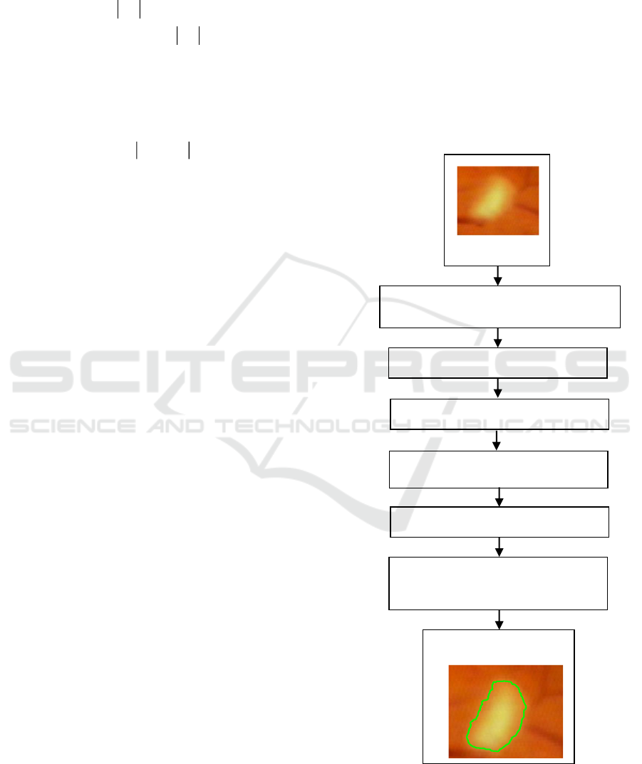
flowchart of the proposed method is shown in Figure
3. The proposed method has several steps. First, the
fundus image is inputted. Furthermore, the image in
the RGB (Red, Green, Blue) color space is
converted to the CIE L*a*b color space. Image in
CIE L * a * b color space has 3 components which
are the L (Luminance), a (reddish-greenish) and b
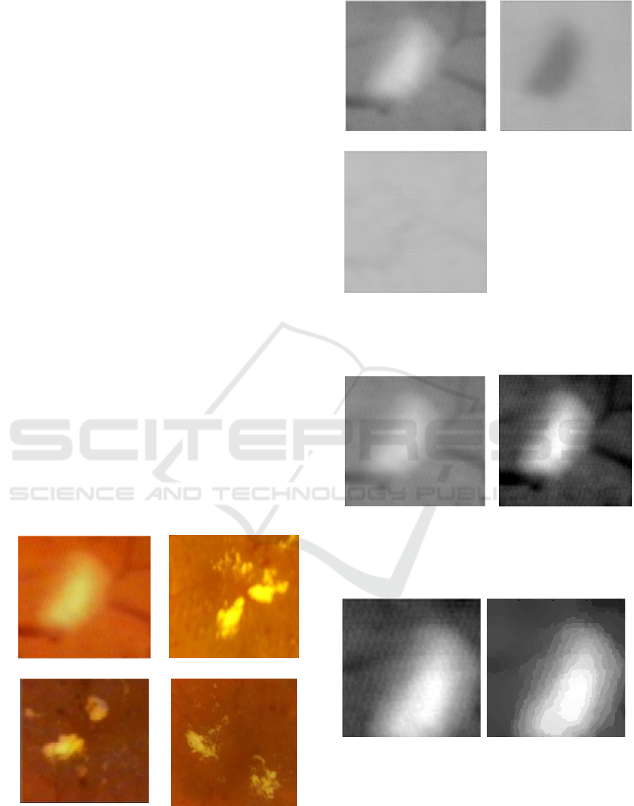
(yellowish-bluish) components. From Figure 5, it
can be seen that the reddish-greenish component is
better than the yellowish-bluish and luminance
components of the format CIE L*a*b for
representing the exudate areas and other areas,
therefore the reddish-greenish component is used for
the next step.
Since the reddish-greenish component has low
contrast as shown in Figure 6 (a), the contrast
enhancement is necessary to be done. After the
image contrast enhancement, the image is better to
visual the exudate, it can be seen in Figure 6.
However, the noise in the image also increases. For
this reason, the noise in the image should be
reduced.
This proposed method uses the PMD filter and
the K-mean algorithm to reduce the noise. Firstly,
the formulation of the PMD filter in the equation (5)
is used for reducing the noise. The initial condition
of the PMD filter in the equation (4) uses the
reddish-greenish component of the image after
applying the image contrast enhancement. The
image result after applying the PMD filter can be
seen in Figure 7. The PMD filter significantly
reduces the noise. However, some noises still exist.
For this reason, the K-means algorithm in Algorithm
1 is used to remove the noise. After applying the K-
means algorithm, the noise disappears, and the
image becomes smooth as shown in Figure 8 (b).
Figure 3: Flowchart of the proposed method.
Convert Format Image from RGB to
CIELAB and Take Green
Component from RGB Image
K-Means ClusterAlgorithm
Segmented
Exudates
Extract the Exudates from
Fundus Image by Using the
Level Set Method
Take Red-Green Component
Enhance the Contrast
Filter the Image by the PMD Filter
Input Image
ICMIs 2018 - International Conference on Mathematics and Islam
112
0 50 100 150 200 250 300
50
100
150
200
250
300
0
50
100
150
200
250
The last step of the proposed method is that the
image resulted by the K-means method is used as
input for the level set method. The equation of the
level set as shown in equation (9) is used to extract
the exudate areas in the fundus image. The final
result of this method can be shown in Figure 8(b).
The exudate areas are the areas inside the green
curve and the other areas are the areas outside the
green curve.
4 RESULTS AND DISSCUSSIONS
For evaluating the proposed method performance,
we use the four various test images of the fundus
with the exudates as shown in Figure 4. The test
images used have different colors, brightness levels,
and contrast levels, this is intended to evaluate the
robustness of the developed method. The test images
also have different levels of clearness on the object
boundaries. The image of Figure 4 (a) has the
unclear object boundaries, while the level brightness
of image of Figure 4 (c) is darker if it is compared
to the other test images. For evaluating the
performance of the proposed method, it is compared
to the standard level set. The different of the standar
level set and the proposed method is in the
calculation of edge stopping function. The standard
level uses the gaussian filter to reduce the noise,
while the proposed method uses the PMD filter and
K-means method.
(a)
(b)
(c)
(d)
Figure 4: Fundus image data set used for evaluating the
proposed method: (a) Image 1
st
, (b) Image 2
nd
, (c) Image
3
th
, (d) Image 4
th
.
(a)
(b)
(c)
Figure 5: Component of CIE L*a*b image, (a) Reddish-
greenish component, (b) Yellowish-bluish, (c) Luminance.
(a)
(b)
Figure 6: (a) The image before the contrast enhancement.
(b) The image after the contrast enhancement.
(a)
(b)
Figure 7: (a) The image before applying the PMD filter.
(b) The image after applying the PMD filter.
H&E image
H&E image
Hybrid of the PMD Filter, the K-Means Clustering Method and the Level Set Method for Exudates Segmentation
113
(a)
(b)
Figure 8: (a) The images which is resulted by the K-means
algorithm. (b) The images which is segmented by the level
set algorithm.
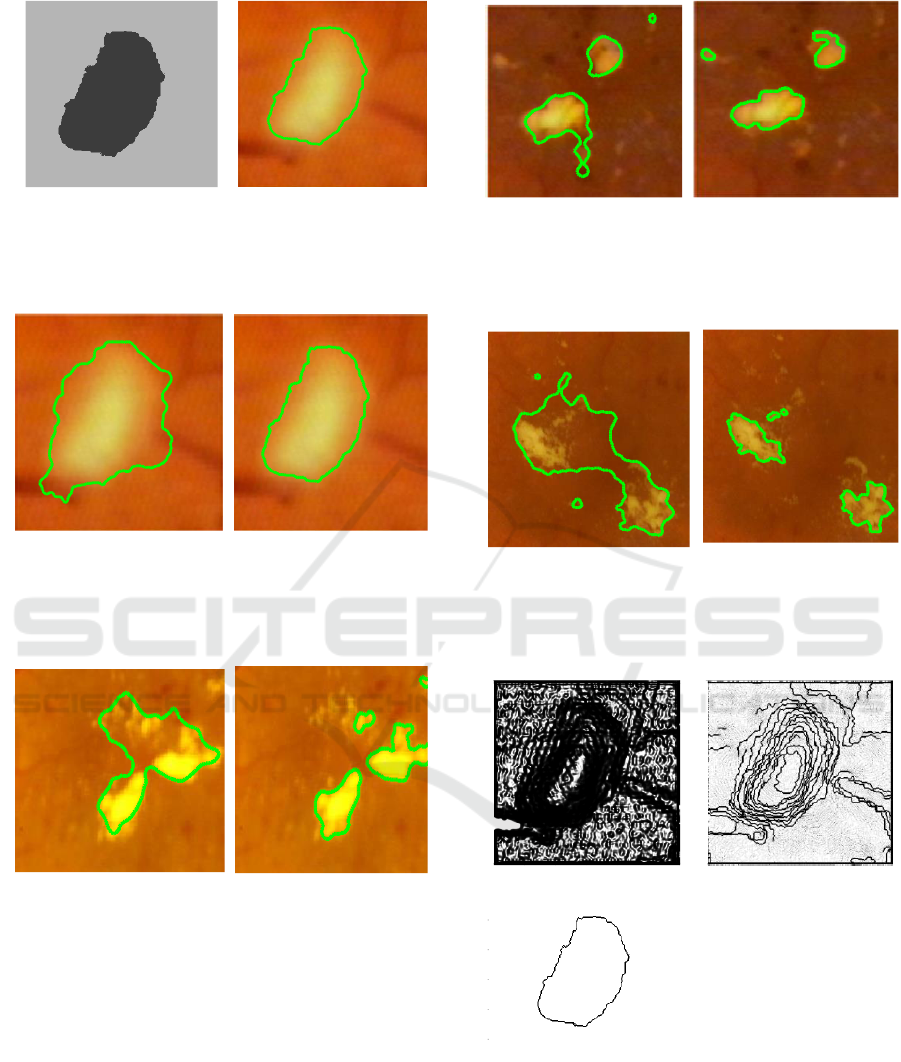
(a)
(b)
Figure 9: Exudates extraction results for image 1st. (a)
Exudates extraction results by the standard level set
method. (b) Exudates extraction results by the hybrid of
the K-means and level set methods.
(a)
(b)
Figure 10:. Exudates extraction results for image 2nd. (a)
Exudates extraction results by the standard level set
method. (b) Exudates extraction results by the hybrid of
the K-means and level set methods.
The images of Figure 9 (a), Figure 10 (a), Figure 11
(a) and Figure 12 (a) show the segmentation results
by using the standard level set, while the images of
Figure 9 (b), Figure 10 (b), Figure 11 (c) and Figure
12 (d) show the segmentation results by the hybrid
of the PMD filter, the K-means and level set
methods (proposed method). The proposed method
is more successfully separates between the exudate
areas and other areas for almost all images if it is
compared to
(a)
(b)
Figure 11: Exudates extraction results for image 3th. (a)
Exudates extraction results by the standard level set
method. (b) Exudates extraction results by the hybrid of
the K-means and level set methods.
(a)
(b)
Figure 12: Exudates extraction results for image 4th. (a)
Exudates extraction results by the standard level set
method. (b) Exudates extraction results by the hybrid of
the K-means and level set methods.
(a)
(b)
(c)
Figure 13: (a) The value of edge stopping of the reddish-
greenish component after applying gaussian filter.b) The
value of edge stopping of the reddish-greenish component
after applying PMD filter. (b) The value of edge stopping
of the reddish-greenish component after applying PMD
filter and K-means algorithm.
objects in cluster 1
0 50 100 150 200 250 300
50
100
150
200
250
300
0.1
0.2
0.3
0.4
0.5
0.6
0.7
0.8
0.9
1
0 50 100 150 200 250 300
50
100
150
200
250
300
0.1
0.2
0.3
0.4
0.5
0.6
0.7
0.8
0.9
1
0 50 100 150 200 250 300
50
100
150
200
250
300
0.1
0.2
0.3
0.4
0.5
0.6
0.7
0.8
0.9
1
ICMIs 2018 - International Conference on Mathematics and Islam
114

the standard level set. The reason is that the value
edge stopping function of the image in Figure 8 (a)
(the proposed method) is high in other areas and
small in the boundary areas as shown in Figure 13
(c). The level set contour will move from outside to
inside when the edge stopping function has the high
value and the level set contour will stop at the
boundary areas. Figure 13 (a) shows the edge
stopping function value of the standard level set. It
takes low not only in the boundary areas but also in
the other areas. It causes the level set contour stop
prematurely in the evolution curve. This condition
results unsatisfactory segmentation as shown in
Figure 9 (a), Figure 10 (a), Figure 11 (a), Figure 12
(a). If the image is only filtered by the PMD filter,
this problem also is happened. For this reason, the
K-means needs to be run after applying the PMD
filter.
However, the proposed method fails to differ the
exudate areas and other areas in several areas as
shown in Figure 9 (b), Figure 10 (b), Figure 11 (c)
and Figure 12 (d). It is caused by the K-means
algorithm cannot works well to differ the exudate
areas and other areas. Since some exudate areas
have similar color intensity with the non-exudate
areas. To solve this problem, it needs to try other
operation to enhance the quality of the fundus
image.
5 CONCLUSIONS
It can be concluded that the hybrid of the PMD
filter, the K-means and level set method works better
in extracting the exudate areas on the fundus image
than the standard level set method. In the evolution
process of the level set, the curve of the level set
stopped prematurely can be avoided by the hybrid of
the PMD filter, the K-means and level set methods
for almost all images used.
ACKNOWLEDGEMENTS
We would like to say our greatest thanks to the
Directorate of Research and Community Service,
Republic of Indonesia who has funded this research
through the “Penelitian Dasar Unggulan Perguruan
Tinggi (PDUPT)” in 2018.
REFERENCES
Airouche, M., Bentabet, L. Zelmat, M., 2009. Image
segmentation using active contour model and level set
method applied to detect oil spills. Proceedings of the
World Congress on Engineering vol. I, pp. 1 - 3.
Anam, S., Uchino, E., Misawa, H., Suetake, N., 2013.
Automatic bone boundary detection in hand
radiographs by using modified level set method and
diffusion filter. Proceedings of the IEEE 6th
International Workshop on Computational
Intelligence and Applications, Hiroshima, Japan, pp.
51-55.
Anam, S., Uchino, E., Misawa, H., Suetake, N., 2014a.
Combining PSO and fuzzy inference for calculation of
coronary plaque boundary in IVUS image.
International Journal of Biomedical Soft Computing
and Human Sciences, vol. 19, no. 1, pp. 51-59.
Anam, S., Uchino, E., Misawa, H., Suetake, N., 2014b.
Retina vessels detection in fundus images by new
morphology operation and the anisotropic diffusion
filter. Proceedings of the Annual Conference of
Biomedical Fuzzy Systems Association, pp. 39-40.
Barca, J. C., Rumantir, G., 2007. A modified K-means
algorithm for noise reduction in optical motion capture
data. Procceeding of the 6th IEEE/ACIS International
Conference on Computer and Information Science
(ICIS 2007). doi:10.1109/icis.2007.29
Dhivya, A., Anitha, D., 2014. Detection of tumor region
using fast fuzzy clustering algorithm. International
Journal of Research in Computer Applications and
Robotics, vol. 2, no. 4, pp. 145-149.
Li, C., Xu, C., Gui, C., Fox, M. D., 2010. Distance
regularized level set evolution and its application to
image segmentation, IEEE Transactions on Image
Processing, vol.19, pp.3243-3254.
Li, Y. F., Zhu, Q. S. , Cao, Y. K. & Wang, C. L., 2005. A
leaf vein extraction method based on snakes
technique. Proceedings of International Conference
on Neural Networks and Brain, pp. 885-888.
Madhukar, M., Manjula, D.Ujwal, Y., Deepthi, J.,
Seshacharan, S., 2017. Exudate extraction: a review.
Perspectives in Communication, Embedded-Systems
and Signal-Processing, vol. 1, no. 2, pp 8-9.
Mazid, K., 2013. Segmentasi citra daun tembakau
berbasis deteksi tepi menggunakan algoritma canny.
Thesis, Dian Nuswantoro University, Indonesia.
Osher, S., Sethian, J., 1998. Fronts propagating with
curvature-dependent speed: algorithms based on
Hamilton-Jacobi formulation. Journal of
Computational Physics, vol. 79, pp. 12-49.
Pandey, P., Bhadauria, S. S., 2016. Removal of high
density impulse noise using modified K-mean
algorithm for digital image. International Journal of
Advance Research in Science and Engineering, vol. 5,
no. 05. pp. 465-473.
Perona, P. and Malik, J., 1990. Scale-space and edge
detection using anisotropic diffusion. IEEE
Transactions on Pattern Analysis and Machine
Hybrid of the PMD Filter, the K-Means Clustering Method and the Level Set Method for Exudates Segmentation
115

Intelligence, vol.12, pp.629 – 639.
Russ, J. C., 2006. The image processing handbook, 5th ed.
Academic Press, New York, 2006.
Santhi, M.V.B.T., Sai Leela, V.R.N.S.S.V., Anitha, P.U.
Nagamalleswari, D., 2011. Enhancing K-means
clustering algorithm. International Journal of
Computer Science and Technology, vol. 2, no. 4., pp.
73-77.
Semeraro, F., Cancarini, A., Dell’Omo, R., Rezzola, S.,
Romano, M., and Costagliola, C., 2015. Diabetic
retinopathy: vascular and inflammatory disease.
Journal of Diabetes Research, pp. 1-16.
Soille, P., 1999. Morphology image analysis: principles
and applications. Springer-Verlag, Telos.
Tarr, J. M., Kaul, K., Chopra, M., Kohner, E. M. And
Chibber, R., 2013. Pathophysiology of diabetic
retinopathy. International Scholarly Research Notices
Ophthalmology, vol. 2013, pp. 1-13.
Tomasi, C., and Manduchi, R., 1998. Bilateral filter for
gray and color images. Proceeding of the 6th
international Conference on Computer Vision, pp.
839 – 846.
Weng, J. and Hu, G. 2017. Diabetes: leveraging the
tipping point of the diabetes pandemic. Diabetes, vol.
66, pp. 1461-1463.
ICMIs 2018 - International Conference on Mathematics and Islam
116
