
Extraction, Identification, and Gel Formulation of Mangiferin from
Mango (Mangifera indica L.) Leaves Extract
Rudi Afrinanda, Yusa Ristiawati, Muhammad Shoufi Islami, Deasy Vanda Pertiwi
Faculty of Pharmacy, Ahmad Dahlan University
Keywords: Mangiferin, Mangifera indica, gel, wound healer, ulcus diabetic
Abstract: Mango (Mangifera indica L.) leaves contain flavonoid which has anti-inflammatory and antioxidants effects
that are beneficial on healing diabetic ulcers. The extract made in gel formulation because it was easy to
dry, forming the washable film layer that provides a cool sensation on the skin. Gel components influence
the stability of formula. To ensure gel quality, safety, and benefits, physical stability test was needed to
fulfill the specifications and stability during storage. This study aimed to extract and identify the mangiferin
as an active compound in mango leaves and to formulate Mangifera indica leaves extract gel as a wound
healer. Extraction of Mangifera indica leaves used soxhlet method with ethanol 70% and determined using
TLC-densitometry method. The optimum formula of the gel was determined by variations of CMC-Na
concentration as gel base, and the compliance of the gel characteristics. The analysis of characteristics
included spreadability test, homogeneity test, adhesivity test, and pH test. The result of extraction was
determined by TLC-Densitometry as 330,52 mg/gram of viscous extract. The formula with 5% CMC-Na gel
base complied with the required characteristics and was the optimum formula, which stability analysis did
not show any changes in pH, colour, consistency, adhesivity and spreadability during storage.
1 INTRODUCTION
Diabetes Militus (DM) is a disease characterized by
the occurrence of hyperglycemia and carbohydrate,
fat, protein metabolism disorders, as a result of
disorder or insulin deficiency by β Langerhans cells
of the pancreas gland, or caused by the lack of
responsiveness of body cells to insulin. One of the
complications of DM occurring is diabetic ulcers or
diabetic lesions, which are skin lesions caused by
high blood glucose levels resulting in vascular
resuscitation and further vascular neuropathy
(Fatimah, 2015, Sarwono, 2009). Based on the data
of the Indonesian Ministry of Health (2014), the
prevalence of diabetic ulcer wounds in Indonesia
reaches 54%. This disease is often found in
developing countries; Indonesia was ranked seventh
with a number of 10 million diabetic patients in
2015 (IDF, 2015).
Using antioxidants as a treatment on diabetic
wounds is the most effective approach related to
wound healing of diabetes. One of the types of
plants that is potential as a wound healer of diabetes
is mango (Mangifera indica). Mango leaves contain
active compound of mangiferin that acts as
antioxidant and capable of lowering blood sugar
levels in diabetes therapy. Moreover, extracts of
mangiferin has a potential for the healing of wounds
in diabetes (Fithriyani et al., 2014, Khandare, 2016).
Mangiferin total from ethanol extracts of Mangifera
indica is 102 mg/gram of mangiferin compounds.
This plant grows a lot in the community and only the
fruit are commonly consumed, not yet optimally
utilized in increasing the value of use (Fithriyani et
al., 2014).
Utilization in the community is seen as not
optimal yet because it has not been processed into
useful drugs; therefore, it needs formulations to form
products, i.e. preparations in the form of a gel. The
gel is a semi-solid material consisting of a
suspension made of inorganic particles that are small
or large organic molecules including penetration by
a liquid. The gel preparation is chosen because it is
easy to dry out, forming a layer of film that is easily
washable and provides a cool sensation on the skin
(Ansel, 2008, Panjaitan et al., 2012).
Formulation of gel in this study used CMC-Na as
the gel agent. CMC-Na is a polymer derivate
cellulose that quickly expands when supplied with
hot water and neutral, clear crystal and has a strong
138
Afrinanda, R., Ristiawati, Y., Islami, M. and Pertiwi, D.
Extraction, Identification, and Gel Formulation of Mangiferin from Mango (Mangifera indica L.) Leaves Extract.
DOI: 10.5220/0008240701380142
In Proceedings of the 1st Muhammadiyah International Conference on Health and Pharmaceutical Development (MICH-PhD 2018), pages 138-142
ISBN: 978-989-758-349-0
Copyright
c
2021 by SCITEPRESS – Science and Technology Publications, Lda. All rights reserved
bond between molecules (Aponno et al., 2014). In
this study, the variation of gel base CMC-Na was
analyzed to find the optimum formula. It is specified
based on gel characterized, i.e. spreading test,
adhesion test, homogeneity, consistency, and pH.
CMC-Na has advantages over Carbopol; pH of
CMC Na is higher than carbopol, the spreading
power of CMC-Na is greater than carbopol gel, and
also the extraction into CMC-Na does not affect the
spreadability, while the gel of carbopol decreased
power of scatterplot (Maulina and Suhigartini,
2015).
2 MATERIALS AND METHOD
2.1 Materials
Mango leaves (Mangifera indica), 70% Ethanol,
ethyl acetate, glacial acetic acid, formic acid,
methanol, CMC Na, Tragakan, propilenglycol,
Carbopol, glycerin, Methyl paraben, and Aquadest.
2.2 Methods
2.2.1 Preparation of Ethanolic Extract of
Mango Leaves
Mango leaves from the area of Sleman, Yogyakarta
were picked and dried under the blazing sun and
previously washed beforehand; they were covered
with black cloth in the process to avoid direct
contact with sun rays. To obtain even drying, leaves
were then moved into oven for 2-3 hours at a
temperature of 500-600 °C, before being ground to
make powder leaves and sieved with sieve mesh no.
40.
About 625 grams of leaves powder was
transferred into a soxhlet tool and then added with
1500 mL ethanol as solvent. Extraction was
performed for 48 hours (Sachin et al., 2014). The
extract obtained was collected and concentrated on
evaporator to evaporate in a waterbath until viscous
extract was obtained.
=
ℎ
ℎ
100%
(1)
2.2.2 Preparations and Determination of the
Optimum Formula of Mango Leaf
Extract Gel
Mango leaves extract (MLE) gel was formed from
MLE and excipients; the composition of the gel
formulated by a trial-error method in the
preformulation step. According to Adnan (2016),
MLE gel with CMC-Na as gel base has the
composition as in Table 1.
These formulas compared and evaluated to
choose the optimum one. The evaluation includes
organoleptic test and homogeneity, consistency, pH,
adhesive test and spreading test. The results obtained
are indicated in Table 2.
2.2.3 Organoleptic Test and Homogeneity
The organoleptic test was performed by directly
observing the colour and smell. Homogeneity test
carried out by applying the gel on a piece of glass
(Maulina and Sugihartini, 2015).
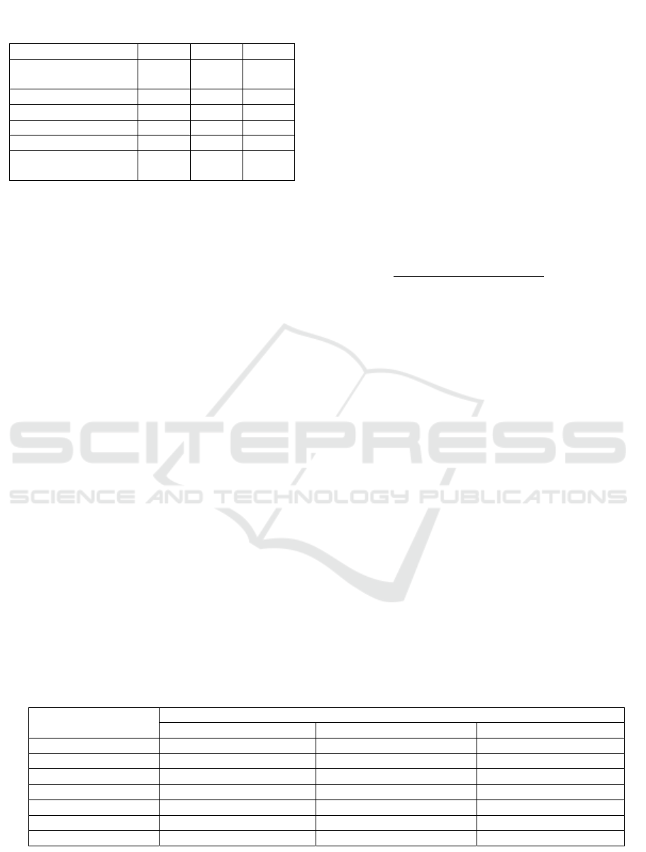
Table 1: Formula of mango leaves extract (MLE)
gel.
Component F1 F2 F3
Mango leaf extract
(MLE)
4% 4% 4%
CMC-Na 5% 5% 5%
Gliserin 5% 5% 5%
Pro
p
ilen
g
likol 2,5 % 2,5 % 2,5 %
Ni
p
a
g
in 0,25% 0,25% 0,25%
Aquadest ad 50
grams
50
grams
50
grams
Table 2: Physical evaluation of MLE gel.
Evaluation Concentration
FI F2 F3
Or
g
anole
p
tic Colou
r
Ver
y
thick brownish
g
reen Thick brownish
g
reen
b
rownish
g
reen
Smell Ver
y
stron
g
Stron
g
Less
Homogeneity Homogenous Homogenous Homogenous
p
H 5 5 5
Sp
r
eadibility 8,117 c
m
6,4 cm 4,13 c
m
Adhesivit
y
3,4 secon
d
5,4 secon
d
56,7 secon
d
Consistenc
y
Stable Stable Stable
Extraction, Identification, and Gel Formulation of Mangiferin from Mango (Mangifera indica L.) Leaves Extract
139
2.2.4 pH Testing
pH test was done using pH paper universal that was
dipped into the diluted samples. Colour change on
the pH paper was compared with the universal
standard (Maulina and Sugihartini, 2015).
2.2.5 Spreadability Test
About 0.5 grams of gel was placed on the glass and
another round glass was placed on top of it, and left
for 1 minute. After that, 150 grams of weight was
added and left for 1 minute. The diameter was then
measured (Astuti et al., 2010).
2.2.6 Adhesivity Test
About 0.25 grams of sample was placed between
two glass objects; given a load of 1 kg for 5 seconds,
which was then lifted and changed with 80 gram of
weight. The time of the release of the gel determined
as adhesivity of gel (Miranti, 2009).
2.2.7 Consistency Test
Consistency test was done using centrifugation test.
Gel samples were centrifuged at 3000 rpm for 5
minutes, and then observed of physical changes
(Djajadisastra et al., 2009).
2.2.8 Data Analysis
The data obtained was processed in statistics using
SPSS 24 software. Normality test (Shapiro-Wilk)
and homogeneity test (Levene) were performed on
the data. To see the relationship between the
treatment groups, the variations were analyzed in
one way (ANOVA) if the data was distributed
normally and homogenously. If the data was not
normal, Gaussian and then Kruskal-Wallis analysis
were done.
3 RESULTS AND DISCUSSION
Mashed dried mango leaves formed the smooth
brownish-green colour with a distinctive odour. Ten
pounds of dry mango
leaves produced 0.8
kilograms of powder. The water content in the dried
mango leaves was 8% ± 0.5, which met the standard
IE (<10%). If water content >10%, it can cause the
onset of enzymatic processes and cause damage due
to microbes, which can change the chemical content
of it.
Mango leaf extract obtained by the soxhlet
method, using ethanol 70% as the solvent, was left
for 24 hours and the ethanol 70% was vaporized on
the waterbath to obtain extracts. The extract
obtained had a bitter taste and a distinctive smell.
Yield and extract obtained was 12.5%.
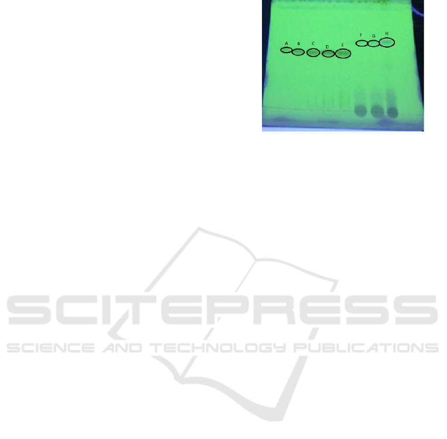
The identification of the xanthan group carried
out was qualitative and quantitative. Qualitative
analysis was done by FeCl3 5% and HCl 1M. The
colour that was changing to brownish blackish
colour proved that the extract contained compound
of the xanthan (mangiferin), positive. Quantitative
analysis was done with the Densitometry method to
obtain TLC mangiferin levels using the stationary
phase of GF 254 and mobile Phase Chloroform:
Methanol: comparison with Formiat Acid 90:10:3.
Test results are presented in Figure 1.
TLC densitometry analysis aimed to determine
levels of mangiferin and quite economical since it
used relatively little motion phase and relatively
TLC densitometry analysis aimed to determine
levels of mangiferin and quite economical since it
used relatively little motion phase and relatively
short time and the measurement of levels of samples
simultaneously. From the analysis, the obtained
concentration of mangiferin was 330.52 mg/gram of
extract.
In the formulation gel, MLE was the active
ingredient. CMC-Na served as gel base, while
propilenglycol and Glycerin as humectant that
increased the stability of formula. CMC-Na was
used as the gel base in the formula because it has
good stability in acid and alkaline condition (pH 2-
10). Propilenglikol in gel formula was used as a
humectant to maintain the stability while keeping the
moisture content in the material properties of the gel.
This material can be stable at pH 3-6. The most
influential factors in the physical quality of the gel
preparations are the base and humectant. The base
gel will form a structure which is an important factor
in the gel formulation. Humectant serves to keep the
Figure 1: TLC plate of MLE in UV 245.
MICH-PhD 2018 - 1st Muhammadiyah International Conference on Health and Pharmaceutical Development
140
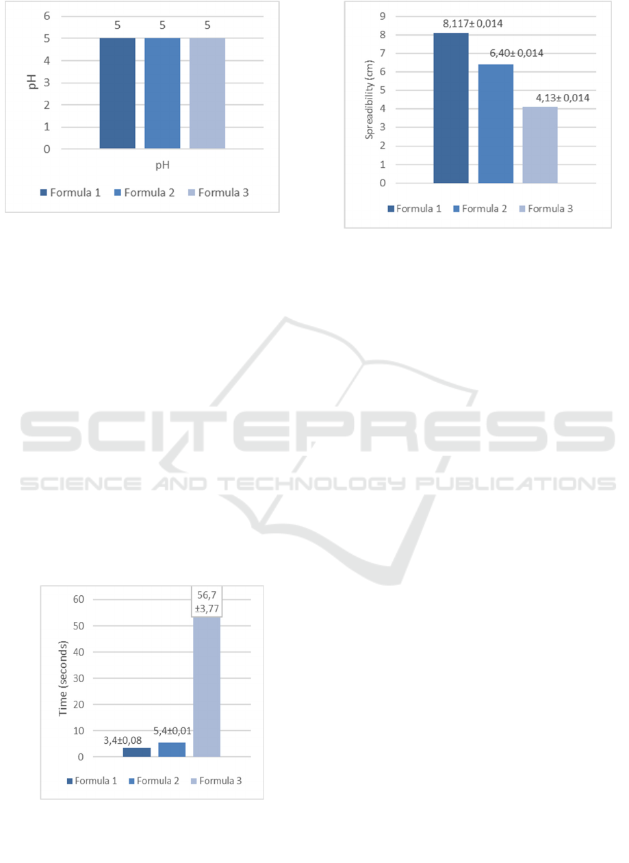
Figure 2: pH diagram with variations of the base
formula of CMC Na 2,5%% (FI), 5% (FII), dan 7,5%
(FIII).
gel formulation by absorbing moisture and reduce
evaporation of water from the formulation. Methyl
paraben was used as a preservative because gel has a
high moisture content that can lead to the occurrence
of microbial contamination.
The physical properties test of the gel are
required to guarantee the quality of gel formulation.
Test results from the third formula shown in the
Table 2. pH of topical formulation requirement is 5-
7 because the normal pH skin IE 4.5 – 6.5
(Martin,1983). Test results are presented in Figure 2.
The gel in this research has pH of 5, which is
appropriate for skin pH. If the pH is not a match for
the skin, it will irritate the skin and reduce comfort
when applied to the skin.
Spreading power test done to learn the ability of
the gel to spread on the skin. The terms of the power
of spread in the topical formulation are 5-7 cm
(Ulaen 2012, et al.). Increasing spreadability of
topical formulation will simplify in the application.
Figure 3 presented the test results.
In this research, the spreading power that can
qualify in specified. The higher concentration of gel
base will cause the smaller area of spreading. It is
because in the high concentration of base gel, it
contains the lower of water. It makes increasingly
viscous gel consistency so that the spreading power
decline. Statistic analysis for the third formula, there
are F1, FII and FIII, normal distributed data and
homogeneity of data that show a different result. It is
indicated by one way ANOVA statistics with value
p = 0,200 or > 5% of the value that is assigned. So
that the third formula meets the requirements.
To see the ability of the gel in attaching to the
skin, adhesivity test was performed. Power
requirement for the material to latch onto the skin is
no shorter than 4 seconds (Ulaen, et al., 2012).
Figure 4 presented the test results.
The formulas that met the requirements were
formula II and III. It is because the lower
consistency made shorter time in contact with skin.
In addition, the increase of concentration caused a
thick consistency that also was increasingly
adhesive. Statistic analysis of normal distributed and
homogeneity by ANOVA retrieved result of the sig
(5%; p < p = 0.00) and the data is not homogeneous
(5%; p < p = 0.04), so then the Kruskal Wallis test
produced a significant value of (p 5%; < p = 0,026).
It means the difference of concentration affected
adhesivity of the gel. Consistency test showed that
segregation in gel formulation did not occur. It
indicated that the gel formula were stable in storage.
Figure 3: Spreadability diagram with variations of the
base formula of CMC Na 2,5 %(FI), 5% (FII), dan 7,5%
(FIII).
Figure 4: Adhesivity diagram with variations of the base
formula of CMC Na 2,5 %(FI), 5% (FII), 7,5% (FIII).
Extraction, Identification, and Gel Formulation of Mangiferin from Mango (Mangifera indica L.) Leaves Extract
141

4 CONCLUSION
The results showed the MLE gel formulation were
green-brown with a distinctive smell, homogeneous,
pH 5, spreading 4.13-8.117 cm, latching for 3.4-56.7
seconds, and stable in storage. The most optimal
formula is gel with a concentration of CMC Na 5%.
ACKNOWLEDGEMENT
The authors would like to thank The Ministry of
Research Technology and Higher Education for
providing the grant used in this study.
REFERENCES
Adnan, J. 2016. Fomulasi Gel Ekstrak Daun Beluntas
(Pluchea indica L.) dengan Na-CMC sebagai Basis
Gel. Journal of Pharmaceutical Science and Herbal
Technology. 1(1).
Ansel, H.C. 1989. Pengantar bentuk sediaan farmasi edisi
keempat. Jakarta:UI Press, pp. 107-513.
Aponno, J. V, Yamlean, P.V.Y. & Supriati, H.S., 2014.
Uji Efektivitas Sediaan Gel Ekstrak Etanol daun
Jambu Biji (Psidium guajava Linn) Terhadap
Penyembuhan Luka yang Terinfeksi Bakteri
Staphylococcus aureus pada Kelinci (Orytolagus
cuniculus), Jurnal Ilmiah Farmasi, 3(3), pp.279–286.
Astuti I. Y., D. Hartanti, dan A. Aminiati. 2010.
Peningkatan Aktivitas Antijamur Candida albicans
Salep Minyak Atsiri Daun Sirih (Piper bettle L.)
melalui Pembentukan Kompleks Inklusi dengan β-
siklodekstrin, Majalah Obat Tradisional. (15): 94-99.
Djajadisastra, J., Mun’im, A., Desi, N. P. 2009. Formulasi
Gel Topikal Dari Ekstrak Nerii folium Dalam Sediaan
Antijerawat, Jurnal Farmasi Indonesia 4 (4): 210-216.
Fatimah, R.N. 2015. Diabetes mellitus tipe II. J Majority 4
(5): 93-101.
Fithriyani, L., Dhadhang, W., Eka, P. 2014. Formulasi
Tablet Mukoadesif Ekstrak Etanol Daun Mangga
Bapang (Mangifera indica ‘Bapang’) sebagai
Antidiabetes Menggunakan Matriks Guar Gum. Jurnal
Ilmu Kefarmasian Indonesia, 12(5): 176-182.
Kemenkes R.I. 2014. Buletin Jendela Data dan Pusat
Informasi Diabetes, Jakarta.
Martin,A., Swarbick,J., Cammarata, A. 1993. Farmasi
Fisik. Jilid II edisi ke-3 terj. dari Physical
Pharmacy oleh Joshita. Jakarta: UI Press. Halaman:
566-572.
Maulina, L dan Sugihartini, N. 2015. Formulasi Gel
Ekstrak Etanol Kulit Buah Manggis (Garcinia
mangostana L) Dengan Variasi Gelling Agent Sebagai
Sediaan Luka Bakar. Pharmaciana. 5(1): 43-52.
Miranti, L. 2009. Pengaruh Konsentrasi Minyak Atsiri
Kencur (Kaempferia galanga) Dengan Basis Salep
Larut Air terhadap Sifat Fisik Salep dan Daya Hambat
Bakteri
Staphylococcus aureus Secara In Vitro,
Skripsi, Fakultas Farmasi Universitas Muhammadiyah
Surakarta, Surakarta.
Sachin, S., Shinde, dan Chavan, A. 2014. Isolation of
Mangiferin from Different Varieties of Mangifera
indica Dried Leaves. Internatioal Journal of Scientific
& Engineering Research. 5(6): 928-934.
Sarwono. 2009. Komplikasi Kronik Diabetes: Mekanisme
Terjadinya, Diagnosis dan Strategi Pengelolaan.
Dalam: Sudoyo AW, Setiyohadi B, Alwi I. Buku Ajar
Ilmu Penyakit Dalam. Jilid 3 Edisi V. Pusat Penerbit
Ilmu Penyakit Dalam FK UI, Jakarta.
Ulaen, SPJ., Banne,Y., Suatan,RA., 2012. Pembuatan
salep Anti Jerawat Dari Ekstrak Rimpang
Temulawak (Curcuma xanthorriza Roxb). Jurusan
Farmasi Politeknik Kesehatan Kemenkes Manado.
MICH-PhD 2018 - 1st Muhammadiyah International Conference on Health and Pharmaceutical Development
142
