
The Efficacy of Platelet-rich Plasma Combined with Platelet-rich
Fibrin in the Treatment of Multiple Chronic Venous Leg Ulcer: A
Case Report
Primadhanty Bhadra, Indah Julianto, Mardiana, Adniana Nareswari, Halim Perdana, Endra Yustin
Ellista Sari
Department of Dermatovenereology Medical Faculty of Sebelas Maret University / Dr.Moewardi General Hospital
Surakarta, Central Java
Keywords: chronic Leg ulcer, platelet-rich plasma, platelet-rich fibrin, intralesional PRP injection.
Abstract : Chronic leg ulcer (CLU) is a defect in the skin below of knee persisting for more than six weeks and shows
no tendency to heal after three or more months. Leg ulcers occur in 1% of the adult population and 3.6% in
people over 65 years. It may increase with the onset of aging, people with atherosclerotic occlusion such as
smoker, obese and diabetic. A 55 years old woman, came with multiple ulcer in her left leg since 5 years
ago. There were various size of ulcers in her left leg, the largest size was 12x 9x 0. 1 cm and the smallest
one was 6x 6x 0. 1 cm with erythema base. On examination ankle brachial index (ABI) was 0. 9. Patient
was diagnosed with deep vein thrombosis (DVT) since 5 years ago, with no satisfying treatment.
Compression therapy is still the basic treatment for venous ulcers, but in our case, compression therapy was
not given satisfactory result. Platelet-rich plasma (PRP) and platelet-rich fibrin (PRF) autologous are some
of the therapies that we can use in chronic wounds. Autologous PRP and PRF are contain many growth
factors (GF), that can help the healing process in chronic wounds, the treatments are easy to use, painless
and safe. In our case we used combined therapy of PRP and PRF for treating CLU and we also used
intralesional PRP injection for the initial treatment and results in satisfying improvement after 13 weeks
observation.
1 INTRODUCTION
Chronic leg ulcer (CLU) is characterized with a
chronic wound in the leg without healing after 3
months with proper treatment or not recovering
optimally at 12 month. Chronic leg ulcer (CLU)
common in adults and the symptoms usually include
pain, friable granulation tissue, foul odor, and
wound breakdown instead of healing. This result
social distress, considerable healthcare and personal
costs
(Shubhangi, 2013). In Chronic wound as in
CLU the levels of platelet derived growth factor
(PDGF), basic fibroblast growth factor (bFGF),
epidermal growth factor (EGF), and transforming
growth factor (TGF-) is reduced
(Sebastian, 2007).
Venous ulcer is the most common type of
chronic ulcer, with the incidence of 1-2 % in the
population. It is 75-80% of all vascular ulcers
(Braund, 2007). Heredity, age, female sex, obesity,
pregnancy, prolonged standing, and greater height
are the risk factors for the venous disease. At early
presentation, tenderness, edema, hyperpigmentation,
and varicose veins are typical features, at later stage,
atrophy blanched, lipodermatosclerosis and venous
ulcers
(Burkhart, 2008). Ankle Brachial Index
(ABI) score is usually used for assessing patient
with lower extremity venous insufficiency, the lower
score, means the more severe the arterial obstruction
(Sebastian, 2007). The conventional treatments of
vascular ulcers are leg elevation, compression, and
wound care. Topical steroids, aspirin, and surgery
are as second-line therapies. Autologous platelet-
rich plasma (PRP) is one of many treatment for
chronic leg ulcer, and it is promising therapy
(Braund, 2007).
PRP is a concentration of platelets required from
the patient's own blood which has been in
centrifugation. It contains fibrin and high
concentrations of growth factors
(Yotsu, 2015). A
second-generation in platelet concentrate is PRF, it
is prepared from centrifuged blood, during
preparation we can find a fibrin clot rich in platelets
510
Bhadra, P., Julianto, I., Mardiana, ., Nareswari, A., Perdana, H. and Sari, E.
The Efficacy of Platelet-rich Plasma Combined with Platelet-rich Fibrin in the Treatment of Multiple Chronic Venous Leg Ulcer: A Case Report.
DOI: 10.5220/0008160905100513
In Proceedings of the 23rd Regional Conference of Dermatology (RCD 2018), pages 510-513
ISBN: 978-989-758-494-7
Copyright
c
2021 by SCITEPRESS – Science and Technology Publications, Lda. All rights reserved
without addition of thrombin, platelet-derived
growth factor and TGF-β has been identified in PRF.
Platelet-rich plasma (PRP) and PRF speed up wound
healing by promoting the healing process secondary
to its GF. These include plateletderived GF (αα,
ββ, and αβ) fibroblast GF, vascular endothelial GF,
epidermal GF, insulinlike GF, and transforming
GF, which are needed in chronic wound healing
(Suthar, 2018).
2 CASE
55 years old woman presented to the
Dermatovenereology Outpatient of Dr. Moewardi
General Hospital Surakarta with chief complaint of
multiple ulcer in her left leg since 5 years ago. Itchy
red spot appeared on her left leg, which later became
small wound and her leg skin changed into black.
The wound then enlarged as the time went by. She
then seeked medical help and was diagnosed with
stasis dermatitis. After 2 months there was no
improvement, she came back to the same hospital
and she was treated by internist as she was
diagnosed with DVT comfirmed USG Doppler, after
3 months of treatment with elastic bandage
(compression), aspilet
R
and venosmil
R
without
improving, a new small wound appeared in the same
leg. Because the wound enlarger, the doctor referred
patient to vascular surgery, and she had
debridement. She came back to her internist and she
was received additional therapy with simarc
R
,
amlodipine and candesartan
R
. However, the therapy
did not give satisfactory result, which made her to
came to our clinic.
This patient works as a shopkeeper who required
to stand for a long time. Physical examination
revealed high blood pressure 160/100 (hypertension
grade 1), Body Mass Index (BMI) 26. 6 (Obesity I),
the dorsalis pedis artery pulse was well palpable and
there was no specific finding in the laboratory
examinations.
The dermatological examination demonstrated,
multiple ulcer with the largest size of 9.5x 13x 0. 1
cm and the smallest one 6. 5x 6. 5x 0. 1 cm, at her
left leg varicose vein, hemosiderin and
lipodermatosclerosis also seen.
We treated the patient with PRP injection 0.
2cc/cm2 for initial therapy, after that we used the
topical PRP in antioxidant gel and we combined
with PRF for maintenance. We observed the patient
in every 3 days for 8 weeks months after that we
once a week. After 13 weeks of observation there
was satisfactory improvement.
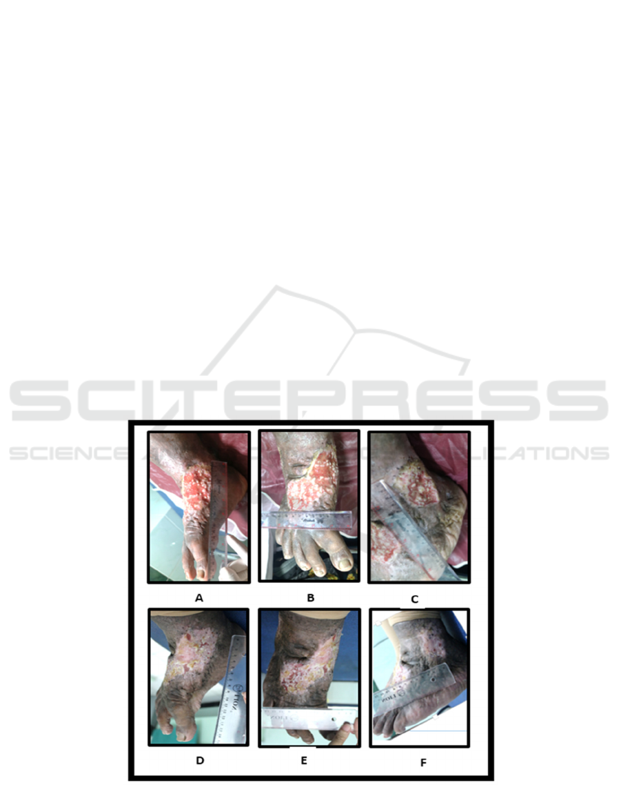
Figure 1. (A-C). Multiple ulcer with the bigger size is 9. 5x 13x 0. 1 cm and the smallest size is 6. 5x 6. 5x 0. 1 cm. (D-F)
13 weeks of therapy with PRP combined PRF given every 3 days for 8 weeks, after that we once a week monthly.
The Efficacy of Platelet-rich Plasma Combined with Platelet-rich Fibrin in the Treatment of Multiple Chronic Venous Leg Ulcer: A Case
Report
511

3 DISCUSSION
One of the up to date modalities for CLU is PRP
(Hafner, 2012). Platelet-rich plasma (PRP) research
has long been studied for its the molecular
interaction of platelets and plasma secretion with
different cell phenotype. Platelet-rich plasma (PRP)
is promising therapy in wound care, as it increases
cell migration, proliferation, angiogenesis, and tissue
anabolism. Safe because free from antibody
formation, graft versus host’s disease and infectious
diseases such as hepatitis and HIV, easy and cost-
effective method with good results in the
management of chronic non healing ulcers are an
advantages of autologous PRP
(Alavi et al, 2016).
Aside from PRP we can use PRF, it also contains
fibroblast growth factor (GF), vascular endothelial
GF, angiopoitin and platelet-derived GF. These
speed up the wound healing
(Andiessen, 2017). He
L, et al in the statistical analysis levels reported that
TGF-β and PDGF-AB released from PRF or PRP at
different time points. PRP released the highest
amounts of TGF-β and PDGF-AB at the first day,
followed by significantly decreased release at later
time points PRF released the highest amount of
TGF-β at day 14
th
and the highest amount of PDGF-
AB at day 7
th.
(He, 2009). Choukroun et al. is the
first who develop PRF in France to be used in oral
and maxillofacial surgery, PRF belongs to a new
generation of platelet concentrates with simply
preparation and without any biochemical blood
handling
(Nagaraju, 2017). The intralesional
injection of platelet concentration constitutes a
valuable alternative in the treatment of chronic
ulcers
(Suthar, 2018).
Handling all possible systemic and local factors
which can interrupt healing process is the first step
in treating ulcers
(Hafner, 2012).Next eliminating or
treating the causes of precipitation, such as, surgical
intervention, until promotes circulation and
improves venous return, for example, compression
therapy. Wound care, lifestyle changes and symptom
management are needed for healing. Self-hygiene
promote preventive care is very important in
preventing its recurrence. Current treatments for
CLU include surgery, sclerotherapy, compress
therapy (conventional therapy), and adjuvant
pharmacotherapy
(Shubhangi, 2013). The mainstay
treatment for patients with venous leg ulcers is
compression therapy, there are 3 different techniques
in compression: (1) bandage system, (2) stockings
/socks, or (3) intermittent compression device,
however there an contraindications in the use of
compression therapy high compression of 40 mm Hg
should considered for a person with an adequate
vascular supply indicated by an ankle brachial
pressure index of 0.8 to 1.2 and in patients with
mixed arteriovenous ulceration (with an ankle
brachial pressure index [0.5 and an absolute ankle
pressure [60 mm Hg), inelastic compression \40 mm
Hg does not impede arterial perfusion and treats
impaired venous return
(Somani, 2017),
(Suryanarayan, 2014), (Moneib, 2017).
Our patient has multiple chronic wounds in her
left leg since 5 years ago, without any improvement
in the treatment, she was even treated with oral
therapy, compression therapy and debridement. The
diagnosis of DVT from this patient was confirmed
with physical examination, USG Doppler and ABI.
Hard to heal wounds are defined as those with
granulation tissue which fail to epithelialize despite
two months of conservative treatment
(Burkhart,
2008). In this case, we can used PRP and PRF for
the treatment, as PRP and PRF can improved wound
healing through promotion of the healing process by
the presence of growth factor which are important in
modulating mesenchyme cell recruitment,
proliferation, and extracellular matrix synthesis
during the healing process
(Alavi et al, 2016). Beside
PRP and PRF we used injection PRP for the initial
therapy, this method accelerates the healing process
in the majority of non-healing wounds and
contributes to either the complete healing or the
preparation of the wound bed for a final
reconstructive procedure
(Dionyssiou, 2012).
Platelet Rich Plasma (RPP) combined PRF given
every 3 days for 8 weeks, after that we once a week
monthly for 13 weeks. We are not use the
compression therapy because our patient felt pain
when using the bandage.
4 CONCLUSION
We acknowledge that this study was not funded by
any organization. There was no conflict of interest in
this study.
REFERENCES
Alavi, A., Sibbald, R. G., Phillips, T. J., Miller, O. F.,
Margolis, D. J., Marston, W., ... & Kirsner, R. S.,
2016. What's new: Management of venous leg ulcers:
Treating venous leg ulcers. Journal of the American
Academy of Dermatology, 74(4),pp. 643-664.
Andriessen, A., Apelqvist, J., Mosti, G., Partsch, H.,
Gonska, C., & Abel, M., 2017. Compression therapy
RCD 2018 - The 23rd Regional Conference of Dermatology 2018
512

for venous leg ulcers: risk factors for adverse events
and complications, contraindications–a review of
present guidelines. Journal of the European Academy
of Dermatology and Venereology, 31(9), pp. 1562-
1568.
Braund, R., Hook, S., & Medlicott, N. J., 2007. The role of
topical growth factors in chronic wounds. Current
drug delivery, 4(3), pp. 195-204.
Burkhart, C.N., Adigun, C., Burton, C.S., 2008. Cutaneous
changes in peripheral venous and lymphatic
insufficiency. In Wolff K, Goldsmith LA, Katz SI,
Gilchrest BA, Paller AS, Leffell DJ, editor.
Fitzpatrick’s dermatology in general medicine, 8thed.
New York: McGraw Hill, 1831-1835.
Dionyssiou, D., Demiri, E., Foroglou, P., Cheva, A.,
Saratzis, N., Aivazidis, C., & Karkavelas, G., 2013.
The effectiveness of intralesional injection of
platelet‐rich plasma in accelerating the healing of
chronic ulcers: an experimental and clinical
study. International wound journal, 10(4), pp. 397-
406.
Hafner, A., Sprecher, E., 2012. Vascular disorder: Ulcers,
Bolognia JL, Jorizzo JJ, Schaffer JV, Callen JP,
Cerroni L, Heymann WR et al. Dermatology, 3rd
edition. London: Elsevier, 1729-1746.
He, L., Lin, Y., Hu, X., Zhang, Y., & Wu, H., 2009. A
comparative study of platelet-rich fibrin (PRF) and
platelet-rich plasma (PRP) on the effect of
proliferation and differentiation of rat osteoblasts in
vitro. Oral Surgery, Oral Medicine, Oral Pathology,
Oral Radiology, and Endodontology, 108(5), 707-713.
Moneib HA, Youssef SS, Aly DG, Rizk MA,
Abdelhakeem YI. Autologous platelet-rich plasma
versus conventional therapy for the treatment of
chronic venous leg ulcers: A comparative study. J
Cosmet Dermatol2017; 00:1–7.
Nagaraju, U., Sundar, P. K., Agarwal, P., Raju, B. P., &
Kumar, M., 2017. Autologous platelet-rich fibrin
matrix in non-healing trophic ulcers in patients with
Hansen's disease. Journal of cutaneous and aesthetic
surgery, 10(1), pp. 3.
San Sebastian, K. M., Lobato, I., Hernández, I., Burgos-
Alonso, N., Gomez-Fernandez, M. C., López, J. L., ...
& Andia, I., 2014. Efficacy and safety of autologous
platelet rich plasma for the treatment of vascular
ulcers in primary care: Phase III study. BMC family
practice, 15(1), 211.
Shubhangi, A.V., 2013. Chronic leg ulcers: Epidemiology,
aetiopathogenesis and management, Hindawi
Publishing Corporation Ulcers, 1-9.
Somani, A., & Rai, R., 2017. Comparison of efficacy of
autologous platelet-rich fibrin versus saline dressing in
chronic venous leg ulcers: A randomised controlled
trial. Journal of cutaneous and aesthetic
surgery, 10(1), 8.
Suryanarayan, S., Budamakuntla, L., Khadri, S., &
Sarvajnamurthy, S., 2014. Efficacy of autologous
platelet-rich plasma in the treatment of chronic
nonhealing leg ulcers. Plastic and Aesthetic
Research, 1(2), pp. 65-65.
Suthar, M., Gupta, S., Bukhari, S., & Ponemone, V., 2017.
Treatment of chronic non-healing ulcers using
autologous platelet rich plasma: a case series. Journal
of biomedical science, 24(1), pp. 16.
Yotsu, R.R., Hagirawa, S., Okochi, H., Tamaki, T., 2015.
Case series of patients with chronic foot ulcers treated
with autologous platelet-rich plasma. J Dermatol 2015;
42: 1–8Hafner A, Sprecher E, Vascular disorder:
Ulcers, Bolognia JL, Jorizzo JJ, Schaffer JV, Callen
JP, Cerroni L, Heymann WR et al. Dermatology, 3rd
edition. London: Elsevier, 2012:1729-46
The Efficacy of Platelet-rich Plasma Combined with Platelet-rich Fibrin in the Treatment of Multiple Chronic Venous Leg Ulcer: A Case
Report
513
