
An Indistinguishable Case of Zinc Deficiency: Acquired Zinc
Deficiency or Acrodermatitis Enteropathica?
Putri Reno Sori, Reiva Farah Dwiyana, Yuri Yogya, Inne Arline Diana, R. M. Rendy, Ariezal Effendi
Department of Dermatology and Venereology, Faculty of Medicine, Universitas Padjadjaran - Dr. Hasan Sadikin Hospital,
Bandung 40161 Indonesia
Keywords: acrodermatitis enteropathica, , acquired zinc deficiency, zinc
Abstract: Zinc (Zn) is an essential micronutrient in growth and development in children. In some condition, zinc
deficiency occurred due to genetic or non-genetic caused. Skin disorders due to Zn deficiency are
differentiated into acrodermatitis enteropathica (AE) and acquired zinc deficiency (AZD). These conditions
are often indistinguished, since they have same skin disorders. However, AE is caused by genetic disorder
that need long-life Zn supplementation, and the other hand, AZD occurred by imbalance intake of Zn,
especially from breast milk. Therefore, further examination for baby and mother are required. Here, we
describe a case report of a 3-month old boy with zinc deficiency. Skin disorders evolved since the patient
was 2-month old as symmetrical dermatitis in perioral, acral, and skin folds area, accompanied with
diarrhea. Patient was born prematurely at 34-week of gestation and was a breast-fed infant. The diagnosis of
AZD was established based on typical skin lesion, diarrhea, and low serum Zn level. This decreased of
serum Zn also found in his mother. Patient was treated with 10 mg Zn sulphate supplement once daily and
clinical improvement was obtained after five days supplementation. Zinc level in patient remained high after
one month discontinuation of Zn supplement. Clinically, AZD is difficult to distinguish with AE, thus zinc
level examination in baby and mother are required, as well as observation during and after discontinued of
zinc supplementation, to support the diagnosis, both for AE or AZD.
1 INTRODUCTION
Zinc (Zn) is an essential micronutrient in proteins
and nucleic acids. Zn also plays role in cell growth
and division, wound healing, and immune cell
activity (Plum et al., 2010). Zn deficiency in
children results in growth retardation, emotional
disturbance, irritability, depression, gastrointestinal
disorders, hypogonadism, anemia, skin disorders,
and immune system disorders (Ackland et al., 2008).
Skin disorders due to Zn deficiency can manifest as
erythematous macules, vesicles, pustules, crusts,
erosion, and scales in the periorifisium, extremities,
and diaper area with symmetrical distribution (Yang
et al., 2012). These lesions could resemble atopic
dermatitis, seborrheic dermatitis, diaper dermatitis,
candidiasis, and skin disorders from vitamin and
other micronutrient deficiency (Kury et al., 2012;
Kharfi et al., 2010). Theredore, this skin disease
often misdiagnosis.
Zinc deficiency is classified into primary and
secondary (Ackland et al., 2008).Primary deficiency
results from low Zn intake or low bioavailability of
Zn. Secondary deficiency is caused by genetic
disorder or disease that interfere Zn absorption as
well as increased Zn excretion in intestinal.
Acquired zinc deficiency (AZD) and acrodermatitis
enteropathica (AE) are disease that occurs due to Zn
deficiency with very similar clinical manifestation
(Ackland et al., 2008). AE is caused by a genetic
disorder resulting in impaired absorption of Zn in
the intestinal with classic triad of periorifisium
dermatitis, diarrhea and alopecia, while AZD is due
to imbalanced intake and Zn requirement without
intestinal disturbance (Yang et al., 2012; Ruiz-
Maldonado& Orozco, 2008).
Here is one case of AZD that initially
undisguisable to AE because of same skin rashes.
However, serum Zn level examination from patient
and mother showed lower level that indicated to
AZD due to imbalance intake, thus Zn
supplementation was given. The purpose of this case
report is to report a Zn deficiency case that clinically
indistinguishable, but after discontinued of Zn
supplementation, the serum Zn level examination
remained normal, that indicated to AZD.
Sori, P., Dwiyana, R., Yogya, Y., Diana, I., Rendy, R. and Effendi, A.
An Indistinguishable Case of Zinc Deficiency: Acquired Zinc Deficiency or Acrodermatitis Enteropathica?.
DOI: 10.5220/0008160404890492
In Proceedings of the 23rd Regional Conference of Dermatology (RCD 2018), pages 489-492
ISBN: 978-989-758-494-7
Copyright
c
2021 by SCITEPRESS – Science and Technology Publications, Lda. All rights reserved
489

2 CASE
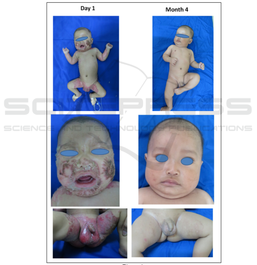
A 3-month-old baby boy presented with 1-month
history of skin eruption, arising initially on his
inguinal area. These lesions spread bilaterally over
his back, hands and feet, then onto his face. The skin
lesion accompanied with diarrhea up to 6 times a
day. He was treated with topical corticosteroids for
skin lesion and antibiotic with other physician and
improvement was not achieved. He was premature,
delivered at 34 weeks, and full breast-fed infant.
Physical examination revealed bright red, eroded
plaques in perioral and extremities, especially folded
ares, as well as vesiculobullous lesions on his hands
and feet (Figure 1). Bacteria was found on Gram
stain and pseudohyphae and blastospore from skin
scrapings with 10% potassium hydroxide (KOH)
solution. Patient had underweight and stunted, and
from laboratory examination, there was
hyponatremia.
From history taking and physical examination,
led to Zn deficiency, which could be AZD and AE.
Therefore, serum Zn examination was needed. The
result were Zn level at patiet was 9 μg/l (reference
range 26–141 μg /l) and Zn level of his mother at 49
μg /l (reference range 60–130 μg /l). This result
confirmed AZD since usually in AE, Zn level of
mother is in normal limit, however, there is
genetically malfunction of Zn absorption in baby,
that lead to Zn deficiency.
The patient was given oral zinc sulphate
supplementation in 10 mg/day dose, and dramatic
improvement of skin lesion had achieved within a
few days. The lesions resolved completely over
following 2 weeks. Zn supplementation still
continued for 3 months, and the normal level of Zn
was achieved as 101 μg /l. After a months of Zn
supementation was stopped, Zn level remain normal
(96 μg /l) and there was no new skin lesion at all
(Figure 1). Based on this condition, the diagnosis of
this patient was AZD, that could be differentiated
with AE from thorough examination and follow up.
3 DISCUSSION
Zinc is an essential micronutrient for growth and
development (Plum et al., 2010). It is required for
the activity of more than 300 enzymes and 1,000
transcription factors, as well as controlling genetic
expression, and plays an important role in protein
and amino acids synthesis, cell replication and
regeneration (Krebs, 2013). Zn sources in infants are
mainly obtained from breast milk. Zn in breast milk
is better absorbed than cow's milk or soy milk,
because it contains zinc-binding ligand (Jen & Yan,
2012). Zn from food will be absorbed in the form of
Zn
+ 2
ions that bind to ZIP4 transporters to be
absorbed in duodenum and jejunum (Deshpande et
al., 2013).
Zinc deficiency characterized by abnormalities in
the skin, gastrointestinal tract, and alopecia (Ruiz-
Maldonado& Orozco, 2008). Skin disorders initially
presented as erythematous macules with scale and
may turn into vesicles, bullae, pustules, and erosion
with symmetrical distribution in periorifisial, and
extremities (Kharfi et al., 2010). Skin lesions may be
accompanied by secondary infections caused by
Candida or bacteria (Kaur et al., 2016). This disorder
is accompanied by diarrhea three to six times per
day which can leads to dehydration and electrolyte
imbalance. In mild cases there is dry and coarse hair,
which become alopecia in severe cases. Other
abnormalities that can be found are anorexia, growth
retardation, irritability, eye abnormalities and
photophobia (Jensen et al., 2008).
Diagnosis of Zn deficiency in this patient
established based on typical clinical features and
laboratory tests (Kury et al., 2012). Zn serum
concentrations was less than 50 μg / l are the gold
standard for the diagnosis of Zn deficiency (Jen &
Yan, 2012).
The skin lesions in this patient are erythematous
macule with scale in symmetrically distributed
between the thighs, buttocks, back and hands and
around the mouth and nose, accompanied by
vesicles and bullae in the legs. Complaints also
accompanied with diarrhea and hair loss. We found
bacteria on Gram stain and pseudohyphae and
blastospore from skin scrapings with 10% KOH
solution. On physical examination, the nutritional
status was underweight and stunted, alopecia, and
from laboratory examination, the patient was
hyponatremia.
Zinc deficiency can be differentiated into congenital
and acquired (Yanagisawa, 2004). AE is a
congenital Zn deficiency arising from a gene
mutation encoding the Zip4 protein, which is a Zn
transporter in the pancreas, so there is no Zn
transport from the duodenum and jejunal lumen to
the epithelium (Azevedo et al., 2008). Protein Zip4
is also produced by the mother's milk glands
secreted into breast milk. Therefore, in AE there is
no Zn deficiency in exclusively breast-fed infants
(Jen & Yan, 2012). In infants with AZD there is an
imbalance between the Zn requirement and the Zn
intake, one of which is due to the low Zn levels in
RCD 2018 - The 23rd Regional Conference of Dermatology 2018
490
the mother.
AZD occured due to low levels of Zn in
breast milk. Low levels of Zn in breast milk can be
caused by uptake Zn uptake in serum by mammary
glands, due to mutations of Zn SLC30A2 (ZnT-2)
transporter (Krebs, 2013). Other studies suggest that
low levels of Zn in breast milk may be due to low
maternal serum Zn levels (Scheplyagina, 2005).
Symptoms of Zn deficiency in AZD usually occur in
first six months of life. At that time there was a rapid
growth in infants so the requirement for Zn also
increased. AZD occurs mostly in premature infants
given breast milk. This is because early in life of
premature infants, Zn absorption in the intestine is
inadequate (Mashhood, 2007), and Zn transfer from
mother to fetus via the placenta occurs mostly in the
last ten weeks of pregnancy (Ackland &
Michalczyk, 2006). In breast-fed infants with AE,
the symptoms of Zn deficiency occur after weaning,
while infants fed formula, symptoms arise more
quickly (Jen & Yan, 2012).
Figure 1: Skin lesion
An Indistinguishable Case of Zinc Deficiency: Acquired Zinc Deficiency or Acrodermatitis Enteropathica?
491

This patient was born prematurely at eight
months of gestation. From birth, the patient received
exclusive breastfeeding from her mother. Clinical
manifestations of typical skin disorders and diarrhea
begin to develop since the patient two months old.
On laboratory examination, patient had low Zn
levels, as well as in her mother. Patient was given by
oral Zn supplementation and the Zn level remain
normal limit after discontinued Zn supplement.
Therefore, diagnose of AZD was established based
on low level of Zn in patient and mother, as well as
the remain of Zn level of patient after discontinued
therapy.
The main therapy for Zn deficiency is
supplementation of Zn sulphate 10-20 mg per day
for up to three to four months. Skin disorders and
diarrhea usually start improving after two to three
days, and skin infections show improvement after
one week (Mashhood, 2007). In AZD, after
discontinuation of Zn supplementation, skin
disorders do not reoccured. However, in AE,
discontinuation of Zn supplementation results in a
decrease in serum Zn levels, and skin disorders will
reappear (Kharfi et al., 2010).
In this case, patient was given by 10 mg/day Zn
sulphate supplements and after one week there were
clinical improvement as skin lesions healed and no
diarrhea. After three months of Zn supplementation,
serum Zn level was 101 μg/l, and after one month
discontinuation of supplement, obtained Zn level
was normal, as 96 μg/l.
4 CONCLUSIONS
It may be concluded from this case report that Zn
deficiency can cause acrodermatitis, perioral
dermatitis, alopecia, and diarrhea, that both found in
AE and AZD. Clinically, AZD is difficult to
distinguish with AE, thus in order to differentiated
them, zinc level examination in mother and baby are
required.
REFERENCES
Ackland, M. L., & Michalczyk, A., 2006. Zinc deficiency
and its inherited disorders-a review. Genes &
nutrition, 1(1), pp. 41-50.
Azevedo, P. M. C., Gavazzoni‐Dias, M. F. R., Avelleira, J.
C. R., Lerer, C., De Sousa, A. S., & Azulay, D. R.,
2008. Acrodermatitis enteropathica in a full‐term
breast‐fed infant: case report and literature
review. International journal of dermatology, 47(10),
pp. 1056-1057.
Deshpande, J. D., Joshi, M. M., & Giri, P. A., 2013. Zinc:
The trace element of major importance in human
nutrition and health. Internationa Journal of Medical
Science and Public Health, 2(1), pp. 1-6.
Jen, M., & Yan, AC., 2012. The skin in systemic disease.
In: Goldsmith LA, Katz SI, Gilchrest BA, Paller AS,
Leffell DJ, Wolff K, editor. Fitzpatrick’s dermatology
in general medicine. 8
th
Edition . New York: McGraw-
Hill, p. 1520–1523.
Jensen, S. L., McCuaig, C., Zembowicz, A., & Hurt, M.
A., 2008. Bullous lesions in acrodermatitis
enteropathica delaying diagnosis of zinc deficiency: a
report of two cases and review of the
literature. Journal of cutaneous pathology, 35, pp. 1-
13.
Kaur, S., Sangwan, A., Sahu, P., Dayal, S., & Jain, V.,
2016. Clinical variants of acrodermatitis enteropathica
and its co-relation with genetics. Indian Journal of
Paediatric Dermatology, 17(1), pp. 35.
Kharfi, M., El Fékih, N., Aounallah‐Skhiri, H., Schmitt,
S., Fazaa, B., Küry, S., & Kamoun, M. R., 2010.
Tropical medicine rounds: Acrodermatitis
enteropathica: a review of 29 Tunisian
cases. International journal of dermatology, 49(9), pp.
1038-1044.
Krebs, NF., 2013. Update on zinc deficiency and excess
in clinical pediatric practice. Annual Nutrition
Metabolism, 62(1), pp. 19-29.
Küry, S., Kharfi, M., Schmitt, S., & Bézieau, S., 2012.
Clinical utility gene card for: acrodermatitis
enteropathica. European Journal of Human
Genetics, 20(3), 1-4.
Mashhood, A. A., 2016. Role of correct dose of zinc
sulphate in the treatment of acrodermatitis
enteropathica in two siblings. Journal of Pakistan
Association of Dermatology, 17(2), pp. 116-121.
Plum, L. M., Rink, L., & Haase, H., 2010. The essential
toxin: impact of zinc on human health. International
journal of environmental research and public
health, 7(4), pp. 1342-1365.
Ruiz-Maldonado, R., Orozco-Covarrubias, L., 2008.
Nutritional disease. In: Bolognia JL, Jorizzo JL,
Rapini RP, editor. Dermatology. 2
nd
Edition.
Philadelphia: Mosby Elsevier, p. 670–671.
Scheplyagina, L. A., 2005. Impact of the mother's zinc
deficiency on the woman's and newborn's health
status. Journal of Trace Elements in Medicine and
Biology, 19(1), pp. 29-35.
Yanagisawa, H., 2004. Zinc deficiency and clinical
practice. Japan Medical Association Journal, 47(8),
pp. 359-364.
Yang, W. L., Hsu, C. K., Chao, S. C., Huang, C. Y., &
Lee, J. Y. Y., 2012. Transient zinc deficiency
syndrome in a breast-fed infant due to decreased zinc
in breast milk (type II hypozincemia of infancy): A
case report and review of the literature. Dermatologica
Sinica, 30(2), pp. 66-70.
RCD 2018 - The 23rd Regional Conference of Dermatology 2018
492
