
Secondary Syphilis with Suspected Retinitis Syphilis in HIV-infected
Patient: A Case Report
Haken Tennizar Toena, Qamariah, Devi Artami Susetiati, Agnes Sri Siswati, Satiti Retno Pudjiati
Department of Dermatovenereology, University of Gadjah Mada, Yogyakarta, Indonesia
Keywords: secondary syphilis, retinitis, ocular syphilis, syphilis HIV
Abstract: Secondary syphilis is a systemic vasculitis which caused by a high level of Treponema pallidum in a blood
and immunologic response. In HIV-infected patient, the course of syphilis could change, usually with more
severe lesions and complications. One of the complication is an ocular manifestation, which usually
happened at secondary and late stage of syphilis. We present a case of a 18-year old Human
Immunodeficiency Virus (HIV)-infected male, presented to Sexually Transmitted Disease Department with
1-month history of redness and scaly skin in both of palms, soles, and scrotum. He also complained about 3-
weeks of progressive deterioration of both of his eyesight and a visual field abnormality especially in the
left eye. Serology test showed results of TPHA + and VDRL 1/64. The patient was given Benzatin
Pennicilin 2,4 million IU injection and was referred to ophtalmologist department, which assesed his eye
complaint as a retinitis syphilis because there’s an inflammation in the left retina.
1 INTRODUCTION
An increase in the incidence of syphilis has been
reported across the world over the last decade. After
the marked decline in syphilis infection rates in the
1980s with the HIV/AIDS epidemic raising safe-sex
awareness, the recent increase is thought to be
primarily due to higher risk sexual behaviour,
particularly among men who have sex with men
(Hughes et al., 2010).
Moreover, syphilis has a variable clinical
presentation, as it can affect many organ systems of
the human body including the skin, heart, blood
vessels, bones, nervous system, and the eye
(Indriatmi, 2017; James and Berger, 2016).
Manifestations of ocular syphilis itself are also
diverse. Patients may complain of eye pain, vision
loss, floaters, flashing lights, eye pressure, or
photophobia. Syphilis has been documented to affect
almost every structure of the eye and may affect the
eye at both in the early and late stages of syphilis in
both HIV-uninfected and HIV-infected patients
(Dutta et al., 2017).
Syphilitic retinitis generally responds well to
intravenous penicillin leading to favorable visual
outcome, thus a high clinical suspicion and
recognition of syphilitic retinitis in HIV-infected
individuals followed by prompt initiation of
treatment are crucial for clinicians even in the
absence of objective evidence of syphilis (Shinha
and Weaver, 2016). Herein we report a case of
suspected syphilitic retinitis in a patient with
secondary syphilis and HIV-positive.
2 CASE
An 18-year-old male, a private employee, came to
the STI clinic in Sardjito General Hospital at July
2017 with a chief complaint of a reddish scaly spots
on the palms, soles, and his scrotum since the last 2
weeks, which is not itchy, nor does it painful. This
complaints was started in his palms, then spread to
the soles and scrotum. When asked about a history
of genital ulcer, he denied it. He also complaint
about visual impairment in both of his eyes since 1
month ago. This deterioration was progressive, he
can only see a light with his left eye, his right eye
still could see clearly but with a slight visual
impairment.
This patient was already diagnosed with HIV-
positive since November 2015, but never received
antiretroviral therapy since then. He previously
experienced similar complaints of reddish and scaly
patches in both of his palms and soles in March
Toena, H., Qamariah, ., Susetiati, D., Siswati, A. and Pudjiati, S.
Secondary Syphilis with Suspected Retinitis Syphilis in HIV-infected Patient: A Case Report.
DOI: 10.5220/0008159704610464
In Proceedings of the 23rd Regional Conference of Dermatology (RCD 2018), pages 461-464
ISBN: 978-989-758-494-7
Copyright
c
2021 by SCITEPRESS – Science and Technology Publications, Lda. All rights reserved
461

2016, was referred to STI clinic in Sardjito General
Hospital, and in there he was diagnosed as
Secondary Syphilis. He didn’t admit that he was
HIV-positive at that time to the doctor in STI clinic.
Benzatin Pennicilin G 2,4 million IU was injected
intramuscularly, and since then, he never came back
for serologic testing after the treatment.
The patient was a consumer of sex worker and
already had a sexual intercourse with more than 4
female sex workers. He also had sexual intercourse
with 1 male partner, usually become the receptive
one. The last time he had intercourse was about 1
year ago with sex worker, after being infected with
HIV.
From the physical examination, the patient was
fully alert and generally in a great condition. On
both of the palms and soles, as well as the scrotum,
there was a defined border erythematous plaque,
with a white scale on the surface of the lesion. Our
differential diagnosis was secondary syphilis,
palmolpantar psoriasis, and tinea manus and pedis.
Skin scraping examination with potassium
hydroxide showed no fungal element. Serologic test
for syphilis was done with results of VDRL 1/64
and TPHA +. Based on clinical and laboratory
examinations, the patient was diagnosed as a
secondary syphilis. The patient was administered
with an injection of Benzathine of Penicillin 2.4
million IU.
After the treatment was given, the patient felt the
reddish plaques improve. Plaques on the scrotum
disappeared, and the lesions on both of the palms
and soles have faded. However the patient
complained of blurred vision in both of his eyes, so
we refer the patient to the ophtalmology department.
Based from examination with ophtalmoscope, there
was an inflammation in the left retina and a keratitis
in the right cornea. Visus for both of his eye were
6/18 for the right eye, and 1/300 for the left one. He
was assesed as retinitis syphilis, with a differential
diagnosis of retinitis CMV, because laboratory
examination showed results of increased level of
IgG Anti CMV (28 UA/mL), but with a normal level
of IgM Anti CMV (0,1 UA/mL). He was given
erythromycin eye drop for his right eye, but no
treatment for the left one. The patient was also
complaining about headache, so we refer the patient
to neurology department and to get CSF
examination. In neurology department, MRI was
done with normal results, but they didn’t do a
lumbal puncture examination. The patient was
assessed as Tension Headache and was given
NSAID to relieve his headache.
3 DISCUSSION
In this report, we described a case of secondary
syphilis with suspected retinitis due to syphilis in a
patient with AIDS. Retinal involvement due to
syphilis has been described in individuals with
advanced HIV infection (Shinha and Weaver, 2016;
Matsuo et al., 2017; Wells et al., 2017; Maves et al.,
2008; Doris et al., 2006). Our case posed a
diagnostic challenge since the fundoscopic findings
were also suspicious for viral retinitis, particularly
CMV. CMV retinitis is characterized by dense
retinal whitening, which can vary in appearance
from “fluffy” to “dry and granular.” Hemorrhage is
frequently present, but in highly variable amounts,
and may be absent (Heiden et al., 2007). In our case,
ophthalmologist department only mention about
inflammation and dilated vessel in the left retina.
Retinitis CMV doesn’t need to be checked for
laboratory examinations, the diagnosis could be
made just from clinical presentation which is typical
(Heiden et al., 2007). Based from the examination
results from the ophthalmologist, we still can’t draw
out a conclusion about the retinitis, is it due to
syphilis or CMV.
Though ocular syphilis is typically thought to
occur in the secondary or tertiary stages of syphilis,
it can occur at any stage. Panuveitis is the most
common complication associated with ocular
syphilis; however, it can affect nearly all ocular
structures. Patients may present with eye pain, vision
loss, floaters or photophobia. The diagnosis of
ocular syphilis includes serologic evidence of
syphilis and clinical symptoms or signs consistent
with ocular disease, but there are almost no eye
findings that are absolutely specific for syphilis. As
ocular syphilis may be associated with
neurosyphilis, a lumbar puncture should be
performed (Powell and Carbo, 2017). The United
States Center for Disease Control and Prevention
recommends performing a lumbar puncture to
evaluate for neurosyphilis in all individuals with
ocular syphilis.
Examination of the CSF is mandatory in patients
with syphilitic optic neuritis to confirm the diagnosis
of neurosyphilis and subsequently to plan treatment.
Major indications of performing a lumbar puncture
in patients with ocular syphilis are: 1) syphilis with
neurological involvement, 2) re-treatment of patients
with a relapse, 3) before treatment with a non-
penicillin regimen, and 4) infants with congenital
syphilis (Dutta et al., 2017). In our case, lumbar
puncture wasn’t performed by neurology
department. This is a weakness in our report,
RCD 2018 - The 23rd Regional Conference of Dermatology 2018
462
because if the VDRL from the cerebrospinal fluid
was positive, we can make a definitive diagnosis of
ocular syphilis. Up to 70% of patients with ocular
syphilis will have evidence of neurosyphilis in
lumbar puncture (Herbort, 2011). Neurosyphilis was
one of the manifestation of tertiary stage of syphilis,
when T. pallidum invade central nervous system. It
usually happened in the interval of 5 – 12 years after
primary infection (Indriatmi, 2017). We should
suspected neurosyphilis when the patient had a
symptoms of headache, neck stiffnes, memory loss,
weakness of extremity, and personality disorder. Our
patient complained about a headache. From MRI
scan, none of any problem was found.
In HIV-infected individuals with syphilis,
atypical clinical manifestations are not uncommon.
More severe clinical manifestations, lack of response
to penicillin therapy and inappropriate antibody
responses, have been described in the literature. The
ocular manifestations of syphilis are diverse since it
can involve any anatomical structures of the eye. In
a study of 22 cases of ocular syphilis in HIV
negative individuals, non-granulomatous anterior
uveitis was the most common presentation (18/22)
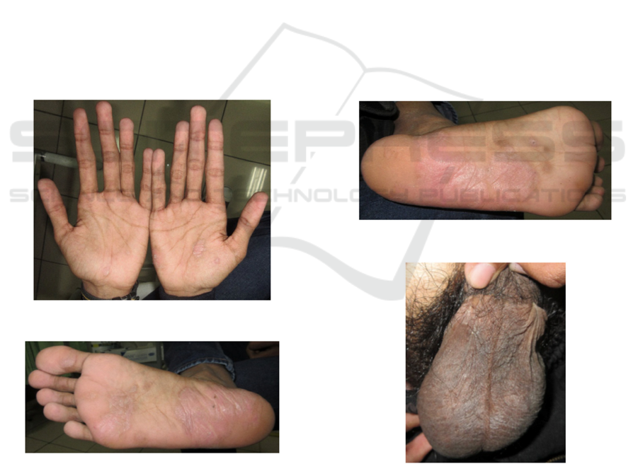
Figure 1: Red patches on both of palms.
Figure 3: Red Patches on right sole.
(Wells et al., 2017). Although anterior uveitis is
common in immunocompetent individuals, posterior
segment involvement has been described more
commonly in HIV-infected individuals with
advanced stages of immunosuppression. Ocular
syphilis may be complicated by central nervous
system involvement, thus investigation for
neurosyphilis should be considered especially for
patients with AIDS. Another study reported a high
proportion of neurosyphilis in HIV-infected patients
with syphilitic uveitis; 7 of 9 patients (77.8%)
demonstrated CSF abnormalities (Herbort, 2011).
Even with no evidence of neurosyphilis, syphilitic
retinitis should be treated with the same regimen for
neurosyphilis; a 10–14 day course of intravenous
penicillin is recommended. Syphilitic retinitis
generally responds well to penicillin therapy with
good visual outcome (Dutta et al., 2017). Another
weakness in our report was this patient wasn’t given
any treatment for his retinitis. If it was caused by
syphilis, we should give the treatment as the same as
neurosyphilis, i.e. intravenous aqueous crystalline
penicillin G 18–24 million units per day for 10 to 14
days (Dutta et al., 2017).
Figure 2: red patches on left sole.
Figure 4: Red Patches on scrotum.
Secondary Syphilis with Suspected Retinitis Syphilis in HIV-infected Patient: A Case Report
463

4 CONCLUSION
We report a case of HIV-infected male with
secondary syphilis lesions in both of palms and
soles, and the scrotum. The lesions improved after
being given Benzatin Pennicilin G 2,4 million IU
intramuscularly. Complaints of visual disturbance,
especially in the left eye, was assesed as retinitis
syphilis by ophtalmologist department. It’s
important for a clinician to suspected ocular syphilis
in a syphilis patient with visual complaint, as ocular
syphilis could be happened in any syphilis stadium.
REFERENCES
Doris, J. P., Saha, K., Jones, N. P., & Sukthankar, A.,
2006. Ocular syphilis: the new epidemic. Eye, 20(6),
703-705.
Dutta Majumder, P., Chen, E. J., Shah, J., Ching Wen Ho,
D., Biswas, J., See Yin, L., ... & Agrawal, R., 2017.
Ocular Syphilis: An Update. Ocular immunology and
inflammation, 1-9, Available from:
https://doi.org/10.1080/09273948.2017.1371765..
Heiden, D., Ford, N., Wilson, D., Rodriguez, W. R.,
Margolis, T., Janssens, B., ... & Sabapathy, K., 2007.
Cytomegalovirus retinitis: the neglected disease of the
AIDS pandemic. PLoS medicine, 4(12), 1845–1851.
Herbort, C.P., 2011. Ocular Syphilis. Retina Inflammation
Eye Disease, 1–7.
Hughes, E. H., Guzowski, M., Simunovic, M. P., Hunyor,
A. P., & McCluskey, P., 2010. Syphilitic retinitis and
uveitis in HIV‐positive adults. Clinical &
experimental ophthalmology, 38(9), pp. 851-856.
Indriatmi, W., 2017. Infeksi Menular Seksual dengan
Penyebab Bakteri - Sifilis. In: Infeksi Menular
Seksual. 5th ed. Jakarta: Fakultas Kedokteran
Universitas Indonesia, p. 103–29.
James, W.D., Berger, T.G. E.D., 2016. Syphilis, Yaws,
Bejel, and Pinta. In: Andrew’s Diseases of the Skin :
Clinical Dermatology. 18th ed. Philadelphia: Elsevier
Inc. p. 18–44.
Matsuo, T., Mori, N., Furukawa, K., & Furukawa, K.,
2017. Ocular involvement with secondary syphilis in a
non-HIV infected man. IDCases, 10, 30-31, Available
from:
http://linkinghub.elsevier.com/retrieve/pii/S22142509
17301257..
Maves, R. C., Cachay, E. R., Young, M. A., & Fierer, J.,
2008. Secondary syphilis with ocular manifestations in
older adults. Clinical Infectious Diseases, 46(12),
e142-e145, Available from:
http://www.ncbi.nlm.nih.gov/pubmed/18462103..
Powell, C. A., & Carbo, A. R., 2018. The Great Imitator:
Visual Changes in a 37-Year-Old Man with HIV. The
American journal of medicine, 131(1), e19-e20,
Available from:
http://linkinghub.elsevier.com/retrieve/pii/S00029343
17308112..
Shinha, T., & Weaver, B. A., 2016. Necrotizing retinitis
due to syphilis in a patient with AIDS. IDCases, 6, 17-
19, Available from:
http://dx.doi.org/10.1016/j.idcr.2016.08.005..
Wells, J., Wood, C., Sukthankar, A., & Jones, N. P., 2018.
Ocular syphilis: the re-establishment of an old
disease. Eye, 32(1), 99, Available from:
http://www.nature.com/doifinder/10.1038/eye.2017.15
5.
Zambon, F., Silva, F. L. N., Cavalcante, A. F. D. S.,
Nakashima, Y., & Helal Jr, J., 2010. Syphilitic retinitis
and panuveitis simulating acute retinal necrosis: case
report. Arquivos brasileiros de oftalmologia, 73(3),
pp. 288-290.
RCD 2018 - The 23rd Regional Conference of Dermatology 2018
464
