
Numerous Basal Cell Carcinoma: A Case Report in a Suspected
Nonsyndromic Patient
Arinia Kholis Putri
1
, Adhimukti T. Sampurna
1
, Larisa Paramitha Wibawa
1
, Roro Inge Ade Krisanti
1
1
Departement of Dermatology and Venereology Faculty of Medicine Universitas Indonesia, Dr. Cipto Mangunkusumo
National General Hospital, Jakarta, Indonesia
Keywords: Basal cell carcinoma, numerous, multiple, nonsyndromic, nonmelanoma skin cancers
Abstract: Basal cell carcinoma (BCC) is the most common cancer in humans. BCC usually occurs as a solitary lesion
on sun-exposed areas. Various syndromes have been defined in which basal cell carcinoma exists in multiple
localizations in a single patient. Multiple BCC is often related with inherited conditions such as Gorlin
syndrome, Rombo syndrome, etc. Multiple BCC without history and clinical examination that are not
consistent with those syndromes is considered as nonsyndromic BCC. It is still unclear what environmental
and genetic factors contribute to the development of multiple nonsyndromic BCC. A case of numerous basal
cell carcinoma with multiple localizations without signs and symptoms which is classified as a syndrome is
described in this case report. Dermoscopic evaluation aids the diagnosis of and evaluation for people with
history of nonmelanoma skin cancer.
1 INTRODUCTION
BCC is the most frequent cutaneous neoplasm, with
slowly progressive nature and locally invasive
behavior, that arise from non-keratinizing cells within
the basal layer of the epidermis (Tcherney et al.,
2017; Carucci et al., 2012; Totonchy and Leffell,
2017). The malignancy accounts for approximately
75% of all nonmelanoma skin cancers (NMSC) and
almost 25% of all cancers diagnosed in the United
States (Carucci et al., 2012).
BCC usually occurs as a solitary lesion on sun-
exposed areas. There have been very few cases
reported in the literature so far of multiple BCC. This
condition occur mostly in several genetic syndromes,
but can also happens as a sporadic feature. Among
several syndromes, the most commonly known is
Gorlin syndrome or nevoid basal cell carcinoma
syndrome, an autosomal dominant trait caused by
PTCH gene mutation (Satolli et al., 2018). It is still
unclear what environmental and genetic factors
contribute to the development of multiple
nonsyndromic BCC (Tcherney et al., 2017). Risk
factors for BCC have been well characterized
(Totonchy and Leffell, 2017). The most significant
risk factor involved in the pathogenesis is ultraviolet
(UV) which triggers mutations in tumor suppressor
genes (Kim et el., 2017). Increasing age, male, white
race, exposure to ionizing radiation, arsenic, and
polyaromatic hydrocarbons have also been correlated
with higher rates of BCC (Totonchy and Leffell,
2017; Kim et al., 2017). Both genetic predisposition
and exposure to environmental risks are involved in
the pathogenesis of the malignant transformation in
BCC (Tcherney et al., 2017). The early detection and
eradication of these tumors are of importance for
treatment effectiveness and quality of life because
BCC could have an aggressive course and behavior
which can lead to severe disfiguration and destruction
(Tcherney et al., 2017; Carucci et al., 2012; Totonchy
and Leffell, 2017; Kim et al., 2017).
BCC comprising several lesions is not
uncommon, but nonsyndromic with numerous lesions
are rare entities. We present a patient, came with
numerous BCC without any signs and symptoms of a
specific syndrome as we discuss the potential
triggering risk and the further appropriate therapeutic
options.
2 CASE
A 56-year-old female patient with Fitzpatrick skin
type IV was admitted to the dermatological unit with
a complaint of three ulcerated pigmented skin lesions
with painless sensation on the face. The patient had
428
Putri, A., Sampurna, A., Wibawa, L. and Krisanti, R.
Numerous Basal Cell Carcinoma: A Case Report in a Suspected Nonsyndromic Patient.
DOI: 10.5220/0008159004280432
In Proceedings of the 23rd Regional Conference of Dermatology (RCD 2018), pages 428-432
ISBN: 978-989-758-494-7
Copyright
c
2021 by SCITEPRESS – Science and Technology Publications, Lda. All rights reserved

previously received Mohs micrographic surgery
(MMS) for a BCC lesion, with diameter
approximately 3 cm, on the upper lip at our hospital 4
years ago. But she was loss to follow up after the
treatment for the flap from plastic reconstruction
surgery unit. The patient was a scavenger and was
constantly exposed to direct sunlight for most of her
lifetime. She denied any history of sunburn, family
history of skin cancer, past history of radiotherapy,
and exposure to chemical substances including
arsenic.
She had hypertension, which is controlled with
medications and showed no regional
lymphadenopathy. The laboratory blood tests and
imaging diagnostic procedures did not revealed any
abnormalities nor signs for systemic involvement. No
symptom and sign such as mandibular cyst, palmar
and plantar pitting, skeletal abnormalities,
keratocystic odontogenic tumors, ectopic
calcification of the dura, macrocephaly, and mental
retardation.
On physical examination of the face, three
ulcerations were observed, one measuring
approximately 1 x 1 x 0,5 cm located on the tip of her
nose. The second and third one measuring
approximately 1,5 x 1 x 0,5 cm and 0,5 x 0,5 x 0,1 cm
were located on the right cheek. Those three lesions
showed pigmented pearl-like edges, bleeding surface,
and irregular borders. These lesions were in
accordance clinically, dermoscopically, and
histopathologically for BCC in different stages of
invasion (figure 1). We also found 21 pigmented
lesions on her face and head, also 54 pigmented
lesions were noted on her neck, which she did not
notice nor have any complaints. All of these lesions
have size ranging from 0,4 mm to 1,5 cm and
demonstrated similar clinical signs such as irregular
borders with pearl-like edges, with dermoscopic
appearance also suggestive for BCC (Figure 2b – g).
Electrosurgery was performed for more than
twenty superficial tumors on the neck. After the
procedure, the wound sites were re-examined using a
dermoscope to ensure there were no visible
pigmented lesions left. Furthermore, the patient is still
regularly come to our clinic to remove her remainder
BCC lesions gradually with excision and
electrosurgery. We plan to do periodic checkup for
early detection of NMSC after all the lesions are
removed.
3 DISCUSSION
BCC is the most common human cancer that usually
occurs as a single lesion. The vast majority of BCC
were located on the head and neck (Carucci et al.,
2012; Kim et al., 2017). Recent reports show that the
number of patients who develop more than one BCC
is increasing (Tcherney et al., 2017). According to the
clinical, dermoscopic, and histopathologic findings,
our patient was diagnosed as having multiple BCC
that mostly located on the scalp, nose, and neck.
Multiple BCCs are not uncommon as there is a 36%–
50% increased risk of development of additional
BCCs after the first lesion within 5 years (Kim et al.,
2017). The total number of NMSC is a risk factor for
recurrence of previous tumor, as well as for the
formation of new ones (Tcherney et al., 2017). This
patient also had a history of MMS for a large BCC on
her upper lip 4 years ago.
UV is thought to be the most important
environmental factor in its pathogenesis, although it
is not always possible to correlate the sites where
exposure is most intense to those where lesions are
most frequently found (Kobacas et al., 2010). An
Italian study indicated the important role of sunburns,
and therefore intense sun exposure, rather than that of
prolonged sun exposure to increase the risk of BCC
(Tcherney et al., 2017). Increasing age, Northern-
European ancestry, male, immunosuppression, and
arsenic exposure are the other established risk factors
(Carucci et al., 2012; Kim et al., 2017). Our patient
was 56 year-old female with Asian descendant.
Although she is a scavenger and was constantly
exposed to direct UV for most of her lifetime, she has
Fitzpatrick skin type IV which rarely burns and tans
easily, and denied any history of sunburn. Other
factors include exposure to ionizing radiation,
arsenic, and polyaromatic hydrocarbons, which
appear to be involved in mutations of regulatory
genes and alterations in immune surveillance
(Carucci et al., 2012; Kim et al., 2017). From history
taking this patient denied exposure of ionizing
radiation, arsenic, and polyaromatic hydrocarbons.
Numerous Basal Cell Carcinoma: A Case Report in a Suspected Nonsyndromic Patient
429
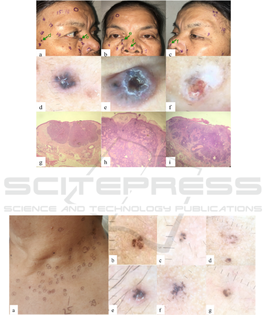
Figure 1. Preoperative photography shows that the lesions (circles) were located on the face (a-c). Dermoscopy appearance
showed ulceration, leaf-like structures, blue-black globules and dots, and arborizing vessels (d-f). Histopathology with
hematoxylin and eosin (H&E) staining with 40x magnification, showed the 3 lesions have basaloid epithelium with peripheral
palisade layer under an ulcerated epidermis, with artefactual cleft, and mucinous stroma deposit on the border and central
tumors (g-i)
Figure 2. Pigmented lesions (circles) were located on the neck (a). Dermoscopy appearance showed leaf-like structures, blue-
black dot, globules, and arborizing vessels (b-g).
Inherited conditions such as the nevoid BCC
syndrome (Gorlin syndrome), Rombo syndrome,
unilateral basal cell nevus syndrome, and Bazex
syndrome are common inherited conditions with
multiple BCC manifestations. Nonsyndromic but
hereditary multiple BCC have been reported in
literatures, but they were ruled out based on the
negative family history (Tcherney et al., 2017; Kim et
al., 2017). In our case, history and clinical
examination were not consistent with these
RCD 2018 - The 23rd Regional Conference of Dermatology 2018
430

syndromes that related to multiple BCC. Also, there
was no sign and symptom of the lesions at a younger
age, nor a positive family history. Thus, this case does
not fit into any of the syndromes seen with basal cell
carcinoma such as Gorlin syndrome or Bazex
syndrome. There was no history of exposure to
arsenic, irradiation, dry ice and no evidence of
keratoacanthoma or xeroderma pigmentosum in this
patient. However, considering the financial status of
the patients, we could not perform polymerase chain
reaction (PCR) assay to rule out this genotype.
Moreover, the treatment plan would not be affected
by the PCR result. Hence, we categorized our cases
as nonsyndromic and nonhereditary type of multiple
basal cell carcinomas worth mentioning.
In some literature, multiple BCC in one patient
increases the risk of recurrence and they often
develop new BCC with similar or different
histological appearance (Tcherney et al., 2017). As
also proven, not all instances of multiple BCC are due
to genetic syndromes. Multiple superficial BCCs
without associated anomalies are distinct from the
Gorlin syndrome and could be explained by an
autosomal dominant phenotype. Alternatively, this
nonsyndromic phenotype might have a polygenic
basis. Furthermore, a recent article has revealed that
multiple BCC can be also part of the BAP 1 mutation
(Satolli et al., 2018). We concluded that our patient
had a high number of basal cell carcinoma lesions
without a syndrome.
Despite the low metastatic potential, local tissue
destruction and disfigurement caused by the tumor
can be enormous if not completely eradicated by early
diagnosis and treatment (Tcherney et al., 2017). Most
basal cell carcinomas can be treated with any of a
number of treatment modalities, including
electrodessication and curettage, cryosurgery,
surgical excision, or MMS. While surgical
interventions such as MMS and surgical excision are
the standard of care and yield the highest cure rates,
the number of non-surgical interventions approved
for the treatment of BCC continues to expand
(Totonchy and Leffell, 2017). Standard surgical
excision with 4-mm margins is the recommended
treatment for BCC with non-aggressive histology,
size of less than 2 cm, and occurrence on low-risk
sites where tissue sparing is not critical (trunk and
extremities). BCC of the face demonstrates high rates
of incomplete excision, and greater efficacy has been
demonstrated using MMS as compared with standard
excision. MMS is recommended in cases of
aggressive histology, recurrent BCC, critical areas of
skin (head, neck, genitalia, hand/feet, nipples) and for
tumors of large size (more than 2 cm) (Totonchy and
Leffell, 2017; Fahradyan et al., 2017).
Current management options are numerous and
focus on tumor eradication while maximizing
cosmetic and functional capacity. The choice of
treatment depends on the tumor type, tumor location,
cost, recurrence rates, and potential cosmetic
disfigurement (Kocabas et al., 2010). Our patient was
treated with 3-4 mm margin excision and performing
histopathologic examination, for lesions that were
bigger than 1 cm on high risk area. But for
approximately 20 small superficial lesions less than 1
cm in size, located not on the high risk area, and
demonstrated with leaf-like structures and arborizing
vessels with the dermoscope, we performed
electrosurgery. After the procedure, the wound sites
were re-examined using a dermoscope to ensure that
there were no visible lesions left. Excision and
histopathology examination for all the BCC lesions
will not be cost-effective for this patient. The early
detection and eradication of these tumors are of
importance for treatment effectiveness and quality of
life (Kim et al., 2017). The patient were asked to
avoid sun-exposure as much as she could possible do.
We plan to do regular checkup for this patient for the
rest of her life to early detection of NMSC.
4 CONCLUSIONS
We have described a patient with multiple
nonsyndromic basal cell carcinoma and had
undergone Mohs micrographic surgery, wide
excision, and electrosurgery. The early detection and
eradication of these tumors are of importance for
treatment effectiveness and quality of life. Our case
illustrates the importance of diagnose and treatment
multiple basal cell carcinoma at early stage.
Performing dermoscopic evaluation will improve in
early detection of BCC.
REFERENCES
Carucci, J.A., Leffell, D.J., Peetersen, J.,2012. Basal cell
carcinoma. In: Goldsmith LA, Katz SI, Gilchrest BA,
Paller AS, Leffell DJ, Wolf K, eds. Fitzpatricks’s
dermatology in general medicine. 8th edition. New
York: McGraw-Hill, p.1294-303.
Fahradyan, A., Howell, A., Wolfswinkel, E., Tsuha, M.,
Sheth, P., & Wong, A., 2017. Updates on the
management of non-melanoma skin cancer (NMSC). In
Healthcare (Vol. 5, No. 4, p. 82). Multidisciplinary
Digital Publishing Institute.
Numerous Basal Cell Carcinoma: A Case Report in a Suspected Nonsyndromic Patient
431

Kim, D.H., Ko, H.S., Jun, Y.J., 2017. Nonsyndromic
multiple basal cell carcinomas, 18(3), pp. 191-196.
Kocabaş, E., Ermertcan, A. T., Bilaç, C., Bilaç, D. B., &
Temiz, P., 2010. Nonsyndromic multiple basal cell
carcinomas successfully treated with imiquimod 5%
cream. Cutaneous and ocular toxicology, 29(4),pp.
300-302.
Pellegrini, C., Maturo, M., Di Nardo, L., Ciciarelli, V.,
Gutiérrez García-Rodrigo, C., & Fargnoli, M., 2017.
Understanding the molecular genetics of basal cell
carcinoma. International journal of molecular sciences,
18(11), pp. 2485.
Satolli, F., Rovesti, M., De Felici, M. B., Zucchi, A.,
Pagliarello, C., & Feliciani, C., 2018. Multiple Basal
Cell Carcinoma Arising in a Verrucous Epidermal
Naevus: Clinical, Histological and Therapeutic
Observations. Acta dermato-venereologica, 98(1-2),
132-133.
Tchernev, G., Pidakev, I., Lozev, I., Lotti, T., & Wollina,
U., 2017. Multiple nonsyndromic acquired basal cell
carcinomas. Wiener Medizinische Wochenschrift,
167(5-6), pp. 134-138.
Totonchy, M., & Leffell, D., 2017. Emerging concepts and
recent advances in basal cell carcinoma.
F1000Research, 6, 1-10.
Verkouteren, J.A.C., 2015. Common variants affecting
susceptibility to develop multiple basal cell carcinomas.
Journal of Investigative Dermatology, 135, 2135–2138.
RCD 2018 - The 23rd Regional Conference of Dermatology 2018
432
