
The Diagnostic Approach to Cutaneous Metastases of
Adenocarcinoma of the Prostate: A Case Report
Peppy Fourina
1
, Riesye Arisanty
2
, Sri Adi Sularsito
1
, Lili Legiawati
1
, Rahadi Rihatmadja
1
, Shannaz
Nadia Yusharyahya
1
1
Department of Dermatology and Venereology, Faculty of Medicine Universitas Indonesia Dr. Cipto Mangunkusumo
National General Hospital, Jakarta, Indonesia
2
Department of Pathology, Faculty of Medicine Universitas Indonesia Dr. Cipto Mangunkusumo National General
Hospital, Jakarta, Indonesia
Keywords: adenocarcinoma of the prostate, cutaneous metastasis, PSA, AMACR
Abstract: The incidence of prostate cancer has increased all over the world, likely due to better detection methods. It
becomes one of the most common cancers among elderly men. Almost all cases of prostate cancer are
adenocarcinoma. The most common sites for metastases are lymph nodes and bone. We present a case of a
67-year-old man with an ulcer on his lower back for the past 6 months. Three years ago, he was diagnosed
with poorly differentiated adenocarcinoma of the prostate with bone metastases. On physical examination,
there was a solitary, 2x2x1 cm ulcer with elevated border and yellowish slough. The ulcer was painful and
surrounded by erythematous and indurated tissues. There is no inguinal lymphadenopathy. Despite
antibiotics, conventional and modern wound dressing, no significant improvement was noted. Considering
the history of malignancy, skin biopsy was performed. Histopathological examination revealed scattered
atypical cells around the blood vessels that stained positively with prostate-specific antigen (PSA) and
alpha-methylacyl-CoA racemase (AMACR), confirming cutaneous metastases of AP. Metastasis of AP to
the skin is rare, and indicates a poor prognosis. Early recognition of cutaneous spread manifesting as ulcer
that does not respond to proper treatment in the background of malignancy is important.
1 INTRODUCTION
Based on the GLOBOCAN 2012 statistics, prostate
cancer is the third most common cancer in
Indonesian men after lung and colorectal, with an
estimated incidence of 13.663 (WHO, 2012). About
all cases of prostate malignancies are
adenocarcinoma (Crawford, 2009). Adenocarcinoma
of the prostate (AP) favors pelvic lymph nodes and
bone for its metastases, while cutaneous metastases
are distinctly rare (Patne et al., 2015; Tengue et al.,
2017). Cutaneous metastases of AP usually appear
as nodule or papule in the abdominal, inguinal
region, anterior thigh, and near the umbilicus
(Pistone et al., 2013; Alcaraz et al., 2012). Although
uncommon, cutaneous metastases usually occur late
and indicate grave prognosis (Patne et al., 2015;
Wang et al., 2008). Dermatologists have to be aware
of the various clinical lesions of cutaneous
metastases because early diagnosis and prompt
management will result in favorable prognosis. We
report a rare case that illustrated the diagnostic
approach to cutaneous spread of AP, presented as a
chronic ulcer on the lower back.
2 CASE
A 67-year-old male patient was consulted from
Internal Medicine Department with a chronic ulcer of
6-month duration located on his lower back. Initially,
it began as pruritic papules that later became
ulcerated. No previous medications were applied.
Past history was noted for diabetes mellitus,
hypertension, and prostatic cancer.
Three years before, he complained worsened
lower back pain followed with weakness on lower
extremities; physical and imaging examination
concluded spinal cord compression due to metastasis.
Based on a markedly elevated prostate-specific
antigen (PSA) and imaging studies, the primary
cancer is adenocarcinoma of the prostate. Prostate
424
Fourina, P., Arisanty, R., Sularsito, S., Legiawati, L., Rihatmadja, R. and Yusharyahya, S.
The Diagnostic Approach to Cutaneous Metastases of Adenocarcinoma of the Prostate: A Case Report.
DOI: 10.5220/0008158904240427
In Proceedings of the 23rd Regional Conference of Dermatology (RCD 2018), pages 424-427
ISBN: 978-989-758-494-7
Copyright
c
2021 by SCITEPRESS – Science and Technology Publications, Lda. All rights reser ved
biopsy confirmed a Gleason score of 4+5=9,
associated with poorly differentiated prostate
adenocarcinoma. Whole abdomen CT scan showed
no paraaorta, parailliac, and inguinal
lymphadenopathy. After bilateral subcapsular
orchiectomy as an androgen deprivation therapy, his
PSA dropped from 215 ng/ml to 0.03 ng/ml. He also
had 30 radiotherapy session, followed by bone
directed therapy using bisphosphonate injections for
24 months.
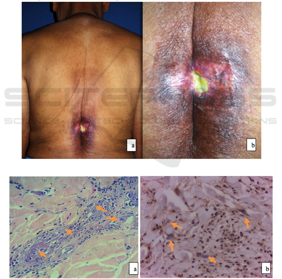
The patient was moderately ill. A solitary, 2x2x1
cm ulcer was found on his lower back corresponding
with lumbar 1-2. It had elevated border and
yellowish slough as its base. The ulcer was painful
and surrounded by erythematous and indurated
tissues (figure 1). No lymphadenopathy was noted.
He was treated as bacterial ulcer with normal saline
dressing and topical fucidic acid 2% cream.
When no significant improvement was achieved
after 2 weeks, the ulcer was treated with cutimed
sorbact gel and cutimed siltech. Still, it has not
healed by two weeks. Cutaneous metastases from PA
was considered along with the differential diagnoses
of bacterial ulcer and squamous cell carcinoma. We
performed bacterial culture and incisional biopsy.
Bacterial culture was positive for Pseudomonas
aeruginosa, sensitive to Levofloxacine.
Figure 1. (a)(b). Solitary ulcer at the lower back
Figure 2. Histopathological and immunohistochemistry examination: (a). scattered atypical cells (H&E 400x), (b). the cells
were positive for AMACR stain
The Diagnostic Approach to Cutaneous Metastases of Adenocarcinoma of the Prostate: A Case Report
425

Table 1. Sensitivity, specificity, positive predictive value, and negative predictive value for PSA and AMACR highly
specific for AP
15
Prostatic
markers
Sensitivity Specificity Positive predictive
value (PPV)
Negative predictive
value (NPV)
PSA 100% 90,6% 89,5% 100%
AMACR 66,7% 77,3% 86,1% 77,3%
Histopathological examination revealed scattered
atypical cells around the blood vessels, suggesting
metastasis (figure 2a). Immunohistochemistry was
positive for PSA and AMACR (figure 2b). These
finding strongly supported the diagnosis of
cutaneous spread of AP.
3 DISCUSSION
Cutaneous metastasis is defined as a spread of
malignant cells from a primary cancer to the skin.
The exact mechanism of metastasis remains unclear.
Malignant cells may spread beyond the prostate
through some hypotheses, such as direct infiltration,
lymphatic, hematogenous, or combination of these
routes (Rattanasirivilai et al., 2011; Rodriguez-Lojo
et al., 2016). A meta-analysis study by Krathen et al.
(2003) found the overall incidence of cutaneous
metastasis in 2003 was 5.3% from 20,380 cases. The
most common tumor which spread to the skin was
breast cancer, with an incidence of 24%. Skin
metastates from AP are rare, with an incidence of
0.7% (Crawford, 2009; Stanko et al., 2007).
AP favors bones and lymph nodes for its
metastases (Tengue et al., 2017; Brown et al., 2014).
The result of whole spine MRI and bone scan
revealed osteoblastic lesions as metastases to spine,
cranium, costae, sacroiliac, and ischium. For this
bone metastases, the patient had 30 radiotherapy
session and followed by bone directed therapy with
bisphosphonate injections once in a month for 24
months. Whole abdomen CT scan showed no para
aorta, para iliac, and inguinal lymphadenopathy.
Cutaneous metastases of PA have more than one
of clinical morphology, they most frequently appear
as nodules or papules in the abdominal wall,
inguinal region, anterior thigh, and near the
umbilicus as Sister Mary Joseph nodules (Mak et al.,
2014; Wang et al., 2008). They usually
asymptomatic and rarely ulcerated (AbAziz et al.,
2013).
Immunohistochemistry might aid to confirm the
origin tumor of cutaneous metastases (Rodriquez et
al., 2016). In our patient, the specimen was positive
for PSA and AMACR, strongly supported the
diagnosis of skin metastases from the patient’s AP.
PSA and AMACR staining is widely used to identify
metastasis of AP. PSA is a serine protease member
of the human glandular kallikrein family which is
highly specific for AP, because it is synthesized in
the prostate ductal and acinar epithelium. AMACR
is a peroxisomal and mitochondrial enzyme that
plays a key role in beta oxidation of fatty acid. It is
identified as being overexpressed in AP cells (Oh et
al., 2016).
4 CONCLUSION
Cutaneous spread of AP is rare, but it happens in
0.7% of all skin metastases cases. Patient’s complain
and physical appearance can vary from one patient
to another. Malignancy should always be kept in
mind when working up on diagnosis of unhealed
skin lesion after adequate local and systemic
treatment done. The combination of clinical history,
physical examination, laboratory tests, routine
pathology, and immunohistochemistry assay can
provide enough information for a diagnosis of
metastatic adenocarcinoma of the prostate.
REFERENCES
AbAziz, A., Mahaletchumy, T., & Chung, J. K., 2013.
Skin Manifestation of Unsuspecting Prostate Cancer
Detected by 18F-FDG PET/CT Performed To Assess
Underlying Multiple Myeloma. Nuclear medicine and
molecular imaging, 47(4), pp.285-288.
Alcaraz, I., Cerroni, L., Ruetten, A., Kutzner, H., &
Requena, L., 2012. Cutaneous metastases from
internal malignancies: a clinicopathologic and
immunohistochemical review. The American journal
of dermatopathology, 34(4), pp. 347-393.
Brown, G. T., Patel, V., & Lee, C. C. R., 2014. Cutaneous
metastasis of prostate cancer: a case report and review
of the literature with bioinformatics analysis of
multiple healthcare delivery networks. Journal of
cutaneous pathology, 41(6),pp. 524-528.
Crawford, E. D., 2009.. Understanding the epidemiology,
natural history, and key pathways involved in prostate
cancer. Urology, 73(5), S4-S10, . Available from:
http://dx.doi.org/10.1016/j.urology.2009.03.001.
RCD 2018 - The 23rd Regional Conference of Dermatology 2018
426

Krathen, R. A., Orengo, I. F., & Rosen, T., 2003.
Cutaneous metastasis: a meta-analysis of
data. Southern medical journal, 96(2), 164-168.
Mak, G., Chin, M., Nahar, N., & De Souza, P., 2014.
Cutaneous metastasis of prostate carcinoma treated
with radiotherapy: a case presentation. BMC research
notes, 7(1), pp. 505.
Oh, W. J., Chung, A. M., Kim, J. S., Han, J. H., Hong, S.
H., Lee, J. Y., & Choi, Y. J., 2016. Differential
immunohistochemical profiles for distinguishing
prostate carcinoma and urothelial carcinoma. Journal
of pathology and translational medicine, 50(5), pp.
345–354, Available from:
https://doi.org/10.4132/jptm.2016.06.14.
Patne, S. C., Naik, B., Patnaik, P., & Trivedi, S., 2015.
Cutaneous metastases of prostatic
adenocarcinoma. Journal of Cytology/Indian Academy
of Cytologists, 32(2), 121-123, Available from:
http://www.pubmedcentral.nih.gov/articlerender.fcgi?
artid=4520044&tool=pmcentrez&rendertype=abstract
Pistone, G., Pistone, A., Aricò, M., & Bongiorno, M. R.,
2013. Multiple cutaneous metastases in the chest from
prostatic carcinoma. Case reports in
dermatology, 5(1), 27-30, Available from:
http://www.pubmedcentral.nih.gov/articlerender.fcgi?
artid=3719122&tool=pmcentrez&rendertype=abstract.
Rattanasirivilai, A., Kurban, A., Lenzy, Y. M., & Yaar, R.,
2011. Cutaneous metastasis of prostatic
adenocarcinoma: a cautionary tale. Journal of
cutaneous pathology, 38(6), 521-524.
Rodríguez-Lojo, R., Castiñeiras, I., Rey-Sanjurjo, J. L., &
Fernández-Díaz, M. L., 2016. Distant Cutaneous
Metastases of Prostate Cancer: A Report of 2
Cases. Actas Dermo-Sifiliográficas (English
Edition), 107(7), pp. 52-56, Available from:
http://www.sciencedirect.com/science/article/pii/S000
1731016000697.
Stanko, C., Grandinetti, L., Baldassano, M., Mahmoodi,
M., & Kantor, G. R., 2007. Epidermotropic metastatic
prostate carcinoma presenting as an umbilical nodule-
Sister Mary Joseph nodule. The American journal of
dermatopathology, 29(3), pp. 290-292.
Tengue, K., Kpatcha, T., Sewa, E., Sikpa, K., Botcho, G.,
Leloua, E., ... & Dosseh, E., 2017. Prostate cancer
revealed by skin metastasis: A case report in black
African man. African Journal of Urology, 23(4), pp.
204-207, Available from:
http://dx.doi.org/10.1016/j.afju.2016.10.003..
Wang, S. Q., Mecca, P. S., Myskowski, P. L., & Slovin, S.
F., 2008. Scrotal and penile papules and plaques as the
initial manifestation of a cutaneous metastasis of
adenocarcinoma of the prostate: case report and
review of the literature. Journal of cutaneous
pathology, 35(7), 681-684.
World Health Organization., 2012. GLOBOCAN 2012:
estimated cancer incidence, mortality and prevalence
worldwide in 2012 [Internet]. [cited 2018 Feb 12].
Available from:
http://globocan.iarc.fr/Pages/fact_sheets_population.as
px
The Diagnostic Approach to Cutaneous Metastases of Adenocarcinoma of the Prostate: A Case Report
427
