
A Very Rare Case of Dissecting Cellulitis of the Scalp in an
Indonesian Man
Rizky Lendl Prayogo
1
, Lusiana
1
, Sri Linuwih Menaldi
1
, Sondang P. Sirait
1
, Eliza Miranda
1
1
Department of Dermatology and Venereology Faculty of Medicine Universitas Indonesia / Dr. Cipto Mangunkusumo
National General Hospital, Jakarta, Indonesia
Keywords: dissecting cellulitis of the scalp, dissecting folliculitis, follicular occlusion tetrad, diagnosis, isotretinoin
Abstract: Dissecting cellulitis of the scalp (DCS), also known as dissecting folliculitis, perifolliculitis capitis abscedens
et suffodiens (PCAS), or Hoffman’s disease, is a primary neutrophilic cicatricial alopecia without clear
etiology. Along with hidradenitis suppurativa, acne conglobata, and pilonidal cyst, they were recognized as
‘follicular occlusion tetrad’. A 43-year-old Indonesian man presented to our department with four years
history of persistent, slightly painful subcutaneous nodules, abscesses, and sinuses that discharged purulent
exudate on vertex and occipital scalp. There was also associated patchy alopecia. He had severe acne during
his adolescence to early adulthood. Trichoscopic evaluation showed yellowish and whitish area lacking of
follicular openings. Histopathological examination showed follicular occlusion, dilatation, and rupture with
mixed inflammatory infiltrates, mainly neutrophils. The diagnosis of DCS was confirmed by clinical,
trichoscopic, and histopathological examinations. Isotretinoin 20 mg daily was given to normalize the
follicular keratinization. Considering its very rare occurrence in an Indonesia man, this case was reported to
emphasize the diagnosis of DCS.
1 INTRODUCTION
DCS, also known as dissecting folliculitis, PCAS,
or Hoffmann’s disease, is a very rare primary
neutrophilic citatricial alopecia without clear etiology
(Badaoui et al., 2016). It was first described by
Spitzer in 1903 who termed the disease “dermatitis
follicularis capitis et perifollicullaris
conglobata”(Spitzer, 1903). Hoffman then termed it
as PCAS in 1908 (Hoffman, 1908). Since the first
description to 2014, the details of only 72 patients
have been published.1 DCS has been considered to be
a part of ‘follicular occlusion triad’, along with
hidradenitis suppurativa and acne conglobata (Otberg
& Shapiro, 2012). Other literature included pilonidal
cyst and altogether they were recognized as ‘follicular
occlusion tetrad’ (Badaoui et al., 2016). Its chronic
relapsing courses resulted in cicatricial alopecia with
hypertrophic or keloidal scars formation (Otberg &
Shapiro, 2012) . Various treatments, such as systemic
antibiotics, intralesional corticosteroid, oral
prednisolone, and isotretinoin showed clinical
improvement (Otberg & Shapiro, 2012; Scheinfeld,
2014). In refractory and more advanced cases, anti
tumor necrosis factor alpha (anti-TNF α) and surgery
should be considered (Otberg & Shapiro, 2012;
Scheinfeld, 2014). Considering its low prevalence in
Indonesia, we are intrigued to report a case
emphasizing the diagnosis of DCS.
2 CASE
A 43-year-old Indonesian man presented to our
department with four years history of persistent,
slightly painful lumps that discharged purulent
material during compression on his vertex and
occipital scalp. There was also associated patchy
alopecia. Those lumps firstly appeared as small red
bumps resembling folliculitis, which then enlarged.
He experienced severe facial acne during his
adolescence to early adulthood. He regularly washed
his hair daily and got his hair cut with scissors
monthly. He often wears a hat, which was washed
once every one or a couple of weeks. There was no
familial history with the same complaints and
previous mechanical trauma. On physical
examination, there were multiple flesh-colored
subcutaneous nodules that fluctuated, sinuses that
discharged purulent exudate, and patchy alopecia.
398
Prayogo, R., Lusiana, ., Menaldi, S., Sirait, S. and Miranda, E.
A Very Rare Case of Dissecting Cellulitis of the Scalp in an Indonesian Man.
DOI: 10.5220/0008158303980401
In Proceedings of the 23rd Regional Conference of Dermatology (RCD 2018), pages 398-401
ISBN: 978-989-758-494-7
Copyright
c
2021 by SCITEPRESS – Science and Technology Publications, Lda. All rights reserved
These lesions were slightly painful (VAS 2-3). The
surrounding tissues were neither erythematous nor
edematous. We also did not observe any tufted hairs.
Trichoscopic evaluation showed yellowish and
whitish area lacking of follicular openings.
Microscopic examination with KOH 20% from the
alopecia patch revealed no fungal elements. Bacterial
culture from the discharge and skin tissues were done
and showed growth of Staphylococcus epidermidis,
which was still sensitive to various antibiotics.
Histopathological examination showed follicular
occlusion, dilatation, and rupture with mixed
inflammatory infiltrates, mainly neutrophils. Based
on the clinical, trichoscopic, and histopathological
examinations, the diagnosis of DCS was confirmed.
Isotretinoin 20 mg daily was then initiated.
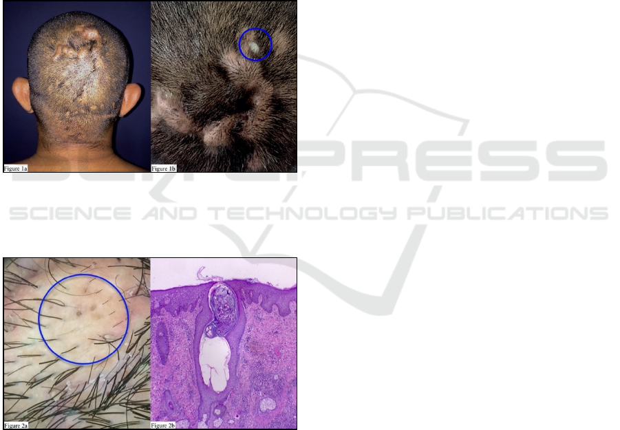
Figure 1. Clinical lesions: A: Fluctuating flesh-colored
nodules in vertes-occipital scalp area; B: draining sinuses
with purulent discharge.
Figure 2. Trichoscopic and histological examination. A:
Yellowish and whitish are lacking of follicular openings; B:
Follicular occlusion and dilatation (H&E40x).
3 DISCUSSION
Dissecting cellulitis of the scalp is a very rare disease
characterized by inflammative nodules, abscesses,
and sinuses, which may progress into scarring
alopecia (Segurado-Miravalles et al., 2017). Along
with hidradenitis suppurativa, acne conglobata, and
pilonidal cyst, DCS forms the follicular occlusion
tetrad (Badaoui et al., 2016;Segurado-Miravalles et
al., 2017). These diseases shared similar
etiopathogenesis involving hyperkeratosis, follicular
occlusion, and subsequent inflammation (Segurado-
Miravalles et al., 2017). Not all patients presented all
these four diseases (Badaoui et al., 2016). A
retrospective study of 51 patients showed that 12%
patients were also presented with hidradenitis
suppurativa, 16% with acne conglobata, and 4% with
coexisting hidradenitis suppurativa and acne
conglobata (Badaoui et al., 2016). No patients were
identified as having pilonidal cyst (Badaoui et al.,
2016). Mechanical trauma as a predisposing factor
was only found in five patients (Badaoui et al., 2016).
Subjective complains are pain or itch (Badaoui et al.,
2016). Eighty six percent patients progressed into
chronic disease (Badaoui et al., 2016). The observed
abscesses were usually sterile (Gaopande et al.,
2015). Bacterial infection can occur secondarily
during the course (Gaopande et al., 2015).
Microorganisms cultured in reported cases include
Pseudomonas species, Staphylococcus epidermidis,
Prevotella intermedia, Peptostreptococcus
asaccharolyticus, and Proprionibacterium acnes
(Gaopande et al., 2015). DCS is predominantly found
in African American men between 20 and 40 years
(Gaopande et al., 2015). Although it rarely affected
Asian, it has been reported in other Asian ethnicities
(Chinese and Indian) (Gaopande et al., 2015; Qi et al.,
2014). A retrospective study by Badaoui et al Badaoui
et al., 2016). showed that the mean age was 26,6
years, with wider range of age between 15-62.
Gaopande et al (2016) even reported the occurrence
of this disease in a 7-year-old boy. A multicenter
study which was conducted in four hospitals in Spain
also reported the occurrence in a 23-year-old Asian
woman (Segurado-Miravalles et al., 2017). The
pathophysiology of DCS remains unclear (Badaoui et
al., 2016). The young age of onset, the occurrence in
patients with dark phototype, and the cases of familial
DCS suggest a genetic predisposition (Badaoui et al.,
2016). The predominance of patients with dark
phototype raises the question of the role of hair type,
which is often coarse and frizzy, as well as traumatic
factor, such as hair shaving (Badaoui et al., 2016).
The male dominance and vertex as the predilection
A Very Rare Case of Dissecting Cellulitis of the Scalp in an Indonesian Man
399

area could also suggest a hormonal risk factor
(Badaoui et al., 2016). Finally, commensal bacteria
may play an essential role as alloantigens in the
pathophysiology of DCS (Badaoui et al., 2016). The
loss of immune tolerance to these alloantigens may
lead to an inflammatory reaction (Badaoui et al.,
2016; Scheinfeld, 2014). To the best of authors’
knowledge, this is the third case of DCS in an
Indonesian man. Although unpublished, Sirait (2015)
reported the occurrence of DCS associated with
hidradenitis suppurativa in 2015. Rahman et al.
(2017) then reported a case of follicular occlusion
tetrad in 2017. This patient is a 43-year-old
Indonesian man with straight hairs and no associated
mechanical trauma. There was also no familial
history with the same complaints. The signs and
symptoms of patient reported in this report are well
suited with DCS despite the lack of associated
predisposing factors except male gender. Other
explanations supporting the diagnosis of DCS are
listed below:
1. Anamnesis: lumps that discharged purulent
material on the predilection areas. History of
severe acne during his adolescence to early
adulthood.
2. Physical examination: observed subcutaneous
nodules, abscesses, and sinuses that
discharged purulent exudate.
3. Trichoscopic examination: yellowish and
whitish area lacking of follicular openings.
These were consistent with trichoscopic
findings explained by Laccarubba et al (2017).
4. Histopathological examination: follicular
occlusion, dilatation, and rupture with mixed
inflammatory infiltrates, mainly neutrophils.
Multiple hair shaft fragments are evident in
pilonidal cyst (Calonje et al., 2012). This
feature is not observed, so that the
histopathology is more suited with DCS than
pilonidal cyst.
DCS must be distinguished with other diseases
involving scalp (Vasant et al., 2014). The tendency of
DCS to cause fluctuating nodules and sinus tracts
helps to distinguish it from acne keloidalis nuchae
(AKN) (Vasant et al., 2014). Folliculitis decalvans,
another differential diagnosis, is characterized by
tufted folliculitis, in which multiple hair tufts emerge
from dilated follicular orifices (Vasant et al., 2014).
Various therapeutic strategies were reported
successful in treating DCS, such as systemic
antibiotics (minocycline, tetracycline, cloxacillin,
erythromycin, cephalosporin, or clindamycin),
intralesional corticosteroid, and oral prednisolone
(Otberg & Shapiro, 2012). The benefits of systemic
antibiotics were considered to be their anti-
inflammatory effects rather than antibacterial (Otberg
& Shapiro, 2012). Isotretinoin 0,5-1 mg/kg/day has
shown prolonged remission (Otberg & Shapiro,
2012). The mechanisms of action of isotretinoin are
normalizing follicular keratinization and reducing the
aberrant immune responses (Scheinfeld, 2014). Anti-
TNF α may be used when isotretinoin fails
(Scheinfeld, 2014). It can also defer the need of a
surgical treatment (Scheinfeld, 2014). Incision and
drainage may be done to painful and resistant nodules
(Scheinfeld, 2014). Marsupialization with curettage
of the cyst wall and total scalp excision followed by
split-thickness skin grafting have been reported, but
these surgical procedures should only be done for
extreme and refractory cases (Otberg & Shapiro,
2012). Badaoui et al. (2016) reported that 78%
patients receiving systemic antibiotics (doxycycline,
pristamycin, rifampicin, or a combination of several
antibiotics) showed moderate improvement.
However, the disease was relapsed in all of those
patients after antibiotic cessation (Badaoui et al.,
2016). Seventy one percent patients had received
systemic retinoid and almost all (92%) showed
complete remission after 3 months (Badaoui et al.,
2016). Isotretinoin is considered as the first line
therapeutic option for DCS (Badaoui et al., 2016;
Fransisco et al., 2017). The efficacy of isotretinoin in
treating DCS has been published (Marquis et al.,
2017). However, the optimal dose, duration of
therapy, and combination with other agents have not
been fully elucidated (Marquis et al., 2017). Marquis
et al.(2017) reported an excellence therapeutic
response after 4 months course of isotretinoin at 0,27
mg/kg/day. Considering the possibility of developing
side effects given at higher dose, this patient received
isotretinoin 20 mg daily (equal to 0,25 mg/kg/day).
He has been receiving isotretinoin for the past two
months and showed improvement.
4 CONCLUSIONS
DCS is a very rare case in Indonesia and should be
recognized as one of differential diagnosis when
seeing nodules, abscesses, and sinuses on the scalp.
The diagnosis can be confirmed by clinical,
trichoscopic, and histopathological examinations.
The therapy can be started immediately after the
diagnosis has been confirmed.
RCD 2018 - The 23rd Regional Conference of Dermatology 2018
400

REFERENCES
Badaoui, A., Reygagne, P., Cavelier‐Balloy, B., Pinquier,
L., Deschamps, L., Crickx, B., & Descamps, V., 2016.
Dissecting cellulitis of the scalp: a retrospective study
of 51 patients and review of literature. British Journal
of Dermatology, 174(2), pp. 421-423.
Calonje, E., Brenn, T., Lazar, A., McKee, P.H., 2012.
Pilonidal sinus. In: Calonje E, Brenn T, Lazar A,
McKee PH, editors. McKee's pathology of the skin with
clinical correlations. 4 ed. Edinburgh: Elsevier
Saunders, p. 1584-5.
Gaopande, V. L., Kulkarni, M. M., Joshi, A. R., & Dhande,
A. N., 2015. Perifolliculitis capitis abscedens et
suffodiens in a 7 years male: A case report with review
of literature. International journal of trichology, 7(4),
pp. 173-175.
Hoffman, E., 1908. Perifolliculitis capitis abscedens et
suffodiens. Dermatol Zeitschrift, 15, pp. 122-123.
Lacarrubba, F., & Micali, G., 2017. Regarding trichoscopy
of dissecting cellulitis of the scalp. Journal of the
American Academy of Dermatology, 76(6), e213.
Marquis, K., Christensen, L. C., & Rajpara, A., 2017.
Dissecting cellulitis of the scalp with excellent response
to isotretinoin. Pediatric dermatology, 34(4), e210-
e211.
Otberg, N., Shapiro, J., 2012. Dissecting folliculitis. In:
Goldsmith LA, Katz SI, Gilchrest BA, Paller AS,
Leffell DJ, Wolff K, editors. Fitzpatrick's Dermatology
in General Medicine. 1. 8 ed. New York: McGraw-Hill;
p. 999-1000.
Qi, S., Zhao, Y., Zhang, X., Li, S., Cao, H., & Zhang, X.,
2014. Clinical features of primary cicatricial alopecia in
Chinese patients. Indian Journal of Dermatology,
Venereology, and Leprology, 80(4), 306-312.
Rahman, A., Soenardi, A., Dharmawan, N., Priyambodo,
Ghaznawie, M., Widhiati, S., 2017. Establishment of
diagnose follicular occlusion tetrad. Journal of Clinical
and Experimental Dermatology Research, 8(3 (Suppl)),
55.
Scheinfeld, N., 2014. Dissecting cellulitis (Perifolliculitis
Capitis Abscedens et Suffodiens): a comprehensive
review focusing on new treatments and findings of the
last decade with commentary comparing the therapies
and causes of dissecting cellulitis to hidradenitis
suppura. Dermatology online journal, 20(5), 22692.
Segurado‐Miravalles, G., Camacho‐Martínez, F. M., Arias‐
Santiago, S., Serrano‐Falcón, C., Serrano‐Ortega, S.,
Rodrigues‐Barata, R., ... & Vañó‐Galván, S., 2017.
Epidemiology, clinical presentation and therapeutic
approach in a multicentre series of dissecting cellulitis
of the scalp. Journal of the European Academy of
Dermatology and Venereology, 31(4), e199-e200.
Sirait, S.P., 2015. Hair disorder. First National Basic and
Advanced Dermatopatholo gy Course. [Presentation].
In press 2015. Unpublished.
Spitzer, L., 1903. V. Dermatitis follicularis et
perifollicularisconglobata. Dermatology, 10(2), pp.
109-120.
Vasanth, V., & Chandrashekar, B. S., 2014. Follicular
occlusion tetrad. Indian dermatology online journal,
5(4), 491-493.
A Very Rare Case of Dissecting Cellulitis of the Scalp in an Indonesian Man
401
