
Paraneoplastic Syndrome Presenting As Giant Porokeratosis in A
Patient with Nasopharyngeal Cancer
Fitri Azizah, Sonia Hanifati, Sri Adi Sularsito,
Lili Legiawati, Shannaz Nadia Yusharyahya, Rahadi
Rihatmadja
Department of Dermatology and Venereology, Faculty of Medicine Universitas Indonesia / Dr. Cipto Mangunkusumo
National General Hospital
Keywords: porokeratosis, giant porokeratosis, paraneoplastic syndrome, nasopharyngeal
Abstract:
Giant porokeratosis is a rare condition in which the hyperkeratotic plaques of porokeratosis reach
up to 20 cm in diameter. Porokeratosis is characterized clinically by hyperkeratotic papules or
plaques with a thread-like elevated border. Although rare, porokeratosis has been reported in
conjunction with malignancies suggesting a paraneoplastic nature. Associated malignancies
reported were hematopoietic, hepatocellular, and cholangiocarcinoma. We report a case of giant
porokeratosis in a patient with nasopharyngeal cancer responding to removal of the primary
cancer by chemoradiotherapy.
1 INTRODUCTION
Porokeratosis is a chronic progressive disorder of
keratinization, characterized by hyperkeratotic
papules or plaques surrounded by a thread-like
elevated border corresponds to a typical histologic
hallmark, the cornoid lamella . O regan, 2012) There
are at least six clinical variants of porokeratosis
recognized with known genetic disorder.
1
Some
clinical variant of porokeratosis has been reported in
the setting of immunosuppressive conditions, organ
transplantation, use of systemic corticosteroids, and
infections, suggesting that impaired immunity may
be permissive in gentically predisposed individuals
(O regan, 2012; Cannavo, 2008) .
Porokeratosis has also been reported in
conjunction with malignancies, such as
hematopoietic, hepatocellular, and
cholangiocarcinoma (Torres, 2010; Wang,2017).
Association of porokeratosis and solid organ tumor
is rare, with only few cases have been reported in the
literature and none was reported in conjunction with
nasopharyngeal cancer. About 30% individuals with
eruptive disseminated porokeratosis had a recently
diagnosed malignancy, with mean of onset of 2.7
months preceding or following the diagnosis of
malignancy (Shoimer, 2014). Porokeratosis
associated with malignancy tended to improve or
regress completely after the treatment of
malignancy, suggestive of paraneoplastic syndrome.
2 CASE
Mr. SS, 68-year-old, was referred for evaluation of
pruritic, slightly erythematous plaques with raised,
hyperpigmented border of one and a half year
duration on the extensor surface of both legs. The
lesions shown minimal response to potent topical
corticosteroids and phototherapy given during the
last 8 months in another hospital. None of family
members had malignancies nor similar condition.
Past medical history was notable for
nonkeratinizing undifferentiated nasopharyngeal
carcinoma diagnosed in November 2016. While
staged for the extent of his carcinoma, topical
treatment and phototherapy was discontinued.
Secondly, he also had yellowish thickened palm and
soles since birth and later on, onychogryphosis
developed. Histopathology examination performed
20 years ago from the palm only revealed chronic
dermatitis. Interestingly, similar condition was also
present in his father, brothers, and son.
We sent the patient for biopsy just before he
started chemotherapy. Histopathology examination
revealed massive orthokeratosis, focal columnar
parakeratosis (cornoid lamella) with perivascular
Azizah, F., Hanifati, S., Sularsito, S., Legiawati, L., Yusharyahya, S. and Rihatmadja, R.
Paraneoplastic Syndrome Presenting as Giant Porokeratosis in a Patient with Nasopharyngeal Cancer.
DOI: 10.5220/0008158103890392
In Proceedings of the 23rd Regional Conference of Dermatology (RCD 2018), pages 389-392
ISBN: 978-989-758-494-7
Copyright
c
2021 by SCITEPRESS – Science and Technology Publications, Lda. All rights reserved
389
lymphocytes infiltrate. He then underwent external
radiation dosing 70 gray in 33 fraction and
accompanied with 150 mg carboplatin for 4 cycles.
One month after radiochemotherapy completion,
improvement of the lesions over limbs was noted,
and full resolution was achieved the following
month. However, the lesions over the palms and
soles did not change although treated by moisturizer
and keratolytic agent.
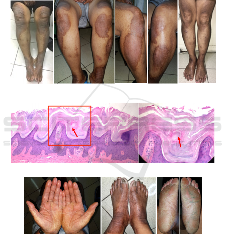
Figure 1. Plaques with elevated borders in November 2016 (A), during radiotherapy in May 2016 (B, C, D), and after
patient had completed chemoradiotherapy in August 2017 (E)
Figure 2. Massive orthokeratosis with column of parakeratosis, 100x (A), 400x (B) (hematoxyllin-eosin)
Figure 3. Yellowish hyperkeratotic plaques on both palms and soles with onychogryphosis
A
B
D
CE
A
B
RCD 2018 - The 23rd Regional Conference of Dermatology 2018
390

3 DISCUSSION
Paraneoplastic dermatoses are skin changes caused
by a malignancy but without intrinsically neoplastic
nature. Cutaneous manifestations of internal
malignancy is a diagnostic enigma both in
determining if it is paraneoplastic in nature and from
which organ the process originates. Once
established, the diagnosis lead to intiate a series of
efforts to locate the presence of the tumor, thereby
allowing prompt intervention. Diagnosis is more
difficult in cases of uncommon dermatosis which
only occasionally reported associated with
malignancy. (Owen,2012) According to Curth’s
postulates established an association between a skin
disease and malignancy, there are correlation
between onset of cutaneous disease and internal
malignancy, parallel course of both skin condition
and malignancy, specific type malignancy associated
with skin disease, statistical evidence of associated
malignancy in specific skin disease compared to
matched controls, and genetic link between a
syndrome with skin manifestations and internal
malignancy.(Shoimer,2014; Owen,2012)
The histopathologic patterns of porokeratosis
consists of hyperkeratotic epidermis, with a thin
column of poorly staining parakeratotic cells
(cornoid lamella), edematous underlying
keratinocytes and striking dermal lymphocytic
pattern. A cornoid lamella is characterized by
vertical column of parakeratosis, marked diminution
of granular layer at the point where the parakeratin
touches epidermal surface, and dyskeratosis and/or
vacuolization of the underlying cells of stratum
spinosum.(O Regan 2012). The epidermis in the
central portion of porokeratosis may be normal,
hyperplastic, or atrophic.
1
Cornoid lamella is not
pathognomonic for porokeratosis and may also be
found in other conditions such as viral warts,
seborrheic keratosis, solar keratosis, squamous cell
carcinoma in situ, lichen planus, and nevus
sebaceous. (O Regan 2012, Biswas, 2015). In our
case, we found hyperkeratotic epidermis, with a
vertical column of parakeratotic cells, cornoid
lamella. Granular layers underlying the vertical
column of parakeratosis were not diminished and
dykeratotic cells and edematous keratinocytes were
not found. Although not typical, clinical appearance
along with histopathological finding of cornoid
lamella support the diagnosis of porokeratosis.
Molecularly, the tumor suppressor proteins p53
and pRb are overexpressed in keratinocytes
immediately beneath and adjacent to the cornoid
lamella, although p53 mutations have not been
identified in porokeratosis. .(O Regan 2012) Other
reported cases of porokeratosis in conjunction with
solid tumor malignancies, share a common
characteristic of p53 protein in their carcinogenesis
(hepatocellular carcinoma, cholangiocarcinoma,
ovarian adenocarcinoma).(Cannavo, 2008) Study by
Lei et al in nasopharyngeal carcinoma stated
expression of tumor suppressor genes p16, p21 and
p53 with positive expression rate of 64.7%, 45.7%,
and 90.5%, respectively.(Lei X, 1999) Similar study
conducted in Istanbul also revealed similar result of
85.4% positive staining for p53 protein in
nasopharyngeal carcinoma patients.
9
There might be
correlation between malignancy and porokeratosis in
terms of p53 pathway, but more studies need to be
done. .(Shoimer, 2014)
In our case, the onset of skin disease preceeded
the finding of nasopharyngeal cancer for seven to
eight months prior. At time he developed skin
manifestations, he only complained of having a flu
followed by bloody runny nose around two or three
months after. It should be taken into account that
cancer might be clinically subtle before detection but
there was good clinical response to
chemoradiotherapy and full resolution of skin
manifestation, two months after he was cleared from
cancer. The patient was informed that reappearance
of skin manifestation could be a hint whether the
primary cancer strikes back and he should came for
reguler checkup to the otolaryngologist.
4 CONCLUSION
To our knowledge, there are no previous reports
associationg porokeratosis with nasopharyngeal
carcinoma. In our case, the clinical appearance, size,
and to some degree, the histopathological feature,
was not highly typical, making diagnosis difficult.
The skin eruption and malignancy ran a parallel
course and good clinical response was achieved after
removal of primary cancer thus we conclude our
case was a paraneoplastic syndrome.
REFERENCES
Agaoglu, F. Y., Dizdar, Y., Dogan, O., Alatli, C., Ayan, I.,
Savci, N., ... & Altun, M, 2004. P53 Overexpression
In Nasopharyngeal Carcinoma. In Vivo, 18(5), pp.
555-560.
Biswas, A., 2015. Cornoid lamellation revisited: apropos
of porokeratosis with emphasis on unusual
clinicopathological variants. The American Journal of
Paraneoplastic Syndrome Presenting as Giant Porokeratosis in a Patient with Nasopharyngeal Cancer
391

Dermatopathology, 37(2), pp. 145-155.
Cannavó, S. P., Borgia, F., Adamo, B., & Guarneri, B. ,
2008. Simultaneous development and parallel course
of disseminated superficial porokeratosis and ovarian
cancer: Coincidental association or true paraneoplastic
syndrome?. Journal of the American Academy of
Dermatology, 58(4), 657-660.
Lei X, Zhou Y, He X, Chen F. The expression of
suppressor gene p16, p21, and p53 in nasopharyngeal
carcinoma. Lin Chuang Er Bi Yan Hou Ke Za Zhi.
1999;13(9):406-408.
O’Regan, G.M., Irvine, A.D., 2012. Porokeratosis. In:
Goldsmith LA, Katz SI, Gilchrest BA, Paller AS,
Leffell DJ, Wolff K, eds. Fitzpatricks’s Dermatology
in General Medicine. Eight. McGraw-Hill, pp. 563-
568.Owen, C., 2012. Cutaneous manifestations of
internal malignancy. Indian Journal of Dermatoogyl,
4(4), 1-53.
Shoimer, I., Robertson, L. H., Storwick, G., & Haber, R.
M., 2014. Eruptive disseminated porokeratosis: a new
classification system. Journal of the American
Academy of Dermatology, 71(2), pp. 398-400.
Torres, T., Velho, G. C., & Selores, M., 2010.
Disseminated superficial porokeratosis in a patient
with cholangiocarcinoma: a paraneoplastic
manifestation?. Anais brasileiros de
dermatologia, 85(2), pp. 229-231.
Wang, Q., Wan, H., Liu, W., & Zhang, L., 2017.
Porokeratosis ptychotropica: A giant lesion in a
Chinese man. Australasian Journal of
Dermatology, 58(3), e149-e150.
RCD 2018 - The 23rd Regional Conference of Dermatology 2018
392
